The expression and function of thymosin beta 10
in tooth germ development
MAHO SHIOTSUKA
1,2,#, HIROKO WADA
1,#, TAMOTSU KIYOSHIMA
1,#, KENGO NAGATA
1,
HIROAKI FUJIWARA
1, MAKIKO KIHARA
1,2, KANA HASEGAWA
1,3, HIROTAKA SOMEYA
1,4,
ICHIRO TAKAHASHI
2and HIDETAKA SAKAI
1,*1Laboratory of Oral Pathology, 2Department of Orthodontics, 3Department of Endodontology and Operative Dentistry and 4Department of Removable Prosthodontics, Faculty of Dental Science, Kyushu University, Fukuoka, Japan ABSTRACT This study presents the expression pattern and functions of thymosin beta 10 (Tb10), a Tb4 homologue during the development of mouse lower first molars. An in situ signal of Tb10 was detected on embryonic day 10.5 (E10.5)-E15.5 mainly in dental mesenchymal cells as well as in dental epithelial cells, while Tb4 was expressed in dental epithelial cells. In the late bell stage, preodontoblasts with strong Tb10 expression and preameloblasts with strong Tb4 expression exhib-ited face-to-face localization, suggesting that an intimate cell-cell interaction might exist between preodontoblasts and preameloblasts to form dentin and enamel matrices. A strong Tb10 signal was found in odontoblasts in the lateral side of the dental pulp and in Hertwig’s epithelial root sheath, thus suggesting that Tb10 participates in the formation of the outline of the tooth root. An inhibi-tion assay using Tb10-siRNA in E11.0 mandibles showed significant growth inhibiinhibi-tion in the tooth germ. The Tb10-siRNA-treated E15.0 tooth germ also showed significant developmental arrest. The number of Ki67-positive cells significantly decreased in the Tb10-siRNA-treated mandibles. The cel-lular proliferative activity was also significantly suppressed in Tb10-siRNA-treated cultured mouse dental pulpal and epithelial cells. These results indicate that developmental arrest of the tooth germ might be caused by a reduction in cell proliferative activity. The stage-specific temporal and spatial expression pattern of Tb10 in the developing tooth germ is indicative of multiple functions of Tb10 in the developmental course from initiation to root formation of the tooth germ.
KEY WORDS:
thymosin beta 10, thymosin beta 4, tooth germ, development, knockdown assay
Introduction
Mammalian tooth development is mediated through sequen-tial and reciprocal epithelial-mesenchymal interactions similar to those observed in other organs, e.g. the hair, glands, lungs, kidneys, etc. A complex multi-step process of gene expression is involved in the early stage of tooth development (Pispa et al., 2003). Tooth germ development occurs via coordinated multi-step molecular interactions between the ectomesenchymal and ectodermal cells (Thesleff 2003). Tooth germ development also occurs via coordinated multi-step molecular interactions between endomesodermal and ectodermal cells (Mina 2001). There are many reports regarding the expression of various genes related to tooth morphogenesis (Cobourne and Sharpe 2003; Thesleff
www.intjdevbiol.com
*Address correspondence to: Hidetaka Sakai. Laboratory of Oral Pathology, Faculty of Dental Science, Kyushu University, 3-1-1 Maidashi, Higashi-ku, Fukuoka
812-8582, Japan. Tel: +81-92-642-6325. Fax: +81-92-642-6329. E-mail: [email protected]
# Note: The indicated authors have contributed equally to this paper.
Supplementary Material (two figures) for this paper is available at: http://dx.doi.org/10.1387/ijdb.120240hs
Accepted: 3 July 2013. Final, author-corrected PDF published online: 27 January 2014. Edited by: Sally L. Dunwoodie
ISSN: Online 1696-3547, Print 0214-6282 © 2014 UBC Press
Printed in Spain
Abbreviations used in this paper: DE, dental epithelium; DM, dental mesenchyme; DP, dental papilla; E, embryonic day; EO, enamel organ; HERS, Hertwig’s epithelial root sheath; GAPDH, glyceraldehyde-3-phosphate dehydrogenase; P, postnatal day; SM, surrounding mesenchyme; Tb4, thymosin beta 4; Tb10, thymosin beta 10; TUNEL, terminal deoxynucleotidyl transferase-dUTP nick end labeling.
2003; Chen et al., 2009). However, the precise molecular signal-ing pathways related to the initiation, growth and differentiation of the tooth germ have not yet been fully elucidated.
Previous studies have demonstrated that thymosin beta 4 (Tb4) is closely involved in the tooth germ development of the
mouse lower first molar (Akhter et al., 2005; Ookuma et al., 2013). Tb10, a Tb4 homologue, showed a quite different expression
intense expression of Tb10 was observed in the dental papilla,
while the expression of Tb4 was localized in the odontogenic
epithelial cells (Akhter et al., 2005), thus suggesting different functional roles for Tb10 and Tb4 in the developmental course of
the tooth germ. Although both Tb10 and Tb4 are actin
monomer-sequestering proteins (Yu et al., 1993), accumulated evidence demonstrates that Tb10 exerts different effects on the cellular
function in comparison to Tb4. Tb10 suppresses angiogenesis
by inhibiting vascular endothelial growth factor (VEGF)-induced endothelial cell proliferation via its interaction with Ras (Lee et
al., 2005), whereas Tb4 is a potent enhancer of angiogenesis
(Philp et al., 2004). An overexpression of Tb10 decreases cell
growth and induces apoptosis in ovarian cancer cell lines (Kim
et al., 2012), while Tb4 mediates the prevention of apoptotic cell
death induced in cardiomyocytes and endothelial progenitor cells (Bock-Marquette et al., 2004; Zhao et al., 2011). Tb10 inhibits
the cell migration of human endothelial cells (Mu et al., 2006). In contrast, Tb4 promotes cell migration by producing matrix
metalloproteinase and downregulating E-cadherin (Wang et al., 2003; Ookuma et al., 2013). In addition, Tb10 is expressed in
embryonic organs and modulates embryological development (Lin
et al., 1990; Hall 1991; Gerosa et al., 2010; Fanni et al., 2011).
Although Tb10 appears to have various functions in different cells,
including embryonic cells, there is little knowledge regarding the detailed expression pattern, possible roles and mechanisms of Tb10 in tooth germ development.
In this study, the expression pattern of Tb10 in the course of tooth development from initiation to root formation was examined to elucidate the possible functional roles of Tb10 in comparison
to Tb4. Furthermore, a Tb10 knockdown assay was performed
in cultured E11.0 mandibles and E15.0 tooth germs using treat-ment with siRNA, to confirm whether Tb10 actually participates
in tooth germ development.
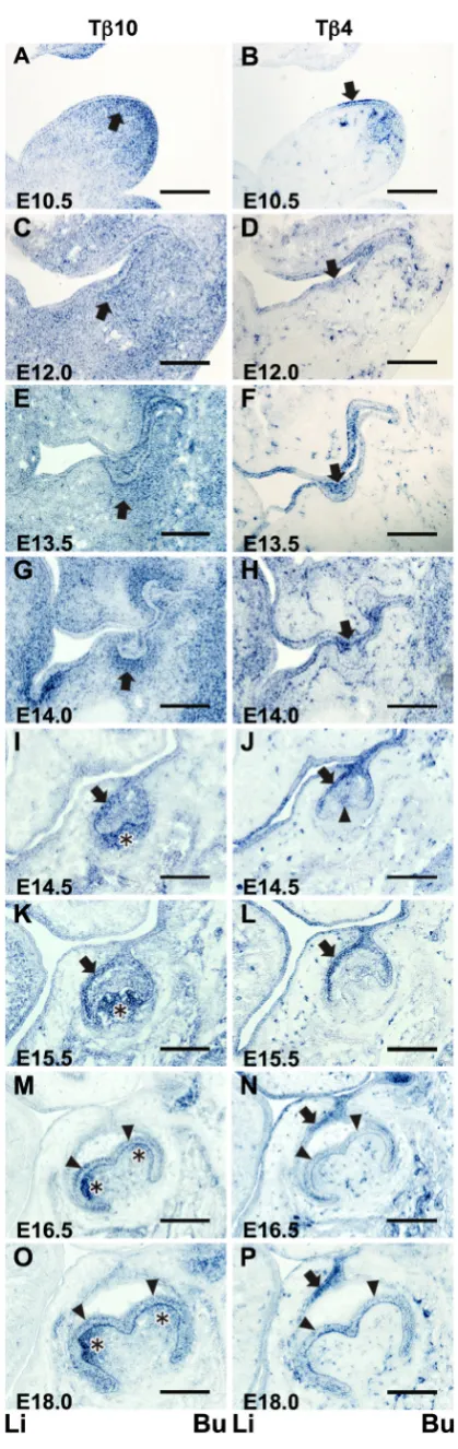
Fig. 1. In situ expression of Tb10 and Tb4 in the tooth germ at fetal
stages. The in situ Tb10 expression was temporally and spatially stage-specific during the tooth germ development (left panels: A,C,E, G,I,K,M,O). The expression pattern was different from that of Tb4 (right panels:
B,D,F,H,J,L,N,P). (A,C) An expression of Tb10 was diffusely observed in the subepithelial mesenchymal cells. Strong Tb10-positive mesenchymal cells were observed to aggregate beneath the mucosal epithelium (arrow) on E10.5 and E12.0. (B,D) On E10.5 and E12.0, a strong Tb4 expression was seen in the oral mucosal epithelial layer at the site where the tooth germ would form (arrow). (E) On E13.5, the Tb10 expression was diffusely found in the enamel organ and the surrounding mesenchymal cells (arrow). (G)
On E14.0, a strong Tb10 expression was seen in the mesenchymal cells surrounding the epithelial tooth bud (arrow), and a weak Tb10 expression was observed in the epithelial cells in the central area of the tooth bud.
Results
In situ hybridization of Tb10 and Tb4 in the developing tooth
germ
Embryonic stages
Initiation (E10.5)
Thickening of the oral mucosal epithelium to form the tooth bud was not apparent on this embryonic day.
A strong expression of Tb10 was diffusely observed in the
subepithelial mesenchymal cells. A weak Tb10 expression was
also detected in the oral mucosal epithelial cells (Fig. 1A). In contrast, a strong Tb4 expression was detected in the oral
mucosal epithelial cells localized at the site where the tooth bud had formed. A weak expression of Tb4 was observed in the blood
vessels (Fig. 1B).
Thickening of the dental epithelium (E12.0)
Local epithelial thickening was observed in the oral mucosal epithelium, indicating the tooth bud formation.
An in situ expression of Tb10 was diffusely observed in the
mesenchymal tissue (Fig. 1C). Strong Tb10-positive mesenchymal
cells were to aggregate around the thickened mucosal epithelium (Fig. 1C, arrow). A weak Tb10 expression was also detected in
the oral mucosal epithelial cells.
The Tb4 expression was observed in the oral mucosal epithelial
cells, including those located at the site of the presumptive tooth bud. The Tb4 expression was also detected in the blood vessels
in the mesenchymal tissue (Fig. 1D).
Bud stage (E13.5 - E14.0)
The thickened dental epithelium invaginated into the mesen-chyme and formed the tooth bud.
A strong Tb10 expression was primarily observed in the
mes-enchymal cells that surrounded the invaginated epithelial tooth bud in the E13.5 mandible. A weak expression was noted in the mesenchymal cells that were diffusely distributed in the mandibular tissue. A weak Tb10 expression was also observed in the epithelial
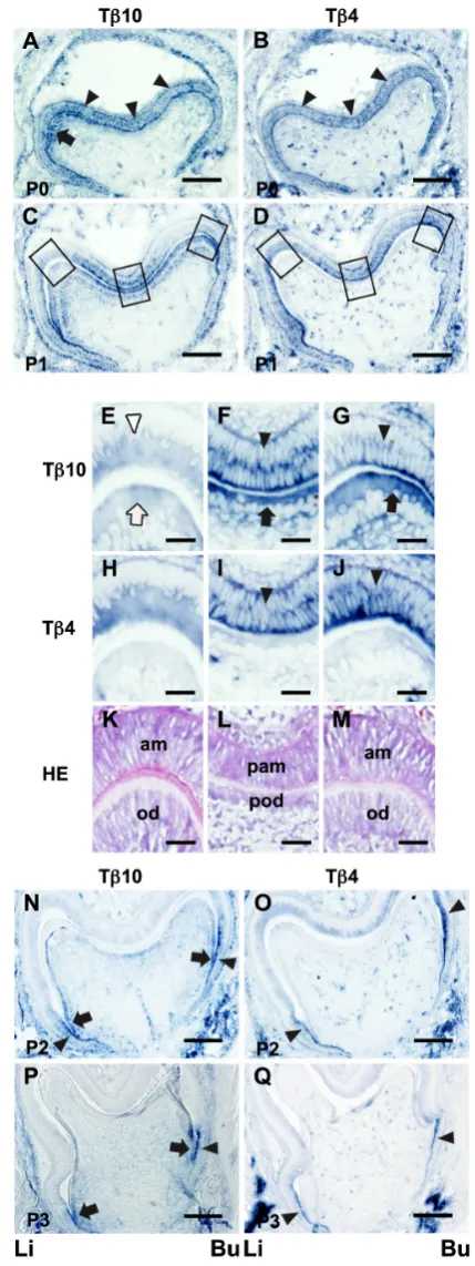
Fig. 2. In situ expression of Tb10 and Tb4 in the developing tooth
germ at the late bell stage. Tb10 expression from P0 to P3 is presented in (A,C,E,F,G,N and P). Tb4 expression is indicated in (B,D,H,I,J,Oand Q).(A) On P0, Tb10 was expressed in the inner enamel epithelial cells (arrowheads) and preodontoblasts in the lingual cusp site of the dental pulp (arrow). (B) A strong Tb4 expression was detected throughout the inner enamel epithelial layer (arrowheads) on P0. (C) On P1, a strong Tb10 expression was detected in the buccal cusp site and the central groove region. (E,F,G) The left, middle and right boxed areas in (C) are shown at a higher magnification, respectively. (E) The Tb10 expression was reduced in the odontoblasts (white arrow) and ameloblasts (white arrowhead) in the lingual cusp where formed matrices were seen. (F) A strong Tb10 ex-pression was detected in the preodontoblasts (arrow) and preameloblasts (arrowhead) in the central groove region (G) A strong Tb10 expression was detected in the preodontoblasts (arrow) and preameloblasts (arrowhead) in
the buccal cusp site. (D) A strong Tb4 signal was demonstrated in the inner enamel epithelial layer only on P1. (H,I,J) The left, middle and right boxed areas in (D) are shown at a higher magnification, respectively. (H) The Tb4 expression was reduced in the ameloblasts in the lingual cusp with matrix formation. (I,J) A strong Tb4 expression was detected in the preameloblasts (arrowhead) in the central groove region and the buccal cusp site. (K,L,M)
The HE-stained images correspond to (E,H), (F,I) and (G,J), respectively. (K) Matrix formation was noted between ameloblasts and odontoblasts.
cells in the central area of the tooth bud (Fig. 1E).
In contrast, an in situ expression of Tb4 was localized in the
invaginated epithelial tooth bud as well as in the mucosal epithelial layer. The Tb4 expression was hardly observed in the
mesenchy-mal cells located around the tooth bud, with the exception of the blood vessels (Fig. 1F).
The in situ expression patterns of both Tb10 and Tb4 in the
E14.0 mandible were coincident to those observed in the E13.5 mandible (Fig. 1 G,H).
Cap stage (E14.5 - E15.5)
The enamel organ showed a cap-shaped structure at this stage. A strong in situ expression of Tb10 was observed in the dental
papilla cells in both the E14.5 (Fig. 1I) and E15.5 (Fig. 1K) man-dibles. A weak in situ signal was detected in the mesenchymal cells of the dental sac. The Tb10 expression was also observed in
the outer enamel epithelium and inner cells of the enamel organ. No signals were detected in the primary enamel knot (Fig. 1I).
A strong in situ expression of Tb4 was demonstrated in the
outer enamel epithelium and primary enamel knot. A strong signal
was also noted in the dental lamina and oral mucosal epithelium. A weak in situ signal was detected in the inner enamel epithelium (Fig. 1J).
The expression patterns of both Tb10 and Tb4 in the E15.5
mandible (Fig. 1 K,L) were almost identical to those at E14.5. However, a higher positive intensity of the in situ Tb10 signal was
detected in the dental papilla cells (Fig. 1K). No Tb4 expression
was found in the enamel knot (Fig. 1L).
Bell stage (E16.5 - E18.0)
The periphery of the enamel organ extended to the mesenchy-mal tissue, resulting in the shape of a bell in the enamel organ. On E16.5, a strong Tb10 expression was demonstrated in the
dental papilla localized at the site where the cusps had formed (Fig. 1M). A weak Tb10 expression was also observed in the inner
enamel epithelium and the dental sac, while there was an undetect-able level of the Tb10 expression in the oral mucosal epithelium
and the outer enamel epithelium in which the Tb10 expression
was detected in the bud (Fig. 1 C,E) and cap stages (Fig. 1 I,K).
Tb4 was strongly expressed in the dental lamina and the outer
enamel epithelium on E16.5. A weak Tb4 expression appeared
in the inner enamel epithelium. No apparent Tb4 expression was
detected in the dental papilla, except for in the blood vessels (Fig. 1N).
The expression patterns of Tb10 and Tb4 on E18.0 (Fig. 1 O,P)
were coincidental with those observed in the E16.5 mandible.
Postnatal stages
Late bell stage (P0 - P3)
The dental pulp cells facing the inner enamel epithelium differ-entiated into preodontoblasts. Neither enamel nor dentin matrices had formed on postnatal day 0 (P0), while the formation of both enamel and dentin matrices between ameloblasts and odontoblasts was seen at the presumptive cusp site in the P1 tooth germ. The cells in the stage just before matrix formation are termed pream-eloblasts and preodontoblasts, respectively (Byers et al., 1990). On P0, Tb10 was expressed in the preodontoblasts localized
at the presumptive cusp sites of the dental pulp. An in situ signal was also found in the inner and outer enamel epithelia (Fig. 2A). A strong Tb4 expression was detected throughout the inner enamel
epithelium on P0. The outer enamel epithelium also exhibited weak positive signals (Fig. 2B).
On P1, strong Tb10 expression was observed in the
preodon-toblasts and preameloblasts localized at the presumptive buccal cusp site and the central groove region (Fig. 2 C,F,G,L,M). Mean-while, the Tb10 signal intensity was reduced in the odontoblasts
and ameloblasts in the presumptive lingual cusp site where matrix formation was detected (Fig. 2 E,K).
Treatment
Stage Ut C Tβ10**
Bud 1 1 10
Cap 7 8 2
Fig. 3. In situ expression of Tb10 and Tb4 in the developing tooth
germ at the tooth root formation stage. The HE staining sections of tooth germs on P5, P7 and P14 are shown (A,D,G). The Tb10 and Tb4 expression patterns are shown in (B,E,H) and (C,F,I), respectively. (B) On P5, the Tb10 expression was observed in HERS cells (arrows). (C) No Tb4 expression was detected in either ameloblasts or odontoblasts at this stage. (E, F) On P7, the Tb10 and Tb4 expression patterns were similar to those observed on P5. (H) On P14, the Tb10 expression was observed in HERS cells, dental pulp cells facing the HERS and periodontal ligament cells (arrows). (I) No Tb4 expression was detected at this stage. Li; lingual side, Bu; buccal side. Scale bars, (A-F) 100 mm; (G-I) 50 mm.
TABLE 1
THE EFFECTS OF Tb10 INHIBITION ON THE DEVELOPMENT
OF THE TOOTH GERM IN CULTURED E11.0 MANDIBLES
An in situ signal of Tb4 was observed through the inner enamel
epithelial layer (Fig. 2D). A strong Tb4 expression was detected in
the preameloblasts localized at the presumptive buccal cusp and the central groove sites where the strong Tb10 expression was
exhibited (Fig. 2 I,J). A weak Tb4 expression was observed in the
ameloblasts located under the formation of the enamel matrix at the presumptive lingual cusp site (Fig. 2 H,K). No in situ signals were found in preodontoblasts or odontoblasts on P1 (Fig. 2 D,H,I,J).
On P2, the Tb10 expression was observed in the odontoblastic
layer (Fig. 2N). A highly strong expression was detected in the lateral sides of the dental pulp. A weak expression was found in the preodontoblasts located at the presumptive cusps and the occlusal
groove sites. A strong expression was also demonstrated in the inner and outer enamel epithelia at the lateral sides of the enamel organ. As a result, preodontoblasts and preameloblasts exhibiting a strong Tb10 expression faced
each other at the lateral side of the tooth germ (Fig. 2N). A strong Tb4 expression was noted in the preameloblasts
located in the lateral enamel organ on P2 (Fig. 2O). A weak
Tb4 expression was also seen in the preameloblasts and
ameloblasts in the inner enamel epithelium.
On P3, the expressions of both Tb10 and Tb4 were
observed in limited areas. Preodontoblasts and preamelo-blasts exhibiting strong Tb10 signals were localized in a
limited area of the lateral edge of the tooth germ (Fig. 2P). The Tb4 expression was observed in the preameloblasts
localized at the lateral edges of the inner enamel epithe-lium only. No in situ signals of Tb4 were detected in the
preodontoblasts or odontoblasts (Fig. 2Q).
Tooth root formation stage (P5 - P14)
Enamel matrix formation was terminated by this stage. The free edge of the enamel organ on the developing tooth germ extended toward the mesenchymal tissue, thus forming a Hertwig’s epithelial root sheath (HERS). Dentin matrix formation further progressed at this stage.
On P5 and P7, a strong Tb10 signal was demonstrated Fig. 4. Schematic illustration of Tb10 and Tb4 expression patterns observed
during tooth development. The different expression patterns of Tb10 and Tb4 observed during tooth development are summarized. An in situ Tb10 signal from the initiation stage to the cap stage was detected primarily in dental mesenchymal cells, while Tb4 was expressed in dental epithelial cells. Interestingly, Tb10-expressing preodontoblasts and Tb4-expressing preameloblasts faced each other in the tooth germ at the late bell stage. The Tb10 expression is indicated by blue dots, while the Tb4 expression is indicated by red dots. The yellow zone indicates the enamel and dentin matrices.
in preodontoblasts in a limited area of the lateral side of the tooth germ (Fig. 3 B,E). These preodontoblasts were considered to have originated before dentin matrix formation, as confirmed by observations of serial sections stained with hematoxylin and eosin (HE)(Fig. 3 A,D). In addition, a strong expression was especially seen in the HERS (Fig. 3 B,E).
There was almost no Tb4 expression in the crown at this stage
(Fig. 3 C,F).
On P14, the root formation was almost completed (Fig. 3G). The Tb10 expression was observed only in the HERS (Fig. 3H).
The Tb10 expression was found in the dental pulp cells facing the Tb10-positive HERS. Periodontal ligament cells were also showed
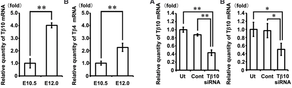
Fig. 5 (Left). Increases in Tb10 and Tb4 mRNA levels in the mandible at E12.0 compared with those observed at E10.5. A real-time PCR analysis showed that theexpression of Tb10 (A) and Tb4 (B) mRNAs were significantly increased in the mandible on E12.0 in comparison to those observed on E10.5. The data represent the mean ± S.D. of more than three samples. **P<0.01; versus E10.5 mandibles.
Fig. 6 (Right). Inhibition of Tb10 gene expression using siRNA treatment. TheTb10 mRNA expression was significantly decreased in the E11.0 man-dible (A) and the E15.0 tooth germ (B) explants treated with Tb10-siRNA (Tb10-siRNA) in comparison to that observed in the untreated explants (Ut), or explants treated with universal negative control siRNA (Cont). The data represent the mean ± S.D. of more than three samples. *P<0.05, **P<0.01.
positive signals. No Tb4 expression was found in the crown and
root (Fig. 3I). The characteristic expression patterns of both Tb10
and Tb4 in each stage are illustrated in Fig. 4.
Quantification of Tb10 and Tb4 mRNA levels in the mandible using real-time PCR
The mRNA level of Tb10 in the mandibles on E12.0 was
in-creased approximately 4-fold in comparison to that observed on E10.5 (Fig. 5A, p=0.0000013). Similarly, a significant increase in the level of Tb4 mRNA in the mandibles on E12.0 was
dem-onstrated in comparison to that observed on E10.5 (Fig. 5B,
p=0.0000091), thus confirming the findings of previous study
(Yamaza et al., 2001).
Inhibition assay for Tb10 gene expression induced by siRNA treatment
The effects of siRNA on the expression of Tb10 were examined
using semi-quantitative real-time PCR methods in the cultured E11.0 mandible and the E15.0 tooth germ on the 8th culture day. The Tb10 mRNA expression levels in both the E11.0 mandible
and the E15.0 tooth germ explants treated with Tb10-siRNA were
significantly decreased to approximately 50% compared with those observed in the untreated explants and the explants treated with universal negative control siRNA (Fig. 6A, p=0.000035 by one-way ANOVA, for each p<0.01 by Tukey-Kramer test; Fig. 6B, p=0.019 by one-way ANOVA, for each p<0.05 by Tukey-Kramer test).
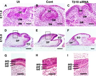
Histological analysis of cultured E11.0 mandibles and E15.0 tooth germs treated with Tb10- siRNA
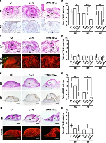
In the controls, the epithelium appeared to form a cap-shaped structure on the 8th culture day. In addition, both dental papilla and dental follicle formed by the surrounding mesenchymal cells were observed (Fig. 7 A,B). The mandibles treated with Tb
10-siRNA exhibited a developmental arrest of the enamel organ and continued to show the bud-like stage on the 8th culture day
(Fig. 7C). The development of the tooth germ was significantly suppressed in the E11.0 mandibles treated with Tb10-siRNA
compared with that observed in the controls (p=0.00059, Table 1). Tooth germs obtained from E15.0 embryos were also cultured for eight days with siRNA treatment. In the controls, the tooth germs appeared to form a bell-shaped structure on the 8th culture day. The formation of a predentin matrix was observed (Fig. 7 D,E). Meanwhile, significant suppression of tooth germ growth was demonstrated in the explants treated with Tb10-siRNA in
comparison to that observed in the controls (p=0.013, Table 2, Fig. 7 D-F). However, treatment with Tb10-siRNA did not influence
the morphology or arrangement of ameloblasts and odontoblasts. Predentin matrix formation was also observed in all of the con-trols and explants treated with Tb10-siRNA (Fig. 7 G-I). These
findings suggest that Tb10 may not affect the differentiation of
ameloblasts or odontoblasts.
Ki67 immunohistochemical and TUNEL staining in cultured E11.0 mandibles and E15.0 tooth germs treated with Tb10-siRNA
Since the development of the tooth germs in the E11.0 man-dibles and the E15.0 tooth germs was disturbed by treatment with
Tb10-siRNA, cell proliferation and cell death were examined in
the tissue specimens.
EFFECTS OF Tb10 INHIBITION ON THE DEVELOPMENT
OF THE DENTAL PAPILLA IN CULTURED E15.0 MANDIBLES
Developmental arrest of dental papilla was significantly demonstrated in Tb10 in comparison to Ut and C (*P<0.05). Ut: untreated explants, C: explants treated with universal negative control siRNA, Tb10: explants treated with siRNA for Tb10, +: positive findings, -: negative findings.
Fig. 7. Suppressed morphogenesis of the tooth germ in cultured E11.0 mandibles and E15.0 tooth germs treated with Tb10 -siRNA. An inhibitory assay for Tb10 was performed using Tb10-siRNA in cultured E11.0 mandibles (A,B,C) and E15.0 tooth germs (D,E,F)
terminal deoxynucleotidyl transferase-dUTP nick end labeling (TUNEL)-positive cells and the total cell numbers were counted in three objective areas: the “dental epithelium (DE)”, the “dental mesenchyme (DM)” and the “surrounding mesenchyme (SM)”. The DM was either the “DP and follicle” in the cultured explants showing the normal cap-like tooth germ or the “odontogenic ectomesenchyme” in the explants exhibiting an inhibition of tooth germ development (Figs. 7 and 8) (Takahashi et al., 2010; Ookuma et al., 2013).
The localization of Tb10-expressing cells corresponded well
with that of the Ki67-positive cells in the cultured mandible and tooth germ of the control and/or untreated samples. Furthermore, the immunohistochemical reactivity for Ki67 in the E11.0 mandibles revealed a significant decrease in cell proliferation activity in the
DE, DM and SM of the Tb10-siRNA-treated explants in
compari-son to that observed in the controls. TUNEL-positive cells were also observed in both the controls and the explants treated with
Tb10-siRNA. However, there were no significant differences in
apoptotic cells among the examined groups.
The cultured E15.0 tooth germs were composed primarily of two areas: the “enamel organ (EO)” and the “dental papilla (DP)” (Figs. 7 and 8). The numbers of cells in these areas were counted. Immunohistochemical reactivity for Ki67 in the E15.0 tooth germs revealed a significant decrease in the cell prolifera-tion activity of the EO and DP in the Tb10-siRNA-treated explants
in comparison to that observed in the controls. In the TUNEL staining, there were no significant differences in apoptotic cells among the examined groups.
Fig. 8. Cell proliferation and cell death activity in cultured E11.0 mandibles and E15.0 tooth germs treated with
Tb10-siRNA. A cell proliferation assay was performed to analyze the involve-ment of Tb10 in the morphogenesis of the cultured E11.0 mandibles (A-D) and E15.0 tooth germs (E-H) on the 8th day. (A,E)
Ki67 immunostaining (lower panel) and HE staining (upper panel) are presented in the untreated explants (Ut) and explants treated with universal negative control siRNA (Cont) or Tb10-siRNA (Tb10-siRNA). (C,G) TUNEL staining (lower panel) and HE staining (upper panel) are presented in the Ut, Cont and Tb10-siRNA. (B) Significant decreases in the ratios of the number of Ki67-positive cells to the total cell number (ratio of Ki67 staining) were observed in the DE, DM and SM areas of the Tb10-siRNA in comparison to those observed in the Ut and Cont. (C)
Effects of Tb10-siRNA on the proliferative activity of mDP and mDE6 cells
To confirm the effects of Tb10-siRNA on cell proliferation activity,
a cell growth assay was performed using mDP and mDE6 cells, which are mouse dental pulpal and epithelial cell lines established from tooth germs, respectively (Tsubakimoto et al., 2007; Yoshizaki
et al., 2008).
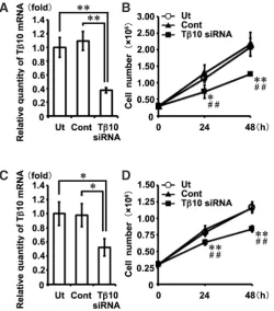
A significant reduction in the Tb10-mRNA expression level was
observed in the mDP cells with respect to Tb10-siRNA-treated
explants in comparison with that observed in the controls (Fig. 9A). The cell proliferation activity level was significantly lower in the mDP cells treated with Tb10-siRNA than in the controls at both 24- and
48-hour culture periods, thus indicating that Tb10-siRNA treatment
affects the cell proliferative activity of tooth germ development (Fig. 9B). Similar effects of Tb10-siRNA on the mRNA expression were
observed in the mDE6 cells (Fig. 9C). Significant suppression of the cell proliferation activity at both 24 and at 48h was also noted in the Tb10-siRNA-treated mDE6 cells in comparison to that
ob-served in the controls (Fig. 9D).
On the other hand, Tb10-siRNA treatment showed no effects
on the expressions of dentin matrix protein-1 (Dmp-1) or dentin sialophosphoprotein (Dspp) in the mDP cells (Supplementary Fig. 1 A,B). The expression of amelogenin (Amel) in the mDE6 cells was not affected by Tb10-siRNA treatment (Supplementary Fig. 1C).
Since both Tb10 and Tb4 are known to be actin
monomer-sequestering proteins (Yu et al., 1993), the influence of Tb10-siRNA
treatment on the change in the ratio of G-/F-actin in the mDP and mDE6 cells was evaluated. Treatment with Tb10-siRNA for 48 h
decreased the ratio of G-/F-actin in the mDP cells, while the ratio of G-/F-actin modestly changed in the mDE6 cells (Supplementary Fig. 2 A,B). The Tb10 expression level in the mDP cells was higher
than that observed in the mDE6 cells, although real-time PCR re-vealed that there was no significant difference between the cells. Meanwhile, the Tb4 expression level in the mDP cells was less than
one-third of that observed in the mDE6 cells. A significant differ-ence was noted between these values (Supplementary Fig. 2 C,D). These results indicate the possibility that Tb10-siRNA
treat-ment induces the arrest of tooth germ developtreat-ment caused by the decreased cell proliferative activity rather than regulating the cell differentiation of both dental epithelial and mesenchymal cells.
Discussion
Several studies have reported the existence of a close rela-tionship between the expression of Tb10 and organogenesis (Lin et al., 1990; Hall 1991; Gerosa et al., 2010; Fanni et al., 2011).
However, there are no articles describing the relationship between tooth germ development and the Tb10 expression.
Previous studies (Akhter et al., 2005; Ookuma et al., 2013) dem-onstrated that Tb4, another member of the beta-thymosin family,
participates in the morphogenesis of the tooth germ. However, the
in situ expression pattern of Tb10 apparently differs from that of Tb4. In this study, the expression of Tb4 was primarily observed in
the epithelial elements. On the other hand, Tb10 was expressed
in the dental papilla in the E15.5 tooth germ (Akhter et al., 2005). We reconfirmed that the in situ expression pattern of Tb10 from
the early to the middle developmental stage (E10.5-E18.0) appar-ently differs from that of Tb4. Carpintero et al., (1996) reported that
the in situ expression of Tb10 in the E9.5 mouse embryo is detected
primarily in the mesenchymal tissue, including the maxilla and mandible. This expression pattern is coincident with that observed in our present study in which Tb10 mRNA was primarily detected
in the mesenchymal tissue of the mandible on E10.5. The Tb10
expression has also been reported to exist in the developing renal proximal and distal tubules (Gerosa et al., 2010), and the acinar and ductal epithelial cells of developing salivary gland (Fanni et al., 2011). Therefore, it may be reasonable to consider that Tb10 may
relate to the growth of mesenchymal tissue as well as epithelial cells in the organogenesis and development of the tooth germ. Indeed, a knockdown assay for Tb10 in the E11.0 mandibles and
the E15.0 tooth germs using siRNA resulted in the developmental arrest of the tooth germ. We further examined whether the devel-opmental arrest of the tooth germ was caused by a disturbance in cell proliferation or an increase in cell death. Ki67 immunostaining showed that treatment with siRNA in the organ cultured E11.0 mandibles resulted in a significantly decreased ratio of Ki67 stain-ing in the dental mesenchymal cells. The cell proliferative activity level evaluated with Ki67 immunostaining was suppressed in the
Fig. 9. Effects of Tb10-siRNA on the cell proliferation of tooth
germ-derived cells. A cell proliferation assay of tooth germ-derived cells, mDP
(A,B) and mDE6 (C,D) cells, was performed with or without Tb10-siRNA treatment. (A) The level of Tb10 transcripts in the mDP cells treated with Tb10-siRNA (Tb10-siRNA) were significantly reduced in comparison to those observed in the untreated explants (Ut) and explants treated with universal negative control siRNA (Cont)(**P<0.01). (B) At 24 and 48h after transfec-tion with siRNA, the cell numbers of Tb10-siRNA were significantly lower than those of Ut (*P<0.05, **P<0.01) or Cont (#P<0.05, ##P<0.01). (C)
Significant decreases in the levels of Tb10 transcripts were also observed in the mDE6 cells treated with Tb10-siRNA (**P<0.01). (D) In the mDE6 cells, similar effects of Tb10-siRNA on the cell proliferation were observed at 24 and 48h after transfection with siRNA (*P<0.05, **P<0.01; versus Ut, #P<0.05, ##P<0.01; versus Cont).
B
A
dental epithelial cells. The Tb10-expressing areas corresponded
to the localization of Ki67-positive cells. Therefore, treatment with
Tb10-siRNA appears to effectively suppress tooth germ
develop-ment in cultured organs, thus resulting in the developdevelop-mental arrest of the tooth germ in the E11.0 mandible and E15.0 tooth germ.
Furthermore, treatment with Tb10-siRNA also showed decreased
proliferative activity in the cultured mDP and mDE6 cells. Meanwhile, no significant increases in the number of TUNEL-positive cells were observed in the cultured E11.0 mandibles following treatment with Tb10-siRNA. Therefore, the developmental arrest of the tooth
germ observed in the cultured E11.0 mandibles might have been caused by a reduced cell proliferation activity rather than by cell death. These results indicate that Tb10 may play important roles in
cell proliferation in odontogenic epithelial and mesenchymal cells in the initiation and development of the tooth germ.
Tb10 is an actin monomer-sequestering protein (Yu et al., 1993),
and therefore changes in the ratio of G-/F-actin were examined. Treatment with Tb10-siRNA changed the ratio of G-/F-actin in the
mDP cells. Tb10-siRNA treatment might effectively suppress actin
sequestering in the mDP cells due to the lower Tb4 expression in
the mDP cells (Supplementary Fig. 2). Santelli et al., (2002) showed that blocking Tb10 using an antisense methodology in human thyroid
carcinoma cells significantly decreases anchorage-independent growth and improves actin filament organization. Thus, Tb10 may
affect cell proliferation via the regulation of actin assembly. In the future study, it is necessary to clarify the detailed mechanisms underlying the actin arrangement and cell proliferation regulated by Tb10 during tooth development.
Interestingly, the expression patterns of both Tb10 and Tb4 just
before matrix formation were quite different in this study. As shown in Fig. 2, a strong expression of Tb10 was observed in
preodonto-blasts as well as in preamelopreodonto-blasts. In contrast, Tb4 was strongly
expressed in preameloblasts only. Furthermore, preodontoblasts with a strong Tb10 expression and preameloblasts with a strong
Tb4 expression exhibited face-to-face localization in the tooth
germ. These findings suggest the presence of an intimate cell-cell interaction between Tb10-expressing preodontoblasts and Tb
4-expressing preameloblasts mediated by two different thymosins in the formation of dentin and enamel matrices. Therefore, other new functions of Tb10 and Tb4 might exist that are not related to
the development or differentiation of the tooth germ. The peculiar expression patterns of both Tb10 and Tb4 observed in the matrix
formation stage indicate that these unknown functions are impor-tant for the growth of the tooth germ. Further examinations are needed to clarify the true functions of Tb10 and Tb4 in tooth germ
development at this stage.
Both Tb10 and Tb4 also showed peculiar expression patterns
in the root formation stage. In this stage, the expression of Tb10
was found in preodontoblasts at the lateral side of the tooth germ. In particular, the expression of Tb10 was localized in the dental
epithelial cells and the dental mesenchymal cells of the dental pulp at the apex site of the enamel organ that formed the HERS after P5. The characteristic expression pattern of Tb10 observed at the
root formation stage suggests that Tb10 may participate in forming
the outline of the tooth root and extending the HERS.
In conclusion, Tb10 and Tb4 exhibit developmental stage-specific
temporal and spatial expression patterns in the tooth germ. These findings suggested that Tb10 and Tb4 might play different roles
in mouse tooth germ development. Previous studies have shown
that the immunolocalization of Tb10 is altered cell by cell in the
developing kidneys and salivary glands (Gerosa et al., 2010; Fanni
et al., 2011). Fanni et al., (2011) reported the existence of a close
relationship between the immunohistochemical expression of Tb10
and salivary gland development, and speculated that alternative immunoreactivity is associated with the organogenesis of the sali-vary glands. Our present study and previous reports indicate that Tb10 and Tb4 could potentially perform multiple functional roles in
tooth germ development. However, the signal transduction path-ways both up- and down-streams of Tb10 and Tb4 in tooth germ
development remain unknown. Further examinations are needed to elucidate the complete functional roles of these beta-thymosins and the interactions between beta-thymosin-associated factors in tooth germ development.
Materials and Methods
Animals
At least three BALB/c embryos and postnatal mice at each develop-mental stage were used in this study. The examined developdevelop-mental stages included E10.5, 11.0, 12.0, 13.5, 14.0, 14.5, 15.0, 15.5, 16.5 and 18.0 after gestation and P0, 1, 2, 3, 5, 7 and 14. Adult BALB/c mice were obtained from Charles River Japan, Inc. (Yokohama, Japan). All experimental proce-dures using mice were performed in accordance with the guidelines of the Animal Care and Use Review Committee of Kyushu University (Fukuoka, Japan). Adult female mice (10-20 weeks) were caged together with male mice. Successful insemination was determined based on the presence of a postcopulatory plug in the vagina 12 h later. The embryonic day E0.5 was defined by the presence of the vaginal plug (Takahashi et al., 2010; Ookuma et al., 2013).
In situ hybridization
To confirm the specificity and sensitivity of the antisense RNA probes for Tb10 and Tb4 mRNAs, membrane hybridization was performed. Membrane hybridization and in situ hybridization methods were carried out according to the protocol described in previous studies (Yamaza et al., 2001). Specific probes for Tb10 and Tb4 mRNAs were designed according to the NCBI Reference Sequences (accession number Tb10: NM_025284.4, Tb4: NM_021278.2) (Akhter et al., 2005). Strong and weak signals were used to describe for the relative evaluation of the signal intensity observed in the same tissue section. The developmental tooth germ process in the embryonic mandible was defined according to previous studies (Akhter et al., 2005; Takahashi et al., 2010).
Semi-quantitative real-time PCR
Total RNA was isolated from mandibles on E10.5 and E12.0, and from tooth germs on E15.0, E18.0, P1 and P5 using the SV Total RNA Isolation System (Promega, WI, USA). Total RNA was also isolated from cultured E11.0 mandibles, E15.0 tooth germs and mDP and mDE6 cells. cDNA was synthesized using the SuperScript® VILOTM cDNA Synthesis System
(Invitrogen, CA, USA) according to the manufacturer’s instructions. Real-time PCR was performed using a Thermal Cycler Dice® Real Time System
with the SYBR Premix Ex TaqTM II (TaKaRa Bio, Shiga, Japan) (Takahashi
melting curve and/or gel electrophoresis (Takahashi et al., 2010; Naher et al., 2012; Ookuma et al., 2013).
Inhibition assay for Tb10 using siRNA on organ culture
The mandibles dissected from the E11.0 embryos and the tooth germs dissected from the E15.0 embryos were cultured for eight days. The culture period of both the E11.0 mandibles and E15.0 tooth germs was determined based on previous studies (Kobayashi et al., 2006; Xie et al., 2007; Oo-kuma et al., 2013). The protocols for organ culture were identical to those used in these studies. In order to inhibit the functions of Tb10 during tooth germ development, cultured explants were treated with siRNAs supple-mented in the culture media according to the manufacturer’s protocol of Lipofectamine RNAiMAX (Invitrogen)(Naher et al., 2012). siRNA for murine Tb10 (Mm_Tmsb10_0122) and a universal negative control siRNA (Sigma-Aldrich, St. Louis, MO, USA) were used as a target and negative control, respectively. 30 pmol of siRNA and 3 ml of Lipofectamine RNAiMAX were contained in 1-ml culture media (siRNA final conc. 30 nM). The culture medium was changed every 24 h.
Histological analysis of the cultured explants
The cultured explants were fixed with 4% paraformaldehyde for 24 h at 4°C and then embedded in paraffin. Paraffin-embedded explants were cut into 5-mm thick sections in the anteroposterior direction, and HE staining was performed. The explants were prepared for the histological analysis as previously described (Kobayashi et al., 2006; Xie et al., 2007; Ookuma et al., 2013).
Cell proliferation and cell death assays in the cultured organs
In order to evaluate the effects of Tb10 on cell proliferation during tooth development, immunohistochemistry using a rabbit polyclonal antibody to Ki67 (Abcam, Cambridge UK) was performed. These sections were nuclear-counterstained with hematoxylin.
TUNEL was applied to detect apoptotic cells using the in situ Apoptosis Detection Kit (TaKaRa). Briefly, terminal deoxynucleotidyl transferase (TdT) enzymes were used on the sections of the cultured organs treated with siRNA, and then the sections were labeled with FITC on the nick sites in the DNA. Propidium iodide was then used for nucleus staining.
The percentages of Ki67-positive or FITC-positive cells were evaluated in the DE, DM and SM areas in the cultured E11.0 mandibles. In the cultured E15.0 tooth germs, the numbers of cells in the EO and DP areas were counted. More than one hundred cells were examined as a population of each area in at least three different explants. The number of stained cells was divided by the total number of stained and non-stained target cells to calculate the ratio of Ki67 or FITC staining. The analyses of cellular pro-liferation and cell death were carried out as described in previous studies (Takahashi et al., 2010; Ookuma et al., 2013).
Cell lines and cell culture
Mouse dental epithelial and pulpal cell lines established from tooth germs (named mDE6 and mDP, respectively) were kindly provided by Professor Satoshi Fukumoto (Tohoku University, Japan) and Associate Professor Masahiro Saito (Tokyo University of Science, Japan) (Tsubakimoto et al., 2007; Yoshizaki et al., 2008). Both types of cells were maintained in D-MEM/F-12 (Invitrogen, CA, USA) supplemented with 10% fetal bovine serum (FBS, Filtron, Brooklyn, Australia), 100 U/ml of penicillin and 100 mg/ml of streptomycin (Invitrogen) in a humidified atmosphere of 5% CO2 at 37°C.
Cell growth assays of dental epithelial and mesenchymal cells treated with Tb10 using siRNA in cell culture
Tb10-siRNA (final conc. 124 nM) was simultaneously transfected into mDP and mDE6 cells using hemagglutinating virus of Japan (HVJ)-liposomes (GenomOne, Ishihara Sangyo Kaisha, Osaka, Japan) when the cells were seeded in triplicate onto 3 x 10⁵ cells/well in 6-well plates. The HVJ-liposome
complex was prepared according to the manufacturer’s instructions. At 24 and 48 h after treatment with siRNA, the numbers of cells in four
different microscopic fields of each well were counted. The average cell number was calculated in triplicate. At least three independent experiments were performed in triplicate.
Statistical analysis
All experiments were independently repeated at least three times. One-way ANOVA with the Tukey-Kramer comparison test or Student’s t-test was used to determine the presence of significant differences in the real-time PCR data, immunostaining-positive ratios and cell growth values. A p-value (p<0.05 or p<0.01) was considered to indicate statistically significant differ-ences. Significant differences in the numbers of explants were assessed using the chi-square test for independence.
Acknowledgements
This work was supported in part by Grant-in-Aid from the Ministry of Educa-tion, Culture, Sports, Science and Technology of Japan, Nos. 23659859 (to T. K.) and 23792106 (H. W.).
References
AKHTER M, KOBAYASHI I, KIYOSHIMA T, MATSUO K, YAMAZA H, WADA H, HONDA JY, MING X, SAKAI H (2005). Possible functional involvement of thymosin beta 4 in developing tooth germ of mouse lower first molar. Histochem Cell Biol 124: 207-213. BOCK-MARQUETTE I, SAXENA A, WHITE MD, DIMAIO JM, SRIVASTAVA D (2004).
Thymosin beta4 activates integrin-linked kinase and promotes cardiac cell migra-tion, survival and cardiac repair. Nature 432: 466-472.
BYERS MR, SCHATTEMAN GC, BOTHWELL M (1990). Multiple functions for NGF receptor in developing, aging and injured rat teeth are suggested by epithelial, mesenchymal and neural immunoreactivity. Development 109: 461-471. CARPINTERO P, FRANCO DEL AMO F, ANADÓN R, GÓMEZ-MÁRQUEZ J (1996).
Thymosin beta10 mRNA expression during early postimplantation mouse develop-ment. FEBS lett 394: 103-106.
CHEN J, LAN Y, BAEK JA, GAO Y, JIANG R (2009). Wnt/beta-catenin signaling plays an essential role in activation of odontogenic mesenchyme during early tooth development. Dev Biol 334: 174-185.
COBOURNE MT, SHARPE PT (2003). Tooth and jaw: molecular mechanisms of patterning in the first branchial arch. Arch Oral Biol 48: 1-14.
FANNI D, GEROSA C, NEMOLATO S, LOCCI A, MARINELLI V, CABRAS T, MESSANA I, FANOS V, CASTAGNOLA M, FAA G (2011). Thymosin beta 10 expression in developing human salivary glands. Early Hum Dev 87: 779-783.
GEROSA C, FANNI D, NEMOLATO S, LOCCI A, MARINELLI V, CABRAS T, MES-SANA I, CASTAGNOLA M, MONGA G, FANOS V, FAA G (2010). Thymosin beta-10 expression in developing human kidney. J Matern Fetal Neonatal Med Suppl 3: 125-128.
HALL AK (1991). Developmental regulation of thymosin beta 10 mRNA in the human brain. Brain Res Mol Brain Res 9: 175-177.
KIM YC, KIM BG, LEE JH (2012). Thymosin b10 expression driven by the human TERT promoter induces ovarian cancer-specific apoptosis through ROS produc-tion. PLoS One 7: e35399.
KOBAYASHI I, KIYOSHIMA T, WADA H, MATSUO K, NONAKA K, HONDA JY, KOYANO K, SAKAI H (2006). Type II/III Runx2/Cbfa1 is required for tooth germ development. Bone 38: 836-844.
LEE SH, SON MJ, OH SH, RHO SB, PARK K, KIM YJ, PARK MS, LEE JH (2005). Thymosin beta10 inhibits angiogenesis and tumor growth by interfering with Ras function. Cancer Res 65: 137-148.
LIN SC, MORRISON-BOGORAD M (1990). Developmental expression of mRNAs encoding thymosins beta 4 and beta 10 in rat brain and other tissues. J Mol
Neurosci 2: 35-44.
MINA M (2001). Regulation of mandibular growth and morphogenesis. Crit Rev Oral
Biol Med 12: 276-300.
MU H, OHASHI R, YANG H, WANG X, LI M, LIN P, YAO Q, CHEN C (2006). Thymosin beta10 inhibits cell migration and capillary-like tube formation of human coronary artery endothelial cells. Cell Motil Cytoskeleton 63: 222-230.
through interleukin-22 in oral squamous cell carcinoma. Int J Oncol 41: 1577-1586. OOKUMA YF, KIYOSHIMA T, KOBAYASHI I, NAGATA K, WADA H, FUJIWARA H,
YAMAZA H, NONAKA K, SAKAI H (2013). Multiple functional involvement of Thymosin beta-4 in tooth germ development. Histochem Cell Biol 139: 355-370. PHILP D, GOLDSTEIN AL, KLEINMAN HK (2004). Thymosin beta4 promotes
angiogenesis, wound healing, and hair follicle development. Mech Ageing Dev 125: 113-115.
PISPA J, MIKKOLA ML, MUSTONEN T, THESLEFF I (2003). Ectodysplasin, Edar and TNFRSF19 are expressed in complementary and overlapping patterns during mouse embryogenesis. Gene Expr Patterns 3: 675-679
SANTELLI G, BARTOLI PC, GIULIANO A, PORCELLINI A, MINEO A, BARONE MV, BUSIELLO I, TRAPASSO F, CALIFANO D, FUSCO A (2002). Thymosin beta-10 protein synthesis suppression reduces the growth of human thyroid carcinoma cells in semisolid medium. Thyroid 12: 765-772.
TAKAHASHI KF, KIYOSHIMA T, KOBAYASHI I, XIE M, YAMAZA H, FUJIWARA H, OOKUMA Y, NAGATA K, WADA H, SAKAI T, TERADA Y, SAKAI H (2010). Pro-togenin, a new member of the immunoglobulin superfamily, is implicated in the development of the mouse lower first molar. BMC Dev Biol 10: 115
THESLEFF, I (2003). Epithelial-mesenchymal signalling regulating tooth morphogen-esis. J Cell Sci 116: 1647-1648.
TSUBAKIMOTO T, KOSAKA K, SAITO M, TERANAKA T (2007). Immortalization
of dental papilla cells differentiating into odontoblast in vitro. Japan J Conserv
Dent 50: 292-301.
WANG WS, CHEN PM, HSIAO HL, JU SY, SU Y (2003). Overexpression of the thymosin beta-4 gene is associated with malignant progression of SW480 colon cancer cells. Oncogene 22: 3297-3306.
XIE M, KOBAYASHI I, KIYOSHIMA T, YAMAZA H, HONDA JY, TAKAHASHI K, ENOKI N, AKAMINE A, SAKAI H (2007). Functional implication of nucleolin in the mouse first molar development. J Biol Chem 282: 23275-23283.
YAMAZA H, MATSUO K, KIYOSHIMA T, SHIGEMURA N, KOBAYASHI I, WADA H, AKAMIME A, SAKAI H (2001). Detection of differentially expressed genes in the early developmental stage of the mouse mandible. Int J Dev Biol 45: 675-680. YOSHIZAKI K, YAMAMOTO S, YAMADA A, YUASA K, IWAMOTO T, FUKUMOTO
E, HARADA H, SAITO M, NAKASIMA A, NONAKA K, YAMADA Y, FUKUMOTO S (2008). Neurotrophic factor neurotrophin-4 regulates ameloblastin expression via full-length TrkB. J Biol Chem 283: 3385-3391.
YU FX, LIN SC, MORRISON-BOGORAD M, ATKINSON MA, YIN HL (1993). Thymosin beta 10 and thymosin beta 4 are both actin monomer sequestering proteins. J
Biol Chem 268: 502–509.
ZHAO Y, QIU F, XU S, YU L, FU G (2011). Thymosin b4 activates integrin-linked kinase and decreases endothelial progenitor cells apoptosis under serum deprivation. J
Molecular signaling at the fusion stage of the mouse mandibular arch: involvement of insulin-like growth factor family
Kazuya Fujita, Yuji Taya, Yoshihito Shimazu, Takaaki Aoba and Yuuichi Soeno Int. J. Dev. Biol. (2013) 57: 399-406
http://dx.doi.org/10.1387/ijdb.120110ys
The zebrafish sf3b1b460 mutant reveals differential requirements for the sf3b1 pre-mRNA processing gene during neural crest development
Min An and Paul D. Henion Int. J. Dev. Biol. (2012) 56: 223-237 http://dx.doi.org/10.1387/ijdb.113383ma
A novel role for Glucocorticoid-Induced TNF Receptor Ligand (Gitrl) in early embryonic zebrafish development
Lynn D. Poulton, Kathleen F. Nolan, Corina Anastasaki, Herman Waldmann and E. Elizabeth Patton Int. J. Dev. Biol. (2010) 54: 815-825
5 yr ISI Impact Factor (2011) = 2.959 http://dx.doi.org/10.1387/ijdb.082841lp
Over-expression of thymosin beta4 promotes abnormal tooth development and stimu-lation of hair growth
Hee-Jae Cha, Deborah Philp, Soo-Hyun Lee, Hye-Sung Moon, Hynda K. Kleinman and Takashi Nakamura
Int. J. Dev. Biol. (2010) 54: 135-140 http://dx.doi.org/10.1387/ijdb.082735hc
Detection of differentially expressed genes in the early developmental stage of the mouse mandible
H Yamaza, K Matsuo, T Kiyoshima, N Shigemura, I Kobayashi, H Wada, A Akamime and H Sakai Int. J. Dev. Biol. (2001) 45: 675-680
http://www.intjdevbiol.com/web/paper/11461004
GDNF and its receptors in the regulation of the ureteric branching
H Sariola and M Saarma
Int. J. Dev. Biol. (1999) 43: 413-418