The allocation and differentiation of mouse
primordial germ cells
TANIA E. TSANG
1, POH-LYNN KHOO
1, ROBYN V. JAMIESON
1, SHEILA X. ZHOU
1, SIEW-LAN ANG
2,
RICHARD BEHRINGER
3AND PATRICK P.L. TAM*
,11Embryology Unit, Children’s Medical Research Institute, University of Sydney, Westmead, Australia, 2Institute of Genetics and Molecular and Cellular Biology, Illkirch, Strasbourg, France and
3Department of Molecular Genetics, MD Anderson Cancer Center, University of Texas, Houston, Texas, USA
ABSTRACT Analysis of the lineage potency of epiblast cells of the early-streak stage mouse embryo reveals that the developmental fate of the cells is determined by their position in the germ layer. Epiblast cells that are fated to become neuroectoderm can give rise to primordial germ cells (PGCs) and other types of somatic cells when they were transplanted to the proximal region of the epiblast. On the contrary, proximal epiblast cells transplanted to the distal region of the embryo do not form PGCs. Therefore, the germ line in the mouse is unlikely to be derived from a predetermined progenitor population, but may be specified as a result of tissue interactions that take place in the proximal epiblast of the mouse gastrula. The initial phase of the establishment of the PGC population requires, in addition to BMP activity emanating from the extraembryonic ectoderm, normal Lim1 and Hnf3β activity in the germ layers. The entire PGC population is derived from a finite number of progenitor cells and there is no further cellular recruitment to the germ line after gastrulation. The XX PGCs undergo X-inactivation at the onset of migration from the gut endoderm and re-activate the silenced X-chromosome when they enter the urogenital ridge. Germ cells that are localised ectopically in extragonadal sites do not re-activate the chromo-some, even when nearly all germ cells in the fetal ovary have restored full activity of both X-chromosomes. XXSxr germ cells can re-activate the X-chromosome in the sex-reversed testis, suggesting that the regulation of X-chromosome activity is independent of ovarian morpho-genesis.
KEY WORDS:
Primordial germ cells, allocation, mutant embryo, X-chromosome activity, sex reversal.
0214-6282/2001/$25.00 © UBC Press
Printed in Spain
www.ijdb.ehu.es
*Address correspondence to: Patrick P.L. Tam. Embryology Unit. Children’s Medical Research Institute, Locked Bag 23, Wentworthville, NSW 2145, Australia. FAX: +61-2-9681-2120. e-mail: [email protected]
Abbreviations used in this paper: BMP, bone morphogenetic protein; ES, embryonic stem cells; ex-ect, extraembryonic ectoderm; PGC, primordial germ cell; PS, primitive streak; TNAP, tissue non-specific form of alkaline phosphatase.
Prologue
Those of us who have had the privilege of working with Anne McLaren or have visited her laboratory in the MRC Mammalian Development Unit at University College London could recall vividly how we were captivated by Anne’s passion for the fascinat-ing biology of germ cell differentiation, regulation of X-chromo-some activity and sex determination. After graduating from Anne’s laboratory, PPLT thought that he would move on to tackle other questions on the developmental biology of the mouse, but soon found that he had to re-visit germ cells and gonad development many times since as a result of either an interesting discovery or mere serendipity. Our work presented in this review is the out-come of the active pursuit of a number of issues relating to the biology of the primordial germ cells at the Children’s Medical Research Institute.
Establishment of the germ cell lineage in the mouse
Clonal analysis of the lineage potency of epiblast cells has revealed that some cells in the proximal epiblast of the pre- to early-streak stage embryo can give rise to TNAP-expressing cells that colonize the allantoic mesoderm and the hindgut endoderm. How-ever, descendants of these proximal epiblast cells do not contrib-ute only to the presumptive PGCs but also to other somatic tissues such as the extraembryonic mesoderm. Furthermore, cells that may contribute descendants to the PGC population are not region-alized to any area in the proximal epiblast. The localization of the precursors of PGCs in the proximal epiblast suggests that only epiblast cells in this region of the early gastrula may possess the germ-line potency. During development of the germ line, a POU domain gene, Oct4, has been shown to be specifically expressed in the PGCs and their descendants in the gonad (Scholer et al, 1990; Pesce et al, 1998). The Oct4 gene is expressed widely in the epiblast of the early gastrula and apparently shows no regionalization to the proximal epiblast. The activity of Oct4 gene, however, is differentially regulated by enhancer elements for general expres-sion in the epiblast and specific expresexpres-sion in the PGCs (Palmieri et al., 1994; Yeom et al, 1996). The lack of restriction of both the PGC potency and the Oct4 expression pattern strongly suggests that in the mouse, there may not be a pre-determined germ line and the epiblast cells either acquire or retain the potency to differentiate into germ cells.
The lack of a pre-determined population of PGC progenitors implies that germ-cell formation is unlikely to be restricted to subsets of epiblast cells. This idea has been verified by testing the ability of epiblast cells that are localized outside the proximal epiblast to form PGCs. Distal epiblast cells that normally display a neuroectodermal fate (Quinlan et al., 1995) were transplanted by micro-manipulation to the proximal region of the epiblast. They
body and other similar sub-cellular structures in fertilized oocytes, blastomeres and germ cells of several mammalian embryos (de Smedt et al, 2000). However, the ability to induce germ cell formation in epiblast cells of diverse prospective fates does not support the concept of a pre-determined germ line in the mouse embryo.
Experimental analysis of the differentiation of germ cells in early postimplantation mouse embryos is hampered by the paucity of reliable lineage markers for PGCs and/or their precursors.
At-TABLE 1
THE COLONIZATION OF EXTRAEMBRYONIC AND EMBRYONIC TISSUES BY THE EPIBLAST AND EXTRAEMBRYONIC ECTODERM
AFTER TRANSPLANTATION TO THE EPIBLAST OF THE PRE- TO EARLY-STREAK STAGE MOUSE EMBRYO
Graft-derived cells Fraction of total graft-derived cells in
Mean/embryo Total Extraembryonic Hindgut Ectodermb Posterior PGCsc
tissuesa mesoderm
Proximal epiblast to proximal site (30 embryos) d
77.2±10.3 2316 93.8% 0.9% 0.7% 0.5% 4.0%
Distal epiblast to proximal site (63 embryos) d
75.2±10.1 5329 90.6% 2.2% 0.9% 2.6% 3.7%
Extraembryonic ectoderm to proximal site (27 embryos)
85.2±12.0 2300 84.6% 4.0% 6.1% 4.7% 0.6%
Distal epiblast to distal site (12 embryos) d
70.5±17.0 846 0% 0% 96.7% 3.5% 0%
Proximal epiblast to distal site (19 embryos) d
57.2±14.6 1086 2.5% 0% 95.6% 1.4% 0.5%
aExtraembryonic mesoderm of the yolk sac, allantoic, chorion and amnion, and ectoderm of the
amnion. bNeural ectoderm and surface ectoderm. clacZ-expressing cells that display the PGC-specific pattern of alkaline phosphatase activity (Ginsburg,et al., 1990; Lawson and Hage, 1994). d Data from Tam and Zhou (1996).
were found to be able to differentiate into cells that show the typical PGC pattern of TNAP activity. In the reciprocal transplan-tation, proximal epiblast, which presum-ably contains cells that can generate PGCs, colonized the host neural tissue but did not form PGC-like cells after they were grafted to the distal epiblast (Tam and Zhou, 1996; Table 1). These results provide compelling evidence that cells in the distal and the proximal epiblast are equally competent to form PGCs provided that they are placed in an environment that is conducive to germ cell formation. This raises the intriguing possibility that PGC formation is subject to local environ-mental influence/s that is/are unique to the proximal epiblast. This mode of germ line specification is consistent with the conjecture that germ cells are formed by the interaction of pluripotent epiblast cells with other non-germ line (i.e. somatic) cells in the early embryo (McLaren, 1981a). The existence of a functionally distinctive population of germ-line cells throughout development has been postulated based on the presence of cytoplasmic inclusions such as the nuage material, the Balbiani
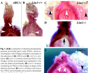
Fig. 1. (A,B) Localization of alkaline phosphatase
tempts to produce a genetic marker for the germ line have been made by introducing a reporter transgene into the locus of the TNAP and Oct4 genes. In the mouse embryo, the TNAP-lacZ allele is expressed in the PGCs only when they are localized in the gut endoderm. In the early gastrula, TNAP-lacZ activity is found unexpectedly in cells localised to the distal region of the
extraembryonic ectoderm (ex-ect) (MacGregor et al., 1995). This raises the question of whether the ex-ect is another potential source of PGCs in addition to the proximal epiblast. To test if the ex-ect may differentiate into PGCs, cells were isolated from the distal ex-ect tissue which contains the TNAP-lacZ expressing cells and transplanted to the proximal epiblast of the host embryo. Following transplantation, descendants of ex-ect cells were found in the extraembryonic and embryonic tissues that are normally colonised by epiblast cells grafted to the proximal epiblast. The most significant departure from the epiblast cells is the lack of differentiation of the ex-ect cells to PGCs as assessed by alkaline phosphatase activity (Table 1; Tam, unpublished). The ex-ect tissue, despite the presence of TNAP-lacZ expressing cells is unlikely to be a source of PGCs in the mouse embryo. It is, however, not clear why the ex-ect cells, which are derived from the polar trophectoderm, could colonize the tissues normally derived from the epiblast and whether the ex-ect descendants differentiate properly into extraembryonic mesoderm of the yolk sac, chorion and amnion.
The introduction of the EGFP transgene into the Oct4 locus results in the expression of green fluorescent protein widely in the epiblast with again no specific restriction to any putative germ cell precursors. The expression of this transgene marks the PGCs that colonize the gut endoderm and the mesentery, but surprisingly not the TNAP-expressing cells that are localized in the yolk sac and allantois (Anderson et al., 2000). The expression of the Oct4-EGFP allele only in a subset of the TNAP-expressing cells raises serious doubts that the TNAP-expressing cells in the extraembryonic tissues are indeed PGCs. It is postulated that these extraembryonic “PGCs” may never re-enter the embryo and contribute to the germ line (Anderson et al., 2000) which seems to be consistent with the finding that allantoic mesoderm is unable to colonize the embryonic mesoderm even after heterotopic transplantation into the primitive streak (Downs and Harmann, 1997). However, the precise fate of the “PGCs” in the allantoic mesoderm has not been tested directly. In transplantation experiments, some cells derived from trans-planted distal and proximal epiblast cells have been shown to express Oct4 as well as TNAP (Tam and Zhou, unpublished). However, it remains to be resolved whether or not they are genuine germ cells. The critical tests for the PGC identity of these graft-Fig. 2. Reduced population of primordial
germ cells in (A) Lim1 and (B) Hnf3β
mu-tant mouse embryos at different
somite-stages. Embryos were collected from
preg-nant mutant mice at 8.0 to 9.5 days post coitum. The somite number was scored and the specimens were processed for TNAP histochemistry using NBT-BCIP reagents. The genotype of the embryo was determined by PCR on samples of the yolk sac or anterior embryonic fragments.
A
B
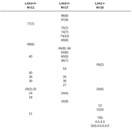
TABLE 2
PRIMORDIAL GERM CELL POPULATION IN 8.5-DAY WILD TYPE AND LIM1 HETEROZYGOUS AND HOMOZYGOUS MUTANT
EMBRYOS
Lim1+/+ Lim1+/-
Lim1-/-N=11 N=17 N=15
98(8) 87(8) 77(7)
75(7) 74(7) 73(10)
69(8) 68(6)
66(8), 66 63(8)
60 60(5)
56(7)
55(2) 53
50
35 35
30 30
27
25(2),25 25(6)
24 24(4)
18
16(9)
13 12(3) 11
7(6) 6,5,4,3 0(4),0,0,0,0,0
The TNAP-positive cells were scored on whole mount specimens with the histochemical reaction revealed by α-naphthyl phosphate and fast TR red reagents.
derived cells would be their ability to contribute to the germ line after re-introduction to the embryo directly or via the derivation of stem cells (embryonic germ cells) or their ability to initiate meiotic differen-tiation in vitro.
Molecular activity that affects the formation and
mainte-nance of the germ line
The search for the molecular control of the specification of the germ line has been focussed on the elucidation of the role of genes that are specifically expressed in the early PGCs. However, the ablation of TNAP function by targeted mutation of this gene has no impact on the formation of the germ cells (MacGregor et al., 1995). The repression of Oct4 activity in the ES cells induces loss of pluripotency and trophectodermal differentiation (Niwa et al., 2000). This may compromise the ability to use Oct4-deficient ES cells to study the impact of the mutation of the Oct 4 gene on germ cell formation. To date, we know of only three genes (Sl, W and Bmp4) in which the loss of their function affects PGCs. The Sl and W genes
encode the Steel factor and its receptor respectively which are known to be essential for mediating cell-cell interaction and migration. In the Sl/Sl and W/W mutant mice, normal numbers of PGCs are present at the primitive streak and gut endoderm in the gastrula- to early-organogenesis stage embryos. But the PGCs fail to migrate and are unviable, leading to a severe reduction of the PGC population and absence of germ cells in the fetal gonad (Buehr et al, 1993a,b). In the pre- to early-streak mouse
embryo, Bmp4 is expressed in the extraembryonic
ectoderm juxtaposing the proximal epiblast and later in the chorion and the extraembryonic mesoderm. The loss of Bmp4 function in the extraembryonic tissues is associated with the absence of TNAP-expressing PGCs in the mutant embryos (Lawson et al, 1999), suggesting that BMP signalling is required for the specification of the germ cell lineage by mediating the inductive tissue interaction between the extraembryonic tissue and the epiblast. How-ever, BMP activity may not be the sole determinant for germ cell formation since not all the descendants of the proximal epiblast will become PGCs. We have tested if an interaction with ex-ect may lead to induction of PGCs in the epiblast by tissue transplan-tation. However, no discernible induction of TNAP-expressing cells is found in the vicinity of the ex-ect tissue that was transplanted to the distal epiblast of the early-streak stage (Tam and Khoo, unpublished). When embryonic fragments containing different segments of the primitive streak were isolated from the late-streak to early-bud stage mouse embryo (nominally at 7.5 days post coitum) and cultured in vitro, TNAP-expressing cells were found only in explants containing the posterior segment of the primitive streak. In some of these explants, the presumptive PGCs were localized in the allantoic mesoderm or congregated to tissues that resemble the gut endoderm (Snow, 1981). This has led to the notion that the specification of the germ cell lineage
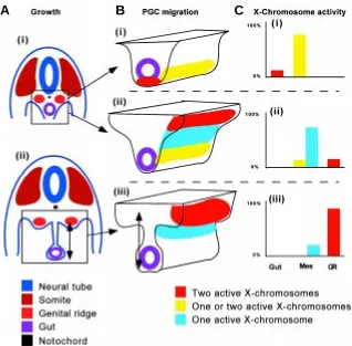
Fig. 3. (A) Schematic transverse section of the mouse embryo at (i) 9-10 days and (ii) 12 days,
showing the increasing separation of the genital ridges from the hindgut due to the expansion of the mesentery (double arrow). (B) The displacement of the primordial germ cells from the hindgut to the genital ridges at (i) 9, (ii) 10 and (iii) 12 days, by active migration and possibly the traction on the cohesive sheet of interconnecting cells which is anchored at the genital ridges as the mesentery expands dorso-ventrally (double arrows). The X-chromosome activity of the primordial germ cells changes at different phases of migration (colour-coded). (C) The relative proportion of the PGC population showing specific status of X-chromosome activity at different points of the migratory path (Gut, Mes, mesentery, GR, genital ridge) at (i) 9, (ii) 10 and (iii) 12 days.
A
B
C
is coincident with the formation of germ layers and that the progeni-tors of the PGCs are co-localized with those of the allantoic meso-derm and hindgut endomeso-derm in the late-gastrula stage embryo.
urogenital mesoderm precursors raises the question of whether the loss of Lim1 activity may also affect other cell lineages such as the primordial germ cells whose progenitors are co-localized with those of the urogenital primordium and the allantoic mesoderm.
To determine the effect of the Lim1 mutation on PGC formation, we have examined mutant embryos obtained from the mating between heterozygous mutant mice for TNAP activity. In the wild type and heterozygous Lim1 embryos, similar numbers of TNAP-positive cells were found at the base of the allantois and the endoderm of the hindgut (Fig. 1 A-C; Table 2). Some TNAP-positive cells in the heterozygous mutant embryos were dispersed in the yolk sac (Fig. 1C). Many gastrula-stage and early-somite stage Lim1-/- embryos were found lacking TNAP-positive PGCs (Fig. 1D) or much fewer PGCs than wild type and heterozygous embryos (Fig. 1E, Table 2). In a few mutant embryos that had normal or above normal numbers of TNAP-positive cells, many of these were scattered ectopically in the yolk sac mesoderm (data not shown). To test if the reduction of “PGCs” in the mutant embryo may be due to retarded development, TNAP-positive cells were scored against the somite number and the results show that PGCs are either not formed or are lost during gastrulation and early organogenesis of the mutant embryo (Fig. 2A). The development of the allantois in Lim1-/- embryos was also studied to reveal if a reduction in PGCs might be correlated with poor allantois development. In 16 Lim1-/- mutant embryos at 8.5 days, only 3 had a normal-length allantois, 6 had severely shortened allantois (e.g. Fig. 1E) and 7 had none. Among the mutant embryos with allantoic defects, 3 were developmentally retarded and the other 3 were at the pre-somite stage, when the formation of the full allantoic bud is expected (Downs and Davies, 1993). Other mutant embryos formed a bulbous allantois that failed to fuse with the chorion even when the embryo had developed to the early-somite stage.
Mutations of several other genes besides Lim1, such as Brachyury (T, Chesley, 1935), Bmp4 (Lawson et al., 1999), VCAM-1 (Gurtner
et al., 1995), Otx2 (Ang et al., 1996) and Hnf3β (Ang and Rossant,
1994), also result in a poorly developed allantois and the failure of chorioallantoic fusion. Among these mutant embryos, only Bmp4-/-embryos are reported to have completely lost PGCs (Lawson et al., 1999). Our preliminary analysis shows that, as in the Lim1 mutant,
the Hnf3β -/- embryos have no or very few PGCs (Fig. 2 A,B), but an
apparently normal number of PGCs are found in the T/T and Otx2-/- mutant embryos (Tsang and Tam, unpublished; Lawson et al.,
1999). In the Lim1 and Hnf3β mutant gastrula-stage embryos, Bmp4
is expressed in the extraembryonic tissues (Kinder et al., 2001).
Therefore, the absence of PGCs in some Lim1and Hnf3β mutant
embryos suggests that BMP4 activity alone is not sufficient to establish or maintain the population. However, the presence of
PGCs in a few Lim1and Hnf3β mutant embryos indicates that Lim1
and Hnf3β activities are probably not critical for the initial
establish-ment of the germ cell lineage but are required for the maintenance of the PGC population. In the Lim1 mutant embryo, the defects in PGCs seem to be closely correlated with abnormal differentiation of the urogenital tissues and the allantois (Tsang et al., 2000). However, in embryos with mutations in other genes, such as eed, the excessive production of extraembryonic mesoderm does not affect PGC forma-tion (Faust et al., 1995; 1998). Mouse embryos that carry a mutaforma-tion of the WT1 (Kreidberg et al., 1993), Emx2 (Miyamoto et al., 1997) or Lhx9 gene (Birk et al., 2000) do not form gonads but PGCs are present in the developing urogenital ridges at 11.5 to 12.5 days. The function of these genes therefore has no apparent impact on the specification of the germ cells or the formation of the gonadal primordium but is probably required for later events of gonad morphogenesis.
Proliferation and migration of the primordial germ cells
The formation of PGCs is accompanied by a lengthening of the cell cycle time as PGCs adopt a doubling time of 12-14 hours in contrast to the characteristic 6-7 hours for epiblast cells (Snow, 1977; Poelmann, 1980; Tam and Snow, 1981; Lawson and Hage, 1994; Power and Tam, 1993). Results of clonal anlayses have produced an estimate of 40 cells in the epiblast as the progenitor population from which the PGCs are formed (Lawson and Hage, 1994). The number of TNAP-expressing PGCs increases from about 150 to 26000 between 8.5 and 13.5 days post coitum, with a doubling of the population at every 12-14 hours (Tam and Snow, 1981). The expansion of the PGC population could be accounted for sufficiently by cell proliferation and does not seem to demand further recruitment to the lineage after gastrulation.
At the early-somite stage, PGCs congregate in the endoderm of the prospective hindgut of the embryo. Subsequently, PGCs are re-located from the ventral to the dorsal aspect of the gut and, over the next 3-4 days, migrate from the hindgut endoderm through the mesentery and colonize the urogenital ridges. The histological and ultrastructural features of the PGCs in the mesentery are consistent with an active migratory behaviour. However, the “migratory” PGCs are found to be interconnected by cellular processes (Gomperts et al, 1994). It is not known whether these cell processes may lead to some form of physical intercellular link among all PGCs in the population. Nevertheless, this discovery raises an intriguing possibility that the migration of PGCs may involve the movement of not just individual cells but the concerted displacement of the population if the cells are physically interconnected in a cell sheet. It is plausible that, because of the proximity of the hindgut to the primordium of the urogenital ridges when it is first formed in the intermediate mesoderm (Fig. 3 A,B), the pioneer PGCs may have entered and anchored themselves in the urogeniral ridge promptly after they have left the gut endoderm (Fig. 3A). Subsequently, the urogenital ridges are separated from the hindgut by the extension of the mesentery (Fig. 3A). If the pioneer PGCs are maintaining a stable connection with later waves of PGCs, the lengthening of the mesentery may generate a traction on the
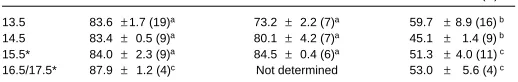
TABLE 3
X-CHROMOSOME ACTIVITY OF GERM CELLS IN THE FETAL OVARY AND AT EXTRAGONADAL SITES AT
13.5-17.5 DAYS POST COITUM (DPC)
Age (dpc) Germ cells in XX ovary (N) Germ cells in XXSxra testis (N) Extragonadal germ cells in XX fetuses (N)
13.5 83.6 ±1.7 (19)a 73.2 ± 2.2 (7)a 59.7 ±8.9 (16) b 14.5 83.4 ± 0.5 (9)a 80.1 ± 4.2 (7)a 45.1 ± 1.4 (9) b 15.5* 84.0 ± 2.3 (9)a 84.5 ± 0.4 (6)a 51.3 ±4.0 (11) c 16.5/17.5* 87.9 ± 1.2 (4)c Not determined 53.0 ± 5.6 (4) c
sheet of interconnected PGCs (Fig. 3B). This traction force may contribute to the displacement of the PGCs towards the urogenital ridges, in addition to active cell migration that is triggered by some homing signals (Tilmann and Capel, 1999; Van Doren et al, 1998).
The tissue environment influences the X-chromosome
activity of the germ cells
The serendipitous creation of a line of transgenic mice that expresses an X-linked lacZ transgene (Tam and Tan, 1992) has provided a unique opportunity to visualize the activity of the X-chromosome in individual germ cells by histochemical means (Tam et al, 1994a,b). Results of our study on transgenic activity have not only reaffirmed that PGCs do inactivate the X-chromosome to down-regulate the X-linked gene dosage like all other somatic cells (Monk and McLaren, 1981), but have provided additional information on the timing of the regulation of X-chromosome activity. Our results show that PGCs in the hindgut maintain two active X-chromosomes and inactivation occurs when PGCs have commenced migration. PGCs re-activate the X-chromosome only after they have gained entry to the genital ridges and re-activation may occur coincidentally with the entry into meiosis of germ cells in the fetal ovary. Several populations of PGCs could therefore be distinguished in the early-organogenesis stage embryo by their location along the migratory pathway and the status of the X-chromosome activity (Fig. 3C, Tam et al., 1994b). That the change in X-chromosome activity may be correlated with the location of the PGCs strongly suggests that the X-chromosome activity of the female germ cells is unlikely to be regulated on a chronological basis but more by mechanisms that are coupled to cellular behaviour and site-specific tissue interactions.
Results of the analysis of X-linked transgene activity show un-equivocally that entry to the genital ridges is an essential trigger for the re-activation of the X-chromosome. PGCs that are chronologi-cally similar to those that have colonized the genital ridge but have not yet arrived in the genital ridge do not re-activate the X-chromo-some (Tam et al., 1994b). PGCs that are found outside the genital ridge after 13.5 days post coitum also do not seem able to re-activate the X-chromosome and display only one active X-chromosome (Table 3). Some of these ectopic germ cells that are localized in the mesonephros and adrenal glands (Francavilla and Zamboni, 1985) will proceed to meiosis at about 16.5-17.5 days despite the absence of the re-activation of X-chromosome (Tam and Zhou, unpublished). These ectopic germ cells, however, will not form functional germ cells and degenerate postnatally (McLaren, 1983; Upadhyay and Zamboni, 1982). Attempts to undergo meiotic differentiation without proper X-chromosome activity may underpin the failure of the ectopic germ cells to accomplish oogenesis. It has been postulated that the trigger to re-activate the X-chromosome acts locally within the developing ovary. Analysis of the X-linked transgene activity of X/XSxr germ cells in the sex-reversed gonad that develops testicular morphology shows that the germ cells can re-activate the X-chromosome (Jamieson et al., 1998; Table 2) and proceed to meiosis (McLaren, 1981b) even in the inappropriate gonadal environment. This sug-gests that the trigger to re-activation is specific to the genital ridge but can be uncoupled from ovarian morphogenesis.
Acknowledgement
This paper is a tribute to Anne’s enormous contribution to the discipline of developmental biology and an appreciation of her constant encourage-ment and generous support of PPLT’s research for the past 20 years. The
research program in PPLT’s laboratory is supported by the National Health and Medical Research Council (NHMRC) of Australia, the Ramaciotti Foun-dation and Mr James Fairfax (PPLT), and the Human Frontier Science Program (SLA, RRB, PPLT and Hiroshi Sasaki). We thank David Loebel and Peter Rowe for reading the manuscript, S.S. Tan for the H253 mice and G.C. Enders for the gift of the GCNA antibody. RVJ was a NHMRC Medical Postgraduate Scholar. PPLT is an NHMRC Senior Principal Research Fellow.
References
ANDERSON, R., COPELAND, T.K., SCHOLER, H., HEASMAN, J. and WYLIE, C. (2000). The onset of germ cell migration in the mouse embryo. Mech. Dev. 91: 61-68.
ANG, S-L., JIN, O., RHINN, M., DAIGLE, N., STEVENSON, L. and ROSSANT, J. (1996). A targeted mouse Otx2 mutation leads to severe defects in gastrulation and formation of axial mesoderm and to deletion of rostral brain. Development 122: 243-252.
ANG, S.-L. and ROSSANT, J. (1994) HNF-3β is essential for node and notochord formation in mouse development. Cell 78: 561-574.
BARNES, J.D., CROSBY, J.L., JONES, C.M., WRIGHT, C.V.E. and HOGAN, B.L.M. (1994). Embryonic expression of Lim1, the mouse homologue of Xenopus Xlim1, suggests a role in lateral mesoderm differentiation and neurogenesis. Dev. Biol. 161: 168-178.
BIRK, O.S., CASIANO, D.E., WASSIF, C.A., COGLIATO, T., ZHAO, L., ZHAO, Y., GRINBERG, A., HUANG, S., KREIDBURG, J.A., PARKER, K.L., PORTER, F.D. and WESTPHAL, H. (2000). The LIM homeobox gene Lhx9 is essential for mouse gonad formation. Nature 403: 909-913.
BUEHR, M., McLAREN, A., BARTLEY, A. and DARLING, S. (1993a). Proliferation and migration of primordial germ cells in We/We mouse ebmryos. Dev. Dyn. 198:182-189.
BUEHR, M., PERACE-KELLY, and McLAREN, A. (1993b). Failure of mouse primordial germ cells to proliferate on fibroblasts from Steel mutant mice in vitro. Biol. Res. 26: 411-415.
CHESLEY, P. (1935). Development of the short-tailed mutant in the house mouse. J. Exp.Zool. 70: 429-453.
DE SMEDT, V., SZOLLOSI, D. and KLOC, M. (2000). The Balbiani body: asymmetry in the mammalian oocyte. Genesis 26: 208-212.
DOWNS, K.M. and DAVIES, T. (1993). Staging of gastrulating mouse embryos by morphological landmarks in the dissecting microscope. Development 118:1255-1266.
DOWNS, K.M. AND HARMANN, C. (1997). Developmental potency of the murine allantois. Development 124: 2769-2780.
ENDERS, G.C. and MAY, J.J. II (1994). Developmentally regulated expression of mouse germ cell nuclear antigen examined from embryonic day 11 to adult in male and female mice. Dev. Biol. 163: 331-349.
FAUST, C., SCHUMACHER, A., HOLDENER, B. and MAGNUSON, T. (1995). The eed mutation disrupts anterior mesoderm production in mice. Development 121: 273-285.
FAUST, C., LAWSON, K.A., SCHORK, N.J., THIEL, B. and MAGNUSON, T. (1998). The polycomb-group gene eed is required for normal morphogenetic movements during gastrulation in the mouse embryo. Development 125: 4495-4506.
FRANCAVILLA, S. and ZAMBONI, L. (1985). Differentiation of mouse ectopic germinal cells in intra- and perigonadal locations. J. Exp. Zool. 233: 101-109.
FUJII, T., PICHEL, J.G., TAIRA, M., TOYAMA, R., DAWID, I.B. and WESTPHAL, H. (1994). Expression patterns of the murine LIM class homeobox gene Lim1 in the developing brain and excretory system. Dev. Dyn. 199: 73-83.
GINSBURG, M., SNOW, M. H. L. and McLAREN, A. (1990). Primordial germ cells in the mouse embryo during gastrulation. Development 110: 521-529.
GOMPERTS, M., GARCIA-CASTRO, M., WYLIE, C. and HEASMAN, J. (1994). Interactions between germ cells play a role in their migration in mouse embryos. Development 120: 135-141.
JAMIESON, R. V., ZHOU, S. X., WHEATLEY, S. C., KOOPMAN, P. and TAM,P.P.L. (1998). Sertoli cell differentiation and Y-chromosome activity: A developmental study of X-linked transgene activity in sex-reversed X/XSxra mouse embryos. Dev. Biol. 199: 235-244.
KINDER, S.J., TSANG, T.E., QUINLAN, G.A., HADJANTONAKIS, A-K., NAGY, A. and TAM, P.P.L. (1999). The orderly allocation of mesodermal cells to the extraembryonic structures and the anteroposterior axis during gastrulation of the mouse embryo. Development 126: 4691-4701.
KINDER, S.J., TSANG, T.E., ANG, S.L., BEHRINGER, R.R. and TAM, P.P.L. (2001). Defects of the body plan of mutant embryos lacking Lim1, Otx2 and Hnf3ß activity. Int. J. Dev. Biol. 1: 347-356.
KREIDGURG, J.A., SARIOLA, H., LORING, J.M., MAEDA, M., PELLETIER, J., HOUSMAN, D. and JAENISCH, R. (1993). WT-1 is required for early kidney development. Cell 74: 679-691.
LAWSON, K.A., MENESE, J.J. and PEDERSON, R.A. (1991). Clonal analysis of epiblast fate during germ layer formation in the mouse. Development 113: 891-913.
LAWSON, K.A. and HAGE, W.J. (1994). Clonal analysis of the origin of primordial germ cells in the mouse. In Germline Development. (Ciba Found. Symp 182) Chichester: Wiley, pp 68-92.
LAWSON, K.A., DUNN, N.R., ROELEN, B.A., ZEINSTRA, L.M., DAVIS, A.M., WRIGHT, C.V., KORVING, J.P. and HOGAN, B.L. (1999). Bmp4 is required for the generation of primordial germ cells in the mouse embryo. Genes Dev. 13: 424-436.
MacGREGOR, G.R., ZAMBROWICA, B.P. and SORIANO, P. (1995). Tissue non-specific alkaline phosphatase is expressed in both embryonic and extraembryonic lineages during mouse embryogenesis but is not required for migration of primordial germ cells. Development 121: 1487-1496.
McLAREN, A. (1981a). Germ Cell and Soma. Yale Univ. Press, New Haven., CT.
McLAREN, A. (1981b). The fate of germ cells in the testis of fetal Sex-reversed mice. J. Reprod. Fert 61: 461-467.
McLAREN, A. (1983). Studies on mouse germ cells inside and outside the gonad. J. Exp. Zool. 228: 167-171.
MONK, M. and McLAREN, A. (1981). X-chromosome activity in foetal germ cells of the mouse. J. Embryol. Exp. Morphol. 63: 75-84.
MIYAMOTO, N., YOSHIDA, M., KURATANI, S., MATSUO, I. and AIZAWA, S. (1997). Defects of urogenital development in mice lacking Emx2. Development 124: 1653-1664.
NIWA, H., MIYAZAKI, J.-I. And SIMTH, A. G. (2000). Quantitative expression of Oct3/ 4 defines differentiation, dedifferentiation or self-renewal of ES cells. Nat. Genet. 24: 372-376.
OZDZENSKI, W. (1967). Observations on the origin of primordial germ cells in the mouse. Zool. Pol. 17: 367-379.
PALMIERI, S.L., PETER, W., HESS, H., and SCHOLER, H.R. (1994). Oct-4 transcrip-tion factor is differentially expressed in the mouse embryo during the establishment of the first two extraembroynic cell lineages involved in implantation. Dev. Biol. 166: 259-267.
PESCE, M., WANG, X., WOLGEMUTH, D.J. and SCHOLER, H. (1998). Differential expression of the Oct-4 transcription factor during mouse germ call development. Mech. Dev. 71: 89-98.
POELMANN, R.E. (1980). Differential mitosis and degeneration patterns in relation to the alterations in the shape of the embryonic ectoderm of early post-implantation mouse embryos. J. Embryol. Exp. Morph. 55: 33-51.
POWER, M.A. and TAM, P.P.L. (1993). Onset of gastrulation, morphogenesis and somitogenesis in mouse embryos displaying compensatory growth. Anat. Embryol. (Berl) 187: 493-504.
QUINLAN, G.A., WILLIAMS, E.A., TAN, S-S. and TAM, P.P.L. (1995). Neuroectoder-mal fate of epiblast cells in the distal region of the mouse egg cylinder: Implication for body plan organization during early embryogenesis. Development 121: 87-98.
SCHOLER, H.R., DRESSLER, G.R., BALLING, R., ROHDEWOHLD, H. and GRUSS, P. (1990). Oc4: a germline-specific transcription factor mapping to the mouse t-complex. EMBO J 9: 2185-2195.
SHAWLOT, W. and BEHRINGER, R.R. (1995). Requirement for Lim1 in head-organizer function. Nature 374: 425-430.
SNOW, M.H.L. (1977). Gastrulation in the mouse: growth and regionalisation of the epiblast. J. Embryol. Exp. Morph. 42: 293-303.
SNOW, M.H.L. (1981). Autonomous development of parts of isolated parts from primitive streak stage mouse embryos. Is development clonal? J. Embryol. Exp. Morphol. 65: 269-287.
TAM, P. P. L. and SNOW, M.H. L. (1981). Proliferation and migration of primordial germ cells during compensatory growth in the mouse embryo. J. Embryol. Exp. Morph. 64: 133-147.
TAM, P.P.L. and TAN, S-S. (1992). The somitogenetic potential of cells in the primitive streak and the tail bud of the organogenesis stage mouse embryo. Development 115: 703-715.
TAM, P. P. L., WILLIAMS, E. A. and TAN, S.-S. (1994a). The expression of an X-linked HMG-lacZ transgene in early mouse embryos: Implications of parental imprinting and lineage-specific X-chromosome activity. Dev. Genet. 15: 491-503.
TAM, P.P.L., ZHOU, S.X. and TAN, S-S. (1994b). X-chromosome activity of the mouse primordial germ cells revealed by the expression of an X-linked lacZ transgene. Development 120: 2925-2932.
TAM, P.P.L. and ZHOU, S.X. (1996). The allocation of epiblast cells to ectodermal and germ-line lineages is influenced by the position of cells in the gastrulating mouse embryo. Dev. Biol. 178: 124-132.
TILMANN, C. and CAPEL, B. (1999). Mesonephric cell migration induces testis cord formation in the mammalian gonad. Development 126: 2883-2890.
TSANG, T.E., SHAWLOT, W., KINDER, S.J., KOBAYASHI, A., KWAN, K.M., SCHUGHART, K., KANIA, A., JESSELL, T.M., BEHRINGER, R.R. and TAM, P.P.L. (2000). Lim1 activity is required for intermediate mesoderm differentiation in the mouse embryo. Dev. Biol. 223: 77-90.
UPADHYAY, S. and ZAMBONI, L. (1982). Ectopic germ cells: Natural model for the study of germ cell sexual differentiation. Proc. Natl. Acad. Sci. USA 79: 6584-6588.
VAN DOREN, M., TARCZY BROIHIER, T., MOORE, L.A. and LEHMANN, R. (1998). HMG-CoA reductase guides migrating primordial germ cells. Nature 396:, 466-469.