Double-Blind Peer Reviewed Refereed Open Access International e-Journal - Included in the International Serial Directories
Mathematical Modeling on the study of Spherical Tumor In Relation To Variation of Temperature in Dermal Layers
V.K. Katiyar1, K.S. Basavarajappa2, Ashwini M Rao3 1. Department of Mathematics, Indian Institute of Technology, Roorkee
2. Department of Mathematics, Bapuji Institute of Engineering and Technology, Davangere 3. Department of Mathematics, Bapuji Institute of Engineering and Technology, Davangere
Abstract
The mathematical study through analytical formulations evaluates the measurement of the size of the tumor due to increase in the metabolic heat rate in relation to skin temperature variation which is caused by the absorption of the varying environmental factors and some specific characters of heredity. The measurement of necrotic core temperature of the spherical tumor suggests the non linearity in the heat conduction. That is the onset of increase of metabolic heat rate from 0.1 to 0.1260 C (10 C=2.98790 F) in the necrotic core. Mathematical modeling concerns the study of spherical growth (heterogeneity) of the tumor by formulating using Bio-heat equation for C-ring shaped tumor for asymptotic approximation. Partial differential equations discretised into the set of ordinary differential equations representing the temperature distribution in three layers are employed in the modeling. Bessel’s series solution method is used in the analysis. The incidence or the initial radius(rI) to the final radius (rF) of the
tumor growth in the necrotic core is considered to study the dynamic growth using Runge-Kutta Fehlberg method. Results suggest that there exists a close relationship between the increase of metabolic heat rate and the spherical growth in the size of the tumor. Numerical computations suggest there is an observable variation in the necrotic core temperature. This causes a slight deviation from the temperature in dermal and epidermal layers.
Key words: bio-heat, spherical tumor, necrotic core, metabolic heat rate
Address for Correspondence:
Dr. K.S.Basavarajappa, Professor and Head,
Department of Mathematics,
Introduction
Tumor is caused by body tissues that grow to form an abnormal mass. This abnormal growth is initiated by irregular cell formations or unbalanced cell division. When tumors are benign, they typically grow at a slow rate. Usually benign tumors are harmless and do not spread to other parts of the body. Detection of benign tumor can also be known by the increase of constant temperature in the dermal layer. Though benign tumors are usually innocuous, their growth can interface with the ability of healthy tissues to grow and thrive. In fact they may grow large enough to apply pressure to vital body organs, resulting in serious illness. When benign tumors become too large, they may require surgical removal for cosmetic purposes or to preserve surrounding tissues. Once removed, benign tumors usually do not return.
Body temperature depends on the balance of heat produced by the metabolically active cellular tissues and the heat lost via skin to the environment. Skin consists of three layers, epidermis, that is cellular but vascular, dermis which is fibrous with small blood vessels along with several epithelial cells and subdermal layer (adjacent to dermal layer). Average adult possess 1.9m2 as surface area with 0.05 to 3.0 mm as thickness and temperature of 34.10 C for epidermis and 36.60 C (10 C = 2.98790 F=9.27270 K) for the deep part of the body. In radial position the temperature surrounding the tumor varies from 250C to 390 C. But at the necrotic core, it is the threshold temperature of 390 C and increases slowly with metabolic heat generation. The thermal interaction between tissue and blood is the subject of the tumor detection and the type of tumor distant from epidermis. The local removal of heat and the arrest of the circulation play a role in detecting the malignant tumor in the human body. The analysis of heat transfer interact with the use of ultrasound and microwave radiation(infrared heat waves of 5 to 20 microns) to produce local hyperthermica in the direction of air for both physical and cancer therapy.
Penner [1] found that the most suitable bioheat equation to describe the tissue and arterial blood pressure in the resting human forearm. Cooper et al. [2] correlated the thermal properties of human tissue with water content. Katiyar et al. [3] analyzed the transient heat transfer analysis for moving boundary transport problems for infinite media. Basavarajappa et al. [4] studied the bio-heat distribution in spherical tissue layers: with reference to an application to thermal spherical tumor. Kai Yue et al. [5] discussed an analytic solution of one-dimensional steady-state Penner bioheat transfer equation in cylindrical coordinates.Yang Yang et. Al [6] described the detection of atherosclerosis through mapping skin temperature variation caused by carotid atherosclerosis plaques.
In view of the study carried out by the researchers, we have made an attempt to understand the thermal effects of different dermal layers. Measurement of variation in the temperature distribution among the dermal layers is approximated using Fisher-Kolmogoroff equation for the asymptotic and nonlinear behavior in the tumor. The Cring shaped tumor is
Double-Blind Peer Reviewed Refereed Open Access International e-Journal - Included in the International Serial Directories
computed for its diameter and it is observed that each ring shape spherical layer will be with the increased diameter.
Therefore heat sensitive tumor with elevated temperature inadequately (420C i.e. 0.1250C to 4.908250C) exhibit enhanced spread and metastasis. Most of the studies (with theoretical and experimental) demonstrate the thermal effects for variable mass flow rate in supplying arteries which supply the perfused blood to the smaller vessels of tissue. In solid tumor, lethal effects may be observed due to pronounced lysosomal activity by a relatively high anaerobic glycolysis that gives rise to a higher intra and extracellular acidity in the tumor tissue. The region is then divided into three natural layers (epidermis, dermis and subdermal tissue). Further subdivisions have been analyzed for normal temperature ranging from 34.10C to 370C and further upto 420C for tumor location.
Formulation
By considering the principle of conservation of energy in an element of tissue, the bio-heat transfer study is based on bio-heat transfer mechanism in tissue which must not only account for conduction but also heat addition or removal by blood supply and the metabolic heat generation.
The blood circulation around the tumor through capillaries is considered to be pulsatile flow and the flow is assumed as non linear, the physiological time in dimensionless form for the blood flow in the micro vessels is employed in the model. Also the variation of the average tumor temperature is evaluated in reference to blood perfusion.
Governing bio heat transfer equation is given by,
2
b b b
T K T m C ( T - T ) + S = ρ C
t
(1)
a
T
K h ( T - T ) + LE η
(2)
Each part of the human body is assumed to be spherical with variable thermal conductivity in biological tissues depending on the blood flow near the skin. Because the biological tissues generate heat uniformly supplied with blood at constant arterial temperature (i.e. convection by blood flow, vascular heat exchange and the tissue metabolism). For normal temperature distribution studies, the parameters T, m, Tb and S are all independent of time.
However, for the situations like benign tumor, malignant tumors and some injuries, the parameters may be considered as time dependent to a smaller extent. Metabolic heat generation and blood mass flow rate are zero at epidermis and constant in subdermal layer. In order to understand the variation in the metabolic heat generation and blood mass flow rate of the three different layers, we study the bioheat equation with variable at dermal layers.
Transforming equations (1) and (2) into spherical polar coordinate system, considering in one –dimensional steady state,
i i
2 i
i b b b i i
2
1
1 i 1 a i i
dT 1 d
K r + M C (T - T ) + S = 0
dr dr
r dT
K = h [T - T ] + L E dr
2 i
i i b i i
2
1
1 1 a
dT 1 d
K r + Q (T - T ) + S = 0
dr dr
r dT
K = h[T - T ] + L E dr
(4)
i i
i b b 3 b
where Q = M C , T = T
For i = 1, the temperature is the same as blood temperature. By convection, radiation and evaporation heat loss takes place when epidermis is exposed to atmospheric temperature. Thermal conductivity is constant in dE (thickness of epidermis), dS (thickness of sub-dermal). Consider
3 2 3 2
3
d T d k d T
K =
d r r d r (5)
D E
S r = d r = d
r = d
1 2 2 3 3 b
[T = T ] , [T = T ] , [T = T ] (6)
dE = d4 - d3, (epidermis 0.5cm), dD = d3 - d2, (dermis 1.0cm) and dS = d2 - d1,
(subdermal 1.2cm)
In general, Ti = Tb [ 1 - ξi ] is taken as dimensionless quantity
i = 1, 2, 3 (epidermis, dermis, subdermis), we get
2 1
1
dξ d
K r = 0 ( For i = 1)
dr dr (7)
22 2 2 2
2 2
1
d ξ dξ m + s
r + r - r ξ = 0 (For i = 2)
dr 2K
dr
(8)
2
3 3
3 2
1
d ξ dξ m + s
r + 2 - r (ξ ) = 0 (For i = 3)
dr 2K
dr
(9)
New boundary conditions are,
b
1 1
a
1 1 1
dξ hξ h L E
= + ξ + (subdermal region , i = 3)
dr k k K T (10)
where b a
a
b
T - T ξ =
T (atmospheric temperature 15
0
C to 330C)
D 3 1 2 1 d K dξ dξ
K = (dermis interface, i = 2)
dr r dr (11)
D
3 2 3
3
d K dξ dξ
= K (epidermis interface, i = 1)
r dr dr (12)
D E
S r = d r = d
r = d
1 2 2 3 3
[ξ ξ ] , [ξ = ξ ] , [ξ = 0] (13)
Analysis
Double-Blind Peer Reviewed Refereed Open Access International e-Journal - Included in the International Serial Directories
1 1 2
1 ξ = A - + A
r
(14)
2 3 0 4 0
ξ = A I (rε) + A K (rε) (15)
and
r ε- r ε
3 5 6
e
ξ = A + A e
r
(16)
where,
1 2
1
m +S ε =
S K
and A1, A2, A3, A4, A5 and A6 are the constants due to the solutions of second
order differential equations (7), (8) and (9)
I0 (rε) and I1( rε ) are the modified Bessel’s functions of the first kind, K0(rε) and K1(rε)
are the modified Bessel’s functions of the second kind. The constant in the Bessel’s modified series is taken as 0.57721 for numerical computations of K0(rε) and K1(rε).
For epidermis, i = 1
1 1
1
1 1 0 2 1 1 2 0 1 2 3 3 4
γ
ξ = [{δ K (rε) + δ K (rε)}I (rε) + K (rε){δ K (rε) + I (rε)}δ + γ {δ + d δ }] r
(17) For dermis, i = 2
1 1
2 2 1 0 2 0 1 0 2 0
ξ = γ [{δ K (rε) + δ K (rε)}I (rε) - {δ I (rε) - δ K (rε)}K (rε) (18) For sub dermal, i = 3
0
rε
- rε 2
3 3 1 0 1 0 1 2 1 1 1
2
4 1 0 1 1 2 1 1 1
e ξ = γ [{δ K (rε) I (rε) + I (rε)K (rε)} + δ {I (rε)K (rε) + (K (rε)) }]
r
e + γ [δ {K (rε)I (rε) + I (rε)K (rε)} + δ {I (rε)K (rε) + (K (rε)) }]
r
(19)
The study of Cring shaped spherical tumor by quantifying the role of heat conduction,
convection and metabolism is analyzed with reference to heat transfer distribution in tumors under the cases of elevated temperature in the dermal layers. Temperature rise in tumors is always due to variation in metabolism as a function of tumor weight (between 2 gm to 20 gm). The corresponding metabolic heat rate (between 0.0 to 5.2) and the temperature difference between (0.01850C to 0.240C) have been employed in the computations. By physiological point of view the temperature in the periphery of the necrotic tumor is higher than that in the central necrotic core. Taking the Cring spherical shape of the tumor growth initially with radius ‘a’ and
with time‘t’ (constant time periods), the varying radius of the tumor is given by,
r = R (t) (20)
Clearly
R (0) = a (21)
The equations for the tumor consisting of pressure P and the nutrient concentration (by anaerobic)are,
Where Sv is the rate of volume loss (model does not include the motivated cell loss mechanism
and shrinkage necrosis)
2ζ 0
[Outside the surface of the tumor layer] (23)
Taking the constant nutrient initially ς and attains maximum ςm at r = R(t). (max)
max
ζ = ζ at R(t) R = r (24)
^
1
n . ζ = μ ζ - ζ at r = R(t) (25) Transforming equation (31) into spherical polar form
2 21 ζ
r 0, r r(t)
r r
r
(26)
Solving equation (28) for radius at initial I and at final F using equations (22), (23), (26), (27), we obtain
rI = 2 Dρ 1/2t 1 − 1
ρt log 4πDt
∂ N0
1/2
(27)
rF = 2 Dρ 1/2t 1 −ρ1t log 4πDtN∂
0
1/2
(28)
Since the tumor growth is considered to be spherical form from the necrotic core towards the outer layer (subdermal dS to epidermis dE), we define the tumor profile as
rA = 1
2 rI+ rF
Where rA is the mean radius of rI and rF
rA = 1
2 2 Dρ 1/2t 1 −
1
ρt log 4πDt
∂
N0
1/2
I
+ 2 Dρ 1/2t 1 − 1
ρt log 4πDt
∂
N0
1/2
F
(29)
Then the mean radial distance of tumor formation from the necrotic core (origin) to the rupture size is modeled using Fisher-Kolmogoroff equation as,
rm = rI+rF
2 =
r0∞ 2c r,t dr
r c r,t dr0∞ = r
2c (r, t)dr
∞
0 (30)
Here c r, t = 2π C0λ ring(r, t, r0)r0dr0 (31)
c r, t = e−r2 4t
1 − e−λ2 4t +r2
4te −r2 4t
1 − e−λ2 4t 1 +λ2
4t + ⋯ (32)
which is called the asymptotic approximation to various temperature distribution values. By employing the expression for cell density in equation (30),
rm = 1 − e−Aλ2 π
4A3/2 + A − Ae−Aλ
2
− A2λ2e−Aλ2 3 π
8A5/2 + ⋯ (33)
Neglecting second and higher terms,
rm = 1 − e−Aλ2 π
Double-Blind Peer Reviewed Refereed Open Access International e-Journal - Included in the International Serial Directories
The temperature rise in the distribution among the three layers is due to metabolism as a function of tumor size, the tumor grows larger and it develops a central necrosis, (the temperature is maximum). Tumor cells are more sensitive to heat than normal cells. Introducing equations (27) and (28) into (19) under the assumption that the spherical tumor can develop adequate nourishment in the necrotic core , then for various values of ξ3 in the radius range 0.25 - 1.05cms we obtain,
3 I 3
3 F
ξ (r ), r = 2.5cm ξ (r) =
ξ (r ), r = 0.5cm
(35)
2 I 2
2 F
ξ (r ), r = 2.5cm ξ (r) =
ξ (r ), r = 0.5cm
(36)
Where rI modeled as 1
AE
I I E E E E E
r = r C Where C = h A (T - T )
Then replacing rI and rF in ξ3 , we compute the values of ξ (r )3 I and ξ (r )3 F . The rise of temperature (nonlinearly in the distribution) in any location describes the progression of the tumor with certain initial radius. This helps in detecting the tumor in the field of thermography using hyperthermia for the minimum size of the tumor (initial stage) when associates with the rise of temperature of the core (i.e. in the subdermal region taken asξ )3 .
Results and Discussion
Mathematical model with Cring shaped spherical tumor predicts the relative metabolic
heat generation in the three spherical layers. Numerical results influence the factors for the improvement on the detection of abnormalities in the temperature pattern in the dermal region. Asymptotic approximation and Numerical scheme suggest the growth of tumor in spherical form due to an increase of metabolic heat rate leads to threshold shift of 0.1250 C. Results have shown that the irregularities between the temperature distributions in the range 1.0 to 1.6 cm at the skin surface will cause the elevation in the temperature of surrounding tumor. The study indicates the variation of Cring shaped tumor due to the variation of
temperature.
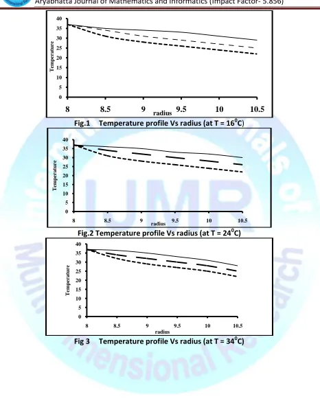
From the figures we observe that the temperature distribution for metabolic heat rate is at 0.029 cal/cm3-min (Fig 1), 0.018 cal/cm3-min (Fig 2), 0.018 cal/cm3-min (Fig 3). Also Fig. (4) shows the increasing of metabolic heat rate in tumor. The difference in the temperature of the necrotic core in the tumor along the spherical layers (towards the decreasing radii of tumor layers) and the subdermal layers is between 0.1270C – 0.2880 C. Therefore the increasing temperature at the necrotic center may indicate the viable tumor in the human mammary epitheliomas and in the surrounding normal tissues.
Referring to Fig 5, we notice that Cring shaped spherical tumor growth becomes
Fig.1 Temperature profile Vs radius (at T = 160C)
Fig.2 Temperature profile Vs radius (at T = 240C)
Fig 3 Temperature profile Vs radius (at T = 340C)
0 5 10 15 20 25 30 35 40
8 8.5 9 9.5 10 10.5
Te
m
p
e
r
a
tu
r
e
radius
0 5 10 15 20 25 30 35 40
8 8.5 9 9.5 10 10.5
Te
m
p
e
r
a
tu
r
e
radius
0 5 10 15 20 25 30 35 40
8 8.5 9 9.5 10 10.5
Te
m
p
e
r
a
tu
r
e
Double-Blind Peer Reviewed Refereed Open Access International e-Journal - Included in the International Serial Directories
Fig4 radius Vs Tumor temperature Fig 5 Time v/s radius of tumor
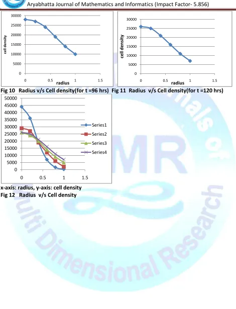
Fig 6 Time v/s mean radius of tumor Fig 7 Radius v/s Cell density(for t = 24 hrs)
Fig 8 Radius v/s Cell density(for t = 48 hrs) Fig 9 Radius v/s Cell density(for t =72 hrs)
0 0.1 0.2 0.3 0.4 0.5 0.6 0.7
0 100 200
ra d iu s o f Tu mo r time 0 0.1 0.2 0.3 0.4 0.5 0.6 0.7
0 50 100
ra d iu s time 0 5000 10000 15000 20000 25000 30000 35000 40000 45000 50000
0 0.5 1 1.5
ce ll d e n si ty radius 0 5000 10000 15000 20000 25000 30000 35000
0 0.5 1 1.5
ce ll d en si ty radius
x- axis: radius, y-axis: Tumor temperature
0 2 4 6 8 10 12 14 16
0.5 1 1.5 2 2.5 3 3.5 4
0 5000 10000 15000 20000 25000 30000
0 0.5 1 1.5
Fig 10 Radius v/s Cell density(for t =96 hrs) Fig 11 Radius v/s Cell density(for t =120 hrs)
x-axis: radius, y-axis: cell density Fig 12 Radius v/s Cell density
0 5000 10000 15000 20000 25000 30000
0 0.5 1 1.5
ce
ll
d
e
n
si
ty
radius
0 5000 10000 15000 20000 25000 30000
0 0.5 1 1.5
ce
ll
d
en
si
ty
radius
0 5000 10000 15000 20000 25000 30000 35000 40000 45000 50000
0 0.5 1 1.5
Series1
Series2
Series3
Double-Blind Peer Reviewed Refereed Open Access International e-Journal - Included in the International Serial Directories
NOMENCLATURE
T – temperature
k – thermal conductivity (cal/cm-sec) mb –blood flow rate (ml/cm2-sec)
cb – specific heat of blood (cal/gm- 0 C)
Tb – blood temperature
S – metabolic heat rate (cal/cm2)
𝜌 - tissue density (gm/cm2)
C – specific heat of tissue (cal/gm-0C) h – heat transfer coefficient
t – time
L – latent heat of evaporation E – rate of evaporation
Ta – atmospheric temperature (0C) 𝜉 - non dimensional temperature dE – thickness of epidermis layer (d4-d3)
dD – thickness of dermis layer (d3-d2)
dS – thickness of subdermal layer (d2-d1)
Qi = mbicbi
ξa = Tb − Ta
Tb
𝐸 = 𝑚 + 𝑆
2𝑘1
1/2
a – radius of the tumor initially R(t) = radius of the tumor at time t
𝛿 - thickness of constriction
𝛾 – sensitivity constant rI – initial radius
rF – final radius
I0 rε , I1 rε - modified Bessel’s functions of first kind K0 rε , K1 rε - modified Bessel’s functions of second kind
D – homogenous dissusion C – cell density
REFERENCES:
1. Penner H H *1948+, ‘Analysis of tissue and arterial blood pressure in the resting human fore arm’, J. Applied physiology, Vol.1, No. 2, 93 - 122.
2. Cooper T E and Trezek G J *1971+, ‘Correlation of thermal properties of some human tissue with water content’, Aerospace medicine, Vol. 42, No. 1, 24 - 27.
3. Katiyar V K and Mohanthy B *1989+, ‘Transient heat transfer analysis for moving boundary transport problems infinite media’, Int. J. Heat and flow, Vol. 10, No.1, 28. 4. Basavarajappa K S *2002+, ‘Bio heat distribution in spherical tissue layers: An application
to thermal spherical tumour’, Ph.D thesis.
5. Kai Yue et al. *2007+, “Analytic solution of one-dimensional steady-state Penner bioheat transfer equation in cylindrical coordinates”.