Conditional activation of the N598R point mutation
in the NRl subunit of the NMDA receptor in
knock-in mice
York Rudhard, Dipl. Biol.
A thesis presented for the degree of Doctor of Philosophy in the
University of London
Wellcome Laboratory for Molecular Pharmacology Department of Pharmacology
University College London
ProQuest Number: U643418
All rights reserved
INFORMATION TO ALL USERS
The quality of this reproduction is dependent upon the quality of the copy submitted.
In the unlikely event that the author did not send a complete manuscript and there are missing pages, these will be noted. Also, if material had to be removed,
a note will indicate the deletion.
uest.
ProQuest U643418
Published by ProQuest LLC(2016). Copyright of the Dissertation is held by the Author.
All rights reserved.
This work is protected against unauthorized copying under Title 17, United States Code. Microform Edition © ProQuest LLC.
ProQuest LLC
789 East Eisenhower Parkway P.O. Box 1346
Abstract
The NMDA-type glutamate receptor is involved in synaptic plasticity. NMDA receptors are heterooligomers of the N R l together with any of the NR2 (NR2A-D) subunit variants. Introduction of the point mutation N598R within the channel-lining segment M2 of N R l abolishes voltage-dependent Mg^^-block and Ca^^-permeability of the NMDA receptor channel.
The NRl N598R mouse model, which had been obtained earlier in the group of Dr. Ralf Schoepfer, carries the N598R point mutation and a floxed neo cassette within intron 18 of the NRl gene. This N Rl (N598Rneo) allele is essentially inactive. However, Cre-mediated excision of the floxed neo cassette yields an active N Rl (N598R) allele.
The presented work has two parts:
1.) ■ Analysis of mice with global expression of the N R l N598R mutation.
Global activation of the mutation - through breeding with the ‘Deleter’ mouse - results in lethality shortly after birth in mice carrying one mutant and one wildtype N R l allele. Thus, the NRl N598R mutation is dominant negative lethal.
The impact of the N R l N598R mutation on the formation of the whisker- related somatotopic map in the brainstem of newborn mice was investigated. A comparative histochemical study involving mice with five different genotypes is presented. A part of this work has been published in abstract form (Rudhard et al.,
2000).
2.) Generation of a Cre knock-in mouse line, in which Cre is co-expressed with the kainate receptor subunit 1 IKAll.
The targeting vector was assembled in yeast by homologous recombination with a Y AC carrying the K A l gene. The following elements were inserted in the 3'UTR of the native KAl gene: internal ribosome entry site (1RES), followed by an EGFP-Cre translation unit, followed by flirted selection markers.
Acknowledgements
I would like to thank the Wellcome Trust for the three-year Wellcome Prize Studentship.
I am grateful for the advice and support of my first supervisor, Dr. Ralf Schoepfer. I would also like to thank my second supervisor, Prof. Steve Moss, for discussion of the first year report and for general advice. Special thanks go to Dr. Matthias Kneussel and Dr. Mohammed Nassar for the N R l mouse models, to Dr. Avtar Roopra for communication of the ‘Targeted Rescue Cloning’ strategy, and to senior ‘man for everything’ of the lab. Dr. Philip Chen. Furthermore, I would like to thank Prof. Stephen Hunt for support in the barrelette study and Dr. Matthias Herkert and Prof. Cord-Michael Becker for NR antibodies. Thanks also to other members of the lab, Raffaella Bosurgi, Isabel Dean (visiting), Christian Specht, Dr. Cezar Tigaret and Dr. Georg Rast, to Tony Langford for excellent lab management and to everybody else at the LMP.
Table of contents
Title...1
Abstract...2
Acknowledgements... 3
Table of contents... 4
List of figures... 10
List of tables... 13
Abbreviations... 14
1
Introduction... 17
1.1 The NMDA receptor... 17
1.1.1 Special properties of the NMDA receptor... 17
1.1.2 Molecular diversity of NMDA receptors...18
1.1.3 Expression patterns of NMDA receptor genes in the CNS during development...21
1.1.4 Structure of NMDA receptors... 22
1.1.4.1 NMDA receptors are heteromeric... 22
1.1.4.2 Transmembrane topology... 23
1.1.4.3 Structural elements determining Ca^"^ -permeability and Mg^”^- block... 24
1.1.4.4 Ligand-binding regions...25
1.1.5 Functional diversity of NMDA receptor channels...26
1.1.5.1 Mg"+-block... 26
1.1.5.2 Kinetics... 27
1.1.5.3 Coincidence detection properties... 28
1.1.5.4 NRl splice variants... 28
1.1.5.5 A role for the NR3A subunit...29
1.2 Synaptic plasticity and the NMDA receptor... 30
1.2.1 Synaptic plasticity and learning...30
1.2.1.1 Long-term potentiation (LTP)...30
1.2.1.3 LTP as a learning mechanism... 31
1.2.2 Developmental plasticity in sensory system s... 32
1.2.2.1 Critical period plasticity in sensory systems... 33
1.2.2.2 Critical period for LTP in sensory cortex... 34
1.3 The rodent trigeminal pathway... 35
1.3.1 Barrels, barreloids and barrelettes... 36
1.3.2 Visualizing barrels, barreloids and barrelettes... 37
1.3.3 Development of the whisker-pathway... 38
1.3.3.1 Development of the whiskerpad...38
1.3.3.2 Pattern formation in the developing whisker-pathway... 39
1.3.3.3 Pharmacological blockade of NMDA receptors...40
1.3.3.4 NMDA receptor mutant mice with impaired trigeminal patterning... 41
1.4 Gene targeting... '... 43
1.5 The N Rl N598R mouse m odel...45
1.5.1 Cre recombinase can activate the N Rl N598R mutation... 46
1.6 The aim of the project is twofold...47
1.6.1 Global activation... 47
1.6.2 Regionally restricted activation... 47
1.6.2.1 Gene targeting strategy...47
2
Materials and Methods...50
2.1 E. coli bacterial strains... 50
2.2 S. cerevisiae yeast strains...50
2.3 Mice ...50
2.4 Cell lines...51
2.5 Mouse Genomic library...51
2.6 Antibodies...52
2.7 Biochemicals... 52
2.8 Drugs ...;... 52
2.9 Radioisotopes... 53
2.10 Oligonucleotides...53
2.11 Vectors ...55
2.13 Buffers and solutions...56
2.14 Media ...56
2.14.1 Media for bacterial culture...56
2.14.1.1 Antibiotic selection... 57
2.14.2 Media for yeast culture... 57
2.14.3 Medium for HEK cell culture... 57
2.15 Preparation of genomic DNA... 58
2.15.1 Small-scale preparation of yeast-DNA...58
2.15.2 Preparation of mouse genomic DNA from tail biopsies... 58
2.15.3 Preparation of mouse genomic DNA from ES-cells...59
2.16 Agarose gel electrophoresis...59
2.17 Polymerase Chain Reaction (PGR)...60
2.17.1 Yeast colony PG R...61
2.17.2 RT-PGR...61
2.18 Pulsed Field Gel Electrophoresis (PFGE)...61
2.18.1 Preparation of high molecular weight yeast D N A ... 62
2.18.2 PFGE of high molecular weight yeast D N A ...62
2.19 DNA sequencing and sequence analysis...63
2.19.1 Automated DNA sequencing...63
2.19.2 Sequence analysis... 64
2.20 Bacterial Cloning ...64
2.20.1 Plasmid DNA digestion...65
2.20.2 Generation of synthetic DNA inserts by PGR (I) using 2 primers .... 66
2.20.3 Generation of synthetic DNA inserts by PGR (II) using 4 prim ers... 66
2.20.4 Other synthetic DNA inserts...67
2.20.5 Creating blunt ends of D N A ...67
2.20.6 Phosphorylation / Dephosphorylation... 67
2.20.7 Ligating DNA-fragments... 68
2.20.8 Preparation of electrocompetent E. coli D H 5 a...68
2.20.9 Transformation of electrocompetent E. coli D H 5a and antibiotic selection... 68
2.20.12 Large-scale preparation and purification of plasmid D N A ... 70
2.20.13 Purification of plasmid DNA by equilibrium centrifugation in CsCl- Ethidium Bromide density gradients... 71
2.21 Cloning in yeast by homologous recombination... 72
2.21.1 Yeast-E.coli shuttle vectors... 72
2.21.1.1 Yip vectors were used for Targeted Rescue Cloning...72
2.21.1.2 YCp vectors were used for plasmid cloning in yeast...73
2.21.2 Preparation of competent yeast cells for transformation with lithium acetate... 74
2.21.3 Transformation of yeast cells (lithium acetate method)...74
2.21.4 Targeted Rescue C loning... 75
2.21.5 Curing yeast of endogenous 2 |xm plasm id...76
2.22 Southern Blot analysis... 77
2.22.1 Genomic DNA digests... 77
2.22.2 Southern Blotting... 77
2.23 RNA preparation...78
2.23.1 Preparation of total RNA... 78
2.23.2 Preparation of polyA^ mRNA...78
2.24 Northern Blot analysis... 79
2.25 Membrane preparation from mouse brain... 79
2.26 SDS-PAGE...80
2.27 Western Blot analysis... 80
2.28 Cell culture...81
2.28.1 Transient transfection...81
2.28.2 X-gal staining... 82
2.29 Histochemistry... 82
2.29.1 Cytochrome C oxidase staining... 82
2.29.2 Nissl staining...83
2.30 Im aging...83
2.30.1 Microscopy...83
3
Results (I) Analysis of whisker-related patterning in brainstem
of NRl N598R mutant mice... 84
3.1 The N598R mutation is dominant negative lethal...84
3.2 Breeding and analysis of N R l mutant genotypes...85
3.3 Analysis of expression of mutant N R l alleles... 89
3.4 Analysis of the whisker-related patterning in brainstem of N R l mutant mice 92
4
Results (II) Generation of KAl-Cre knock-in mice...99
4.1 Isolation of mouse KAl-clones from a YAC-library...99
4.2 Subcloning from KAl-YAC by Targeted Rescue C loning... 102
4.2.1 Targeted Integration of a yeast-£'.co// shuttle vector into KAl-YAC 448B 7... 102 4.2.2 Rescue cloning of KA1 genomic D N A ... 104
4.2.3 Mapping and search for poly-A signals... 105
4.3 Cloning of the ES-cell targeting vector... 108
4.3.1 Bacterial cloning of the targeting cassette... 108
4.3.2 Site-specific integration of the targeting cassette KA1-T5 into a KAl-YAC by homologous recombination in yeast... I l l 4.3.3 Retrieval of the final ES-cell targeting vector in yeast and shuttling into E.coli... 112
4.4 Testing excision of flirted selection cassette in yeast... 114
4.5 Testing IRES-EGFP-Cre-function in HEK-cells... 116
4.6 Transfection of ES-cells with targeting vector YES-1 and screening of antibiotics-selected ES-clones... 119
4.6.1 Transfection of ES-cells with targeting vector YES-1 and antibiotic selection for recombinant ES-clones (Eurogentec)... 119
4.6.2 Southern blot screening of first round ES-cell lysates... 119
4.6.3 Southern blot screening of second round ES-cell lysates... 123
5
Discussion... 130
5.1 The N R l N598R mouse m odel... 130
5.2 Global expression of the N R l N598R mutation is dominant negative lethal. 131 5.3 Expression of N R l, NR2B and NR2D receptor subunits is not altered in NRl mutant mice...132
5.4 Normal NMDA-receptor function is required for whisker-related patterning in the brainstem ...132
5.5 Ways to regionally restricted activation of the N598R allele...134
5.6 Bicistronic knock-in Cre mice... 136
5.7 Generation of KAl/Cre knock-in m ice...136
5.7.1 The KAl-gene... 136
5.7.2 The 1RES EGFP-Cre cassette is functional... 137
5.7.3 Cloning in yeast allows construction of complex knock-in targeting vectors... 138
5.7.4 Flp removes the selection marker cassette from the K A l knock-in D N A ...139
5.7.5 Test-breeding of chimeric mice - the score... 140
5.8 O utlook...140
List of Figures
Figure 1-1 Alignment of members of the ionotropic glutamate receptor gene family
based on amino acid sequence-homology...19
Figure 1-2 Schematic representation of NMDA receptor subunits...20
Figure 1-3 Proposed transmembrane topology of ionotropic glutamate receptor subunits...24
Figure 1-4 Peptide sequence alignment of the M2 region of iGluR subunits... 25
Figure 1-5 Diagram showing the principal components of the rodent trigeminal pathway and their relative sizes and orientations...36
Figure 1-6 Organization of the large whisker representations in the brainstem of a mouse at age P5... 38
Figure 1-7 The NR1^®° allele and the NRl^ allele...45
Figure 1-8 The gene targeting strategy...48
Figure 1-9 Strategy for cloning of the ES-cell targeting vector... 49
Figure 3-1 Generation of NRl mutant mice...87
Figure 3-2 Intron-Exon organisation of gene segments corresponding to the NRl N598R mouse model... 88
Figure 3-3 Southern blot of EcoRV-digested genomic mouse tail DNA from offspring of NRD^^° x NRD^®° matings and offspring of NRD'VCre’^'''^ x NRi+/Ri>eo matings... 89
Figure 3-4 Genotyping of offspring from NRDVCre"^^^ x NRD^^° matings by multiplex PGR...89
Figure 3-5 Northern blot analysis of polyA^ RNA from brains of newborn mice with N R l mutant and wildtype genotypes... 90
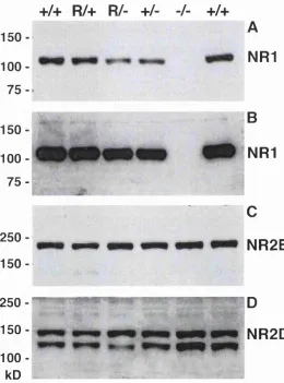
Figure 3-6 Western blot analysis of membrane proteins from whole brains from newborn NRD^^, N R 1^\ NRl^*, NRD^' littermates and from NRT^' and NRD^'^ littermates... 91
Figure 3-7 Development of whisker representations in the brainstem of mice 94 Figure 3-8 Cytochrome C oxidase histochemistry in brainstem trigeminal cortex of N R V \ N R l '- and N R l^ pups at PC...95
Figure 3-10 Neuroanatomy of hippocampus (A, B) and cerebellum (C, D) of NRl^^' (A, C) and N R l^' (B, D) littermates... 97 Figure 3-11 Whisker-related patterns are emerging at PO in nVi of pups which
express the wildtype NRl subunit (NRl'^^^ NRl"*"^', NRl^"^)...98 Figure 3-13 Qualitative differences of staining in nVi...99 Figure 4-1 Southern Blot analysis of KAl-YAC clones isolated from the WI/MTT
mouse YAC library by PCR-screening ...102 Figure 4-2 Integration of KA1-T4-Y403tet into KAl-YAC by transplacement of
K A l- genomic sequences in KAl-YAC 448B7...104 Figure 4-3 Targeted Rescue Cloning... 105 Figure 4-4 Sequence alignment of mouse exonic sequences, subcloned by targeted
Rescue cloning from the KAl-YAC, and rat K A l cDNA... 107 Figure 4-5 Summary of generation and analysis of rescue clones... 108 Figure 4-6 Sequence of bacterial cloning of targeting cassette KA1-T5...110 Figure 4-7 Final cloning step of the targeting cassette KA1-T5 in E.coli... I l l Figure 4-8 Targeted integration of the KA1-T5 cassette into KAl-YAC 448B7
through homologous recombination...113 Figure 4-9 Cloning and analysis of the ES-cell construct YES-1... 114 Figure 4-10 Flp-mediated resolution of the neo-his3 cassette from the targeted KAl-YAC... 116 Figure 4-11 Constructs for testing expression and function of EGFP-Cre...117 Figure 4-12 EGFP-Cre reporter assay...119 Figure 4-13 Gene targeting of KAl-gene with YES-1 and genomic organisation of
wildtype and recombinant KAl-allele... 122 Figure 4-14 Southern blot screen of first round ES-cell lysates... 123 Figure 4-15 Southern blot analysis of Kpnl and Xbal digests of second round
ES-cell DNA with the 3'probe... 124 Figure 4-16 Southern blot analysis of Hpal and Hpal/Clal digests of second round
ES-cell DNA with the 5’probe...125 Figure 4-17 Southern blot analysis of Kpnl and Xbal digests of second round
ES-cell DNA with the neo probe... 125 Figure 4-18 Southern blot analysis of Kpnl and Xbal digests of second round ES-
List of tables
Table 3-1 Phenotypes of N Rl gene-targeted mice... 87
Table 4-1 Test-breeding of chimeras with B1/6J mice...127
Abbreviations
aa AMPA AP5 APV ARS ATP BBS P-gal P-lac B1/6J BLAST BSA BSTC CaMKII acDNA CA CBN ch cir^ CMV CNS CO CSM dATP DG DNA DOB DSB DTT B14 etc. BDTA BGBP BPSC BS BST flirted floxed frt GluR h HBK HGMP HRP HSV ICR amino acid
a-amino-3-hydroxy-5-methyl-4-isoxazole propionic acid see APV
2-Amino-5-Phosphonovaleric Acid autonomously replicating sequence adenosine triphosphate
N, N-bis (2-hydroxyethyl)-2-aminoethansulfonic acid buffered saline
p-galactosidase P-lactamase
C57 Black/6 Jackson inbred mouse strain Basic Local Alignment Search Tool bovine serum albumin
brainstem trigeminal complex
a-calcium-calmodulin-dependent proteinkinase H complementary DNA
cornu ammonis centromere chicken
2 fim plasmid positive Cytomegalovirus central nervous system cytochrome C oxidase
Complete Supplement Mixture deoxy adenosine triphosphate dentate gyrus, gyrus dentatus deoxyribonucleic acid
Drop Out Base downstream element dithiothreitol
embryonal day 14 etc.
ethylene diaminetetraacetic acid enhanced green fluorescent protein excitatory postsynaptic current embryonic stem
expressed sequence tag
flanked by tw o/rf sites in the same orientation flanked by two lax P sites in the same orientation Flp recognition target
glutamate receptor subunit human
human embryonic kidney human genome mapping project horseradish peroxidase
iGluR ionotropic glutamate receptor
10 inferior olive
1RES internal ribosome entry site
KA kainate, kainic acid, kainate receptor subunit
LB Luria-Bertani medium
loxF bacteriophage PI lox site (lox: locus of chrossing over)
LTP long-term potentiation
M l etc. membrane domain letc.
MK-801 (+)-5-methyl-10,1 l-dihydro-5H-dibenzo[a,d]cyclohepten- 5,10-imine
MMLV Moloney-Murine leukaemia virus
MOPS 3-(N-Morholino)propanesulfonic acid
MRC Medical Research Council
m mouse
mRNA messenger RNA
na nucleus ambiguus
no nucleus cuneatus
nh nucleus hypoglossus
NMDA N-methyl-D-aspartate
NMRI naval medical research institute
NR NMDA receptor subunit
nVc subnucleus caudalis of nucleus trigeminus (V)
nVi subnucleus interporalis of V
nVo • subnucleus oralis of V
nVp principal nucleus of V
PAGE polyacrylamide gel electrophoresis
PO, P5 etc. postnatal day
PB phosphate buffer
PBS phosphate buffered saline
PGP phencyclidine
PCR polymerase chain reaction
PEG polyethylene glycol
PFGE pulsed field gel electrophoresis
PKC Protein kinase C
PMA phorbol 12-myristate 13-acetate
PMBSF posteromedial barrel subfield
PMSF phenylmethylsulfonyl fluoride
PVDF polyvinylidene difluoride
r rat
rb rabbit
RNA ribonucleic acid
rpm revolutions per minute
SDH succinic dehydrogenase
SDS sodium dodecyl sulfate
ssDNA single strand DNA
SSPE salt-sodium-phosphate-EDTA
TAE tris-acetate-EDTA
TB Terrific Broth medium
TBE tris-borate-EDT A
TSA temperature sensitive antigen
UTR untranslated region
VB ventrobasal thalamus
X-Gal 5-bromo-4-chloro-3-indolyl P-D-galactopyranoside
YAC Yeast Aortificial chromosome
YCp Yeast Centromere-containing plasmid
YEPD yeast-extract-peptone-dextrose
Yip Yeast Integrative plasmid
1 Introduction
Glutamate is the main excitatory neurotransmitter in the mammalian brain and elicits rapid neuronal responses via glutamate receptors. Glutamate receptors participate in several physiological and pathological processes in the brain, including synaptic plasticity and excitotoxicity.
1.1 The NMDA receptor
The NMDA receptor is a glutamate receptor. Glutamate receptors fall into two functional groups, metabotropic and ionotropic receptors. Metabotropic glutamate receptors transduce signals via coupled G-proteins and are slow-acting, whilst ionotropic glutamate receptors transduce signals via cation-flux through the intrinsic receptor channel and are fast-acting.
The NMDA receptor is an ionotropic glutamate receptor. Ionotropic glutamate receptors can be divided into three subtypes according to their sensitivity to the agonists AMPA (a-amino-3-hydroxy-5-methyl-4-isoxazole propionic acid), kainate (a structural analogue of glutamate) or NMDA (A-methyl-D-aspartate). AMPA and Kainate (AMPA/KA) receptors are collectively called non-NMDA receptors.
NMDA receptors are blocked by a variety of substances, for instance competitively by APV (AP5), and noncompetitively by MK-801, ketamine and PCP.
1.1.1 Special properties of the NMDA receptor
glycine as coagonist (Johnson and Ascher, 1987). And fourth, NMDA receptors have slow activation and inactivation kinetics (msec to sec range) compared to the rapidly activating and desensitising AMPA/KA receptors (msec) (Hestrin et al., 1990; Ishii et al., 1993).
Activation of the NMDA receptor, unlike non-NMDA receptors, requires depolarisation of the membrane, in which the NMDA receptor is embedded. In physiological ion conditions (containing ImM extracellular Mg^"^), Mg^"^ ions, for which the channel is impermeable, are held in position in the channel mouth by the electrical field across the membrane and block the channel. If postsynaptic depolarisation, typically mediated by AMPA/KA receptors, is sufficient to relieve the voltage-dependent Mg^^ -block, then NMDA receptors serve as a gate controlling entry of the second messenger Ca^"^. Moderate increases in intracellular Ca^"^ mediate a variety of physiological processes in developing and mature neurons, whereas immoderate increases resulting from excessive NMDA receptor activation can be neurotoxic (Choi, 1994; McDonald and Johnston, 1990).
1.1.2 Molecular diversity of NMDA receptors
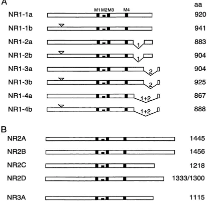
Ionotropic glutamate receptors are formed from a heterooligomeric complex of subunits, which are coded for by individual genes. Up to date, six gene families have been defined by sequence homology: a single family for AMPA receptors, two for kainate receptors and three for NMDA receptors. Two orphan subunits, for which no agonists are known, have also been cloned, i.e. GluR delta 1 and delta 2 (Lomeli et al., 1993; Yamazaki et al., 1992b). Figure 1-1 shows an alignment of the members of the ionotropic glutamate receptor family.
The NMDA receptor gene (NR) family consists of one N R l gene, four variant NR2 genes (NR2A-D) and the NR3A gene.
The N R l cDNA was the first NMDA receptor subunit to be cloned and was identified through expression cloning in Xenopus oocytes (Moriyoshi et al., 1991). Subsequently, the four NR2 subunits were found in rat brain, NR2A-C (Monyer et al., 1992) and NR2D (Ishii et al., 1993). The respective mouse NMDA subunits were named Ç1 and 61-64 (Ikeda et al., 1992; Kutsuwada et al., 1992; Meguro et al., 1992;
0 0 0 ( 7 ) 0 0 0 7 ^ ^
~
c c c c c c c > > 3030333 30
fü» 3 3 3 3 3 3 3 * ^
-»‘I\D |>0C0 4::i.—L o ) ^ C J l ! > U J O O ] >
AMPA kainate
NMDA
F ig u r e 1 - 1 A lign m en t o f m e m b e r s o f th e ion otropic g lu ta m a te r e ce p to r g e n e fam ily b a s e d on a m in o a c id s e q u e n c e - h o m o lo g y (a d a p te d from ( D a s e t al., 1 9 9 8 )). P h a r m a c o lo g ic a l c la ssific a tio n s o f su b u n its are sh o w n u nd er th e align m en t, an d co r re la te w ell with grou p in gs b a s e d on s e q u e n c e sim ilarities.
NR2 subunits share -50% identity with one another but only -15 to 20% identity with N Rl (Ishii et ah, 1993).
An N R l like subunit shares an average identity of 27% with the NRl and NR2 subunits and 23% to non-NMDA subunits and has been renamed NR3A (%-l) (Ciabarra et ah, 1995; Sucher et ah, 1995).
B
NR1-1a
NR1-1b
NR1-2a
NR1-2b
NR1-3a
NR1-3b
NR1-4a
NR1-4b
NR2A
NR2B
NR2C
NR2D
NR3A
M1M2M3 M4
V = "
v = ^
aa
920
941
883
904
904
925
867
888
1445
1456
1218
]
1333/1300
1115
F ig u r e 1 - 2 S c h e m a tic rep resen ta tio n o f NMDA re ce p to r su b u n its. (A) N R l s p lic e variants, (B ) N R 2 su b u n its an d th e N R 3A su b u n it. H yd rop h ob ic s e g m e n t s a r e in d ica ted a s b lack b o x e s within th e g e n e , n um bered M l, M2, M3 an d M4. T h e N-term inal insertion o f 21 am ino a c id s is in dicated by a triangle a b o v e th e g e n e , C -term inal d e le tio n s a r e sh o w n with arab ic n u m b ers (d e letio n 1: 3 7 am in o acid lon g 0 1 c a s s e t t e , d e le tio n 2: 3 8 a m in o acid long 0 2 c a s s e t t e ) . N o te that sp licin g out o f th e 0 2 c a s s e t t e p r o d u c e s a n e w o p e n read in g fram e ( 0 2 ’). P ep tid e le n g th s are indicated (aa). This diagram is not to sc a le !
1.1.3 Expression patterns of NMDA receptor genes in the CNS
during development
Expression of NMDA receptor subunits is subject to developmental regulation. Changing subunit composition of NMDA receptors during development was revealed using subunit-specific antibodies (Laurie et al., 1997; Sheng et al., 1994), in-situ hybridisation studies (Laurie and Seeburg, 1994; Monyer et al., 1994; Toile et al., 1993; Watanabe et al., 1992), RNase protection assays (Goebel and Poosch, 1999; Zhong et al., 1995) and immunohistoblots of brain sections (Wenzel et al., 1997; Wenzel et al., 1996).
In situ studies of mRNAs revealed that from embryonic stages, the N R l gene is expressed in virtually all neurons of the cns during development (Monyer et al., 1994; Toile et al., 1993; Watanabe et al., 1992), as well as in adult forebrain (Moriyoshi et al., 1991), cerebellum (Watanabe et al., 1994c), brainstem (Guthmann and Herbert, 1999; Watanabe et al., 1994b) and spinal cord (Toile et al., 1993; Watanabe et al., 1994a). In contrast, the four NR2 transcripts display distinct spatial and temporal expression patterns. N R l, NR2B and NR2D subunit mRNAs are detectable in the CNS as early as E14, whereas NR2A and NR2C appear only around birth. (Monyer et al., 1994; Watanabe et al., 1992).
Among the NR2 genes, NR2A expression levels increase after birth to moderate levels across the whole brain, with strongest expression in the hippocampus. (Monyer et al., 1994; Watanabe et al., 1992).
NR2B is the predominant NR2 variant in juvenile brain but is low in adult brain with the exception of the hippocampus where it remains highly expressed. Remarkably, NR2B expression ceases in cerebellum. This is complimented by strong upregulation of NR2C which remains largely confined to the cerebellum. Thus, there appears to be a switch in expression from NR2B to NR2C as the predominant subunit in the cerebellum (Monyer et al., 1994; Watanabe et al., 1992).
Similar to the NR2D subunit, the NR3A subunit is expressed primarily during development. In contrast to the NR2D subunit however, the NR3A subunit is expressed in many regions of the immature brain (Ciabarra et al., 1995; Sucher et al.,
1995).
In situ hybridisation studies have indicated that splicing of the N R l transcripts in the brain is regionally regulated (Laurie and Seeburg, 1994). Oligonucleotide probes were employed which were specific for the absence of the N1 cassette, or for the expression of the four possible carboxy termini, containing C l and C2; C2 only. C l and C2’ and C2’ only. Although each splice variant follows a parallel profile of abundance, marked regional differences are observed in splicing at both the 5’ and 3’ sequences. For example, whereas N1-lacking receptors show prominent labelling throughout the brain, other splice variants are expressed to different degree in various brain regions. Relative mRNA expression levels of NRl splice variants have also been quantified using RNase protection assays (Zhong et al., 1995) and RT-PCR (Winkler et al., 1999).
1.1.4 Structure of NMDA receptors
1.1.4.1 NMDA receptors are heteromeric
In mammalian expression systems functional NMDA receptor channels are not formed from any of the NR2 subunits alone. However, when N R l cRNA is injected into Xenopus oocytes, NMDA-gated ion channels are formed, which express many of the properties characteristic of native NMDA receptors (Moriyoshi et al., 1991). In the meanwhile, an endogenously expressed unitary glutamate receptor subunit, XenUl, has been discovered in Xenopus oocytes (Soloviev et al., 1996). When co expressed with N Rl in a mammalian cell line, a unitary receptor was expressed which showed responses to both NMDA and non-NMDA receptor agonists and antagonists (Soloviev et al., 1996).
both in oocytes and in mammalian cells (Ikeda et al., 1992; Ishii et al., 1993; Kutsuwada et al., 1992; Meguro et al., 1992; Monyer et al., 1992).
Co-immunoprecipitation studies with subunit-specific antibodies under conditions that preserve the endogenous subunit associations also implied that NMDA receptors in brain exist as heteromeric complexes (Blahos and Wenthold, 1996; Luo et al., 1997; Sheng et al., 1994). The coexistence of NR2A and NR2B, as well as of two NRl splice variants, in NMDA receptors in brain was shown directly, using a combination of immunoaffinity chromatography and immunoprécipitation (Chazot and Stephenson, 1997).
Immunoprécipitation studies revealed that NMDA receptors in some brain regions contain the NR2D subunit (Dunah et al., 1998). Thus, NR2D is found in a binary complex with NRl in the thalamus but not in the midbrain, where complexes contain either NR2A or NR2B together with N R l.
Final proof, that NMDA receptors require the N R l subunit, was gained from N Rl knockout mouse models (Forrest et al., 1994; Li et al., 1994). In these mice, no NMDA mediated current responses can be recorded, indicating that the N Rl subunit is essential.
1.1.4.2 Transmembrane topology
Ionotropic glutamate receptor subunits have four membrane (hydrophobic) domains and were predicted to have large extracellular N- and C-terminal regions. Contrary to initial models, however, only three and not four domains are crossing the membrane, as shown in Figure 1-3.
1994) and GluR3 (Bennett and D ingledine, 1995). Taken together, these studies
supported the hypothesis that the M2 domain forms a re-entrant hairpin loop if M l
and M3 are a-helical and cross the membrane once.
This model has been confirm ed for N M D A receptors using cysteine-
substitution methods to map residues contributing to the channel pore. Amino-acids
in M2 of N R l and NR2C were cysteine-substituted and considered as channel-lining
if they interacted with sulfhydryl-specific agents (Kuner et al., 1996).
F ig u r e 1 - 3 P ro p o sed tran sm em b ran e top ology o f ionotropic g lu tam ate receptor subunits. M em brane-spanning region s 1, 3 and 4 are cro ssin g the lipid bilayer. M2 is sh ow n a s a re entrant loop entering and leaving the lipid bilayer on th e cy to p la sm ic sid e. At an a n a lo g u e position within M2 the ionotropic glu tam ate recep tors display either a neutral glutam ine (Q), a neutral a sp aragin e (N) or a positively ch arged arginine (R) (N /Q /R -site). T he identity of this am ino acid controls the cation perm eability by constricting th e ion perm eation pathway. N-and C- terminal region s are located extracellularly N-and intracellularly respectively.
1.1.4.3 Structural elements determining -permeability and Mg^-block
At an analogous position within M 2 the ionotropic glutam ate receptor subunits
display either a neutral glutamine (Q), a neutral asparagine (N) or a positively
charged arginine (R) (N/Q/R-site) (Figure 1-3). Site-directed mutagenesis has shown
that an arginine (R) at this position in the GluR2 subunit controls Ca^^-permeability
and NR2 subunits of the NMDA receptor feature an asparagine (N) in this position -
with the exception of the NR3A subunit which features an arginine (R) at adjacent
position (Figure 1-4).
_________ M 2-region________
m i NEGA NR2D NR2C NR2B NR2A KAl KA2 GluRS GluR7 GI11R6
GluRl GluR4 GluR3 GluR2
d e lta s
deltal
L T L S S A M W F S W G V L L ^ S G I F S F S S A L N V C Y A L L F G R T A F T I G K S I W L L W A L V F & S V F T I G K S V W L L W A L V F T O S V F T I G K A I W L L W G L V F M S V F T I G K A I W L L W G L V F ^ S V Y S L G N S L W F P V G G F M ^ Q G S
y t l g n s l w f p v g g f mHq g s
F T L L N S F W F G V G A L M ^ Q G S F T L L N S F W F G M G S L M 0 Q G S
f t l l n s f w f g v g a l m Bq g s
F G I F N S L W F S L G A FmS qGC
f g i f n s l w f s l g a f m ISq g c f g i f n s l w f s l g a f m S q g c f g i f n s l w f s l g a f m S q g c
T T L Y N S M W F V Y G S FvS q GG
a t l h s a i w i v y g a f vËq g g
F ig u re 1 - 4 P ep tide s e q u e n c e alignm ent of the M2 region of iGluR su bunits. T he N /Q /R-site is highlighted. N ote that in the NR3A subunit there h a s a glycin e (G) resid u e at the N/Q/R-site and an arginine (R) residue in adjacent position.
The N to Q mutation in NR I reduces the sensitivity to Mg^^-block and reduces the
Ca^^ -perm eability of heteromeric receptors, w hereas the sam e m utation in NR2
strongly reduces the Mg^^ -block but barely affects the Ca^^'-permeability. In contrast,
the N598R point mutation in N R l alters both key properties of NM DA receptor
channels in a profound manner. Channels of NMDA receptors carrying this mutation
are im permeable to Ca^^-ions and are not blocked by Mg^"^-ions (Bum ashev et al.,
1992; Mori et al., 1992; Sakurada et al., 1993).
1.1.4.4 Ligand-binding regions
Full activation of the NMDA receptor channel requires both glutam ate and glycine
(Johnson and Ascher, 1987; Kleckner and Dingledine, 1988).
Two regions adjacent to the N-terminal of M l and to the C-terminal of M3
(SI and S2) of GluR3, which share structural similarities with the ligand-binding site
agonist binding (Stem Bach et al., 1994). Subsequently, the glycine and glutamate binding sites of NMDA-receptors were mapped to amino acids within S 1 and S2 of NRl (Hirai et al., 1996; Kuryatov et al., 1994; Wafford et al., 1995) and NR2 (Anson et al., 1998; Laube et al., 1997), respectively. Thus, the glycine binding site resides in N Rl and the glutamate binding site resides in NR2.
The structure of the glutamate-receptor ligand-binding core of GluR2 in complex with kainate as ligand has been elucidated by X-ray crystallography (Armstrong et al., 1998). This work confirmed the bilobar structure consisting of the SI and S2 regions which had been proposed (Stem Bach et al., 1994). Thus, the agonist binding site of glutamate receptors is made of the two globular (SI and S2) domains which are forming a clamshell-like stmcture whose closed state is stabilised by binding of agonist, as is the case in bacterial proteins.
1.1.5 Functional diversity of NMDA receptor channels
Native NMDA receptor channels have a voltage-dependent Mg^^-block, high Ca^^- permeability and characteristically slow gating behaviour. These key properties are also observed with recombinant NMDA receptors configured from N Rl and NR2 subunits and differ depending on the particular NR2 subunit co-expressed with NRl (Ishii et al., 1993; Kutsuwada et al., 1992; Monyer et al., 1992).
1.1.5.1 Mg^^-block
While all four NR1/NR2 subunit configurations have com parable Ca^"^- permeabilities, they have differences in the strength of voltage-dependent Mg^"^- block. In physiological concentrations of 1 mM extracellular M g^\ the NR1/NR2C and NR1/NR2D have weaker Mg^^-block than NR1/NR2A and NR1/NR2B receptors, when expressed in mammalian cells (Monyer et al., 1994).
in voltage-dependence. Mg^^-sensitivity is high around birth but decreases after 4 days (Kirson et ah, 1999).
The differences in sensitivity to Mg^^-block between different NR2 subunits were found to depend on three structural elements (Kuner and Schoepfer, 1996). In this study, chimeric NR2 subunits were constructed by replacing segments of the least Mg^^-sensitive NR2C subunit with homologous segments of the most sensitive NR2B subunit. In this way, three small elements within the M1-M4 segment of the NR2 subunit were found to be the major determinants of subtype-specific differences of Mg^^-block in heteromeric NMDA receptor channels.
1.1.5.2 Kinetics
The excitatory EPSCs seen at excitatatory synapses in the CNS are made up of AMP A receptor and NMDA receptor mediated EPSCs (Jahr and Lester, 1992). AMP A receptors activate and deactivate within 3 msec, the AMPA-receptor component of EPSC has a 200 p,msec rise time and a 1-3 msec decay time (Mosbacher et al., 1994). In contrast, NMDA receptors are characterised by much slower activation, the NMDA-receptor component of the EPSC is 20-fold slower and decays over hundreds of milliseconds. Experiments with brief application of glutamate indicated that free transmitter need only be present in the synaptic cleft for short periods. The time course of patch recordings evoked by brief (5 msec) glutamate (100 |xM) pulses mimicks the time course of synaptically stimulated EPSCs. Both types of recordings show 10-90% rise times of around 8 msec and decay phases that can be fitted by two exponentials (90 msec and 260-600 msec) (Lester et al., 1990).
The different kinetics of AMP A and NMDA receptors in the presence of the same agonist are so far explained by the much higher affinity of NMDA receptors for glutamate than AMPA receptors. Low affinity agonists such as aspartate and cysteate produce faster NMDA receptor recoveries than glutamate (Lester and Jahr, 1992).
dominant NR2D subunit have extremely long deactivation times (4.8 sec) (Monyer et al., 1994), as was also analysed on the single channel level using the Xenopus expression system (Wyllie et al., 1998).
1.1.5.3 Coincidence detection properties
The difference between AMPA and NMDA receptor kinetics on the one hand and the difference in kinetics between NMDA receptor subtypes are important for the role of the NMDA receptor in excitatory synaptic transmission. A presumed function of NMDA receptor channels is the detection of synchronous pre- and postsynaptic depolarisation (coincidence detection) (Bourne and Nicoll, 1993). Functional activation of NMDA receptors is linked to the membrane potential by the voltage- dependence of the Mg^^-block, allowing these channels to sense coincidence of synaptic input. Coincidence detection properties depend on the voltage sensitivity of the Mg^^-block and on the gating properties of the channel. Based on their long deactivation kinetics, it was predicted that NR1/NR2B, NR1/NR2C and NR1/NR2D receptors mediate longer lasting excitatory postsynaptic potentials than the NR1/NR2A subtype and would therefore permit the detection of low synchronicity of presynaptic activities and postsynaptic depolarisation (Monyer et al., 1994) In contrast, NR1/NR2C and NR1/NR2D channels have a relatively weak Mg^^-block, and therefore appear to be best suited for detection of synchronicity of presynaptic activity causing relatively small postsynaptic depolarisation (Monyer et al., 1994).
Due to its coincidence detection properties, the NMDA receptor is thought to play a role in the establishment of input-specific connections during the development of the nervous system and in the modulation of neuronal connections during learning and memory (section 1.2).
1.1.5.4 N R l splice variants
Phosphorylation of NMDA receptors by PKC has also been linked to N R l splice variants. A number of PKC-phosphorylation sites have been mapped to the C l cassette of N Rl. However, although splicing-out of the C l cassette removes most of the PKC-phosphorylation sites (Tingley et al., 1993), the splice variant with the shortest C-terminus (i.e. lacking C l), N R l-4, shows maximal potentiation, when assessed with the PKC-activator PMA (Durand et al., 1993). Conversely, the splice variant with both C-terminal exons, N R l-1, exhibits the least potentiation. Not surprisingly then, it was shown recently that protein kinase C potentiation of N- methyl-D-aspartate receptor activity is not mediated by phosphorylation of N- methyl-D-aspartate receptor subunits but rather by associated targeting, anchoring, or signalling protein(s) (Zheng et al., 1999).
Evidence for another possible function of alternative splicing was presented recently. In this study, the splice forms with the longest C-terminal cytoplasmic tail showed the lowest amount of cell surface expression, and the splice forms with the shortest C-terminal tail showed the highest cell surface expression. Hence it was suggested that the splicing of the C-terminal domain of the N R l subunit regulates the cell surface expression of functional NMDA receptors (Okabe et al., 1999).
1.1.5.5 A role for the NR3A subunit
1.2 Synaptic plasticity and the NMDA receptor
Synaptic plasticity is the activity dependent strengthening or weakening of synapses, which entails both changes in synaptic strength and morphological changes in synaptic connectivity. Synaptic plasticity is thought to be the cellular basis for learning and memory (Bliss and Collingridge, 1993; Bourne and Nicoll, 1993), as well as for the reorganization and refinement of developing connections in the nervous system (Goodman and Shatz, 1993).
1.2.1 Synaptic plasticity and learning
Donald Hebb proposed a rule for long-term memory on the basis of the strengthening of the synaptic connection between two neurons (reviewed in (Bailey et al., 2000). The Hebb rule states that the strength of the connection between two neurons is increased for a long period of time when the firing of the presynaptic and postsynaptic neuron are closely correlated in time.
1.2.1.1 Long-term potentiation (LTP)
An experimental form of synaptic plasticity has been termed long-term potentiation (LTP). LTP is a long-lasting increase in synaptic strength (enhancement in synaptic transmission), for example as a consequence to repetitive activation of excitatory synapses, and can be found at synapses in several parts of the brain, notably in the hippocampus where it was originally discovered (Bliss and Lomo, 1973).
1.2.1.2 NMDA-receptor dependent LTP
which is provided largely by permeation of Na"^ and through AMPA receptors, releases the Mg^^-block of the NMDA-receptor (Malenka and Nicoll, 1999). Once the NMDA channel opens, Ca^"^ enters the dendritic spine and initiates a cascade of locally confined intracellular signalling events, which can ultimately lead to changes in gene expression and the reorganisation of the synapse as well as the growth of new synaptic connections (Bliss and Collingridge, 1993; Bourne and Nicoll, 1993).
The exact signal transduction mechanism by which the resulting rise in intracellular Ca^"^ triggers the enhancement of synaptic strength to express LTP remains to be established. Among the few molecules which are thought to form the molecular machinery of LTP evidence is strongest for a-calcium-calmodulin-
dependent proteinkinase II (CaMKIIa) to be a key player. A rise in Ca^"^ induces autophosphorylation of CaMKIIa at Thr^*® in a CaM-dependent manner, keeping CaMKIIa in an active form for a long time after the Ca^"^ signal has returned to baseline. Introduction of a point mutation at Thr^^^ abolishes autophosphorylation of CaMKHa and abolishes LTP in gene targeted mice (Giese et al., 1998). CaMKIIa can directly phosphorylate the AMPA receptor subunit GluRl at Ser^^\ Since the single channel conductance of AMPA receptors containing phosporylated GluRl increases in vitro as well as during LTP, it seems likely that CaMKIIa-dependent GluRl phosphorylation is one mechanism that underlies LTP. This is also supported by the lack of LTP in GluRl-deficient mice (Zamanillo et al., 1999). CaMKIIa is also postulated to influence the subsynaptic localisation of ‘reserve’ AMPA receptors such that more AMPA receptors are recruited to the synaptic membrane from vesicle pools or surrounding plasma membrane areas. In this way ‘silent’ synapses devoid of AMPA receptors might be converted to active synapses (reviewed in (Malenka and Nicoll, 1999)).
1.2.1.3 LTP as a learning mechanism
NMDA receptor antagonist APV (Davis et al., 1992; Morris, 1989; Morris et al., 1986) and more recently from NMDA receptor gene targeting studies (Sakimura et
al., 1995; Tsien et al., 1996b).
Mice lacking the NR2A subunit - which in normal mice is expressed to moderately high levels across the adult brain and confers relatively fast deactivation kinetics - are viable and show significant reduction of the NMDA receptor current and of LTP at the hippocampal synapse in the CA l stratum radiatum. The mice also showed moderate deficiency in spatial learning, as determined using the Morris water maze (Sakimura et al., 1995).
More evidence for the involvement of NMDA receptor dependent activity in hippocampal LTP and in memory comes from a conditional gene targeting study, in which the deletion of the N R l gene was limited to the C A l region of the hippocampus by use of the Cre/lox P recombination system (Tsien et al., 1996; Tsien et al., 1996). In contrast to the lethal global deletion of the N R l gene (Forrest et al., 1994; Li et al., 1994), CAl-restricted N R l knockout mice are viable and grow into adulthood. In these mice, no NMDA receptor mediated synaptic currents and no LTP can be detected at CA l synapses, whereas NMDA receptor dependent LTP can still be induced in the dentate gyrus. These mice show impaired spatial memory as assessed in the Morris water maze (Tsien et al., 1996).
1.2.2 Developmental plasticity in sensory systems
A number of mechanisms are involved in the development of neural connectivity in the nervous system (Goodman and Shatz, 1993). During development of sensory systems such as the visual system and the somatosensory system, afferent and intrinsic axons are first guided by positional cues, such as graded markers, and form exuberant but already topographic projections (Holt and Harris, 1998)\ Then, activity dependent processes between pre-and postsynaptic neurons are thought to refine the synaptic patterning within developing modular representations (e.g. ocular dominance stripes, barrels) (Gierer and Muller, 1995).
A modem articulation of the Hebb rule by Stent (Stent, 1973) suggests a two-part
rule for the activity-dependent modification of young synapses (Constantine-Paton et
ah, 1990):
1. Synaptic contacts between synchronously active pre- and postsynaptic
neurons are selectively reinforced.
2. Synaptic contacts between asynchronously active pre- and postsynaptic
neurons are selectively depressed or eliminated.
The basis for reinforcement and depression of synaptic contacts is thought to be the
ability of the postsynaptic cell to summate the synaptic potentials from converging
synapses. Inputs converging on one postynaptic cell are thought to be reinforced if
they are synchronously or near synchronously active (cells that fire together wire
together). As coincidence detector, the NM DA receptor has been implicated in this
process.
1.2.2.1 Critical period plasticity in sensory systems
During the developm ent of sensory systems, developm ental plasticity of young
synapses has been shown to be limited to so-called ‘critical periods’. The term
‘critical period’ has been coined by W iesel and Hubei to define the time period
during development of the visual system in which the connections between afferents
and their target cells in the primary visual cortex remain plastic. This has been shown
by m anipulations of the natural sensory inputs, such as covering of one eye or
induction of crossed eyes by cutting an ocular muscle (review ed in (Constantine-
Paton et al., 1990)).
Similar to the situation in the primary visual system, plasticity of developing
synapses can also be induced in the som atosensory system o f rodents that is
described in section 1.3. In the barrel cortex, which is the somatotopic representation
of the whiskers at the rodent’s muzzle, ablating whisker follicles in neonatals leads to
anatomical changes in the whisker-related somatotopic map, as first shown by Van
der Loos and W oolsey (Van der Loos and W oolsey, 1973). D am ages to whisker
follicles at later stages has progressively less effect in the cortex and has no effect
when done on postnatal day 6 or later, clearly indicating a critical period for
1.2.2.2 Critical period for LTP in sensory cortex
1.3 The rodent trigeminal pathway
The rodent somatosensory system consists of the trigeminal pathway and the dorsal column pathway, which convey a somatosensory map from the periphery to the cortex by relay stations in the brainstem and thalamus. The dorsal column pathway carries information from the receptor-dense zones of the paws, whereas the trigeminal pathway carries sensory information from the face, in particular from the large mystacial whiskers.
In the mouse and in other rodents the whiskerpad on the muzzle is an important cutaneous sensory organ that serves the animal to investigate the environment in active whisking movements of the entire whisker pad. The receptor units of the whisker pad are the whisker follicles, which are organised in a characteristic, phylogenetically conserved, pattern of 5 rows, A through E, nearly parallel to the bridge of the nose. Each row has 4-7 easily identified large vibrissae, the caudal ones being longer and thicker than the more rostral ones, and within each row the number of whisker follicles is remarkably constant (Woolsey, 1990). In addition, four caudal whiskers ‘straddle’ the rows (a - Ô).
Trigeminal ganglion
BSTC VB PMBSF
midline
nVp
[
nVo^
nVi ^ ,
d
Jt* m
c nVc
^
Face Brainstem Thalamus Cortex
Figure 1 -5 Diagram sh owing the principal c o m p o n e n t s of the rodent trigeminal pathway and their relative s i z e s and orientations (modified from (W oolsey, 1990)). On the whiskerpad there are 5 rows (A-E) with 4-7 large w hiskers ea c h . In addition 4 cau d al w hiskers (a-S) straddle the rows. S e n s o r y information is transmitted to the s o m a t o s e n s o r y cortex via relay stations in the brainstem and in the thalamus. At e a c h station, afferents and their target cells form units (barrelettes in the brainstem, barreloids in the thala m us and barrels in the cortex) that replicate the array of whiskers on the animal’s face. A trigeminal ganglion cell innervates only o n e whisker but projects to all four subdiv isions of the Brainstem Trigeminal Comple x (BSTC). In the BSTC, whisker-related patterns can b e s e e n in tran sver se sections, i.e. in two of the three subnuclei of the spinal trigeminal nucleu s, the s u b n u c le u s cau d alis (nVc) and the s u b n u c l e u s interpolaris (nVi), but not in the s u b n u c l e u s oralis (nVo). In addition, patterning can b e d e t e c t e d in the principal n u c le u s of V (nVp). HRP-injection into the ventrobasal com p lex st ain s mostly cells in the nVp and nVi and little c e lls in nVo and nVc, indicated by the t h ic k n ess of the arrows cr ossin g the midline to the V entrobasal C omple x (VB) in the thalamus. In the cortex the large whisker representatio ns form the posteromedial barrel subfield (PMBSF). C om pass: d = dorsal, m = medial, c = caudal, a = anterior.
1.3.1 B arrels, barreloid s and barrelettes
W oolsey and Van der Loos introduced the term barrel to describe the three
dim ensional structure of m ulticellular units that can be found in layer IV of the
primary somatosensory cortex of mice (Woolsey and Van der Loos, 1970). Each unit
shows a dense ring of cell bodies which has roughly the shape of a circle or ellipsoid.
This ring represents the side of the barrel and surrounds an area of lesser cell density
named hollow. Each barrel is separated by a clear, cell-sparse area (fewer cells than
in the hollow) which is the septum . The hollow consists of neuropil made from
afferent axon arbors and postsynaptic cell dendrites.
In the thalamus and the brainstem there are barrel-equivalent whisker-related
units which are therefore called barreloids and barrelettes respectively (Belford and
There is variation in the details of the structure between species and during development within one species. In the rat for example, prior to the 20th day, the barrels resemble those of the mouse, but mature barrels are relatively indistinct and have a uniformly high cell density, i.e. no hollows (Welker and Woolsey, 1974).
1.3.2 Visualizing barrels, barreloids and barrelettes
S)
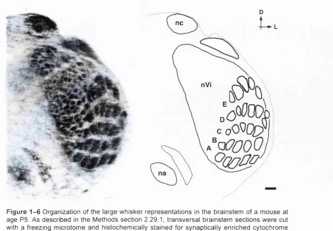
F igu re 1 - 6 Organization of the large whisker representations in the brainstem of a m o u s e at a g e P5. A s described in the Methods sectio n 2.29.1, transversal brainstem se c t io n s w ere cut with a freezing microtome and histochemically stain ed for synaptically enriched cy tochrom e C o x id a s e (CO). Each barrelette and e a c h barrelette row (A-E) s t a n d s in 1:1 relationship to a particular large whisker and whisker row on the face, na, n u c le u s am b igu u s, nc, n u c leu s cu neatus, nVi, su bn ucleu s interpolaris of the nucle u s trigeminus. S c a l e bar: 2 0 0 pm.
1.3.3 D evelop m en t o f the w h isk er-p ath w ay
1.3.3.1 Development of the whiskerpad
In the mouse embryo, the whiskerpad evolves through the m ergence of the lateral
nasal fold with the maxillary arch. At day 11 of gestation ( E l i ) five horizontal ridges
appear in the whiskeipad region. Simultaneously, individual follicles start to develop
along the ridges. At E14 the com plete pattern of the m ystacial follicles on the
whiskerpad is established, consisting of five horizontal rows of them, plus four
follicles adjacent to, and straddling, the caudal elements of each row (reviewed in
1.3.3.2 Pattern formation in the developing vyhisker-pathway
Whisker-related patterns in the rodent trigeminal system are consolidated during the first postnatal week. Pattern formation can be divided into two distinct phases (Jhaveri and Erzurumlu, 1992). In the first phase, the axial framework of the trigeminal neuraxis is laid down in an orderly fashion without guidance from the periphery (Erzurumlu and Jhaveri, 1992). This organisation is hardwired by spatial arrangement of the developing sensory afferents. In the second phase, sensory inputs guide the modular patterning of neuronal elements within the constraints of this axial framework.
Through labelling of fixed rat embryos with carbocyanine dyes (Dil and DiA) it was shown that the peripheral and central processes of trigeminal ganglion cells are spatially ordered when they reach their peripheral target nuclei in the brainstem (Erzurumlu and Jhaveri, 1992). This spatial order is evident before vibrissae rows appear and before the brainstem trigeminal nuclei differentiate, suggesting that trigeminal afferents to the brainstem and whisker pad organise topographically, independent of peripheral or central influences. Thus, in the rat trigeminal ganglion cells are differentiating after E9.5. Their peripheral processes start growing out as a circular bundle of fascicles on E l l and reach the rudimentary whisker field on E l 3 where they fan out. Already at this time point the peripheral processes become labelled with Dil or DiA when applied to the presumptive whisker pad which at this time is still void of follicles. Central processes of the trigeminal ganglion cells enter the CNS on E12 and E l 3 at the level of the brainstem where they bifurcate into ascending and descending branches of the central trigeminal tract. At E14, these axons emit radial collaterals at right angles to the pial surface, to invade all of the newly formed brainstem trigeminal nuclei at roughly the same time. Radial collaterals also emit branches to form axon arbors, and although the arbors have not yet segregated into the disjunctive patches (barrelettes), they are focalised, at least with respect to the dorsonventral axis of the face (Erzurumlu and Jhaveri, 1992).
Finally, afferent-target interactions take place in the perinatal period which lead to formation of whisker-related patterns.
This was done by ablating whisker follicles at different postnatal ages, followed by looking for alterations in the whisker-related patterns along the pathway in the adult animal. Thus, in the mouse follicle damage up to postnatal day 3 alters whisker- related cytoarchitecture in the ventrobasal thalamus (Woolsey et al., 1979) and up to day 5 in the somatosensory cortex (Woolsey and Wann, 1976). For instance, ablating the whisker-follicles of one row causes two major alterations in the thalamus and the cortex, namely (1) fusion of the whisker-representations of that row to a more or less narrow band and (2) ‘com pensating’ increase of the adjacent whisker- representations. In the brainstem no changes in size are seen, indicating that the critical period ends before birth. Instead, zones corresponding to the damaged whiskers are shrunken and pale, regardless of the animal’s age at follicle-damage, probably reflecting the degeneration of the primary sensory afferents in response to follicle damage (Durham and Woolsey, 1984)
1.3.3.3 Pharmacological blockade of NMDA receptors
Two studies have investigated the effect of pharmacological blockade of NMDA receptors on the trigeminal system (Fox et al., 1996; Schlaggar et al., 1993). The first study found that chronic treatment with the NMDA receptor antagonist APV in the somatosensory cortex did not prevent the emergence of a normal barrel pattern (Schlaggar et al., 1993). However, when attemping to prevent the cortical rearrangements that result from damage to a row of whiskers, it was found that the chronic blockade of NMDA receptors by APV substantially reduced the shrinkage of the deprived barrels. Thus, these experiments suggested a partial role of the NMDA receptor activity in the plastic events underlying the formation of the somatotopic patterns as assessed by histochemical stains. Using electrophysiological techniques, a follow-up study investigated whether in the APV-exposed cortex the 1:1 relationship between input and functional unit (i.e. between whisker and barrel) was altered (Fox et al., 1996). The study found APV-treatment does indeed alter the connectivity in the barrel field, i.e. barrel cells responded over a wider distance than normal. Taken together these results suggested a partial role of the NMDA receptor in the formation of a functional somatototpic map.
period, a point in time when the system studied is already partially developed. Thus, the full picture might not be seen. This is not the case with genetic manipulations.
1.3.3.4 NMDA receptor mutant mice with impaired trigeminal patterning
Knockout mice of five NMDA receptor genes have been described. Of these, NRl and NR2B knockout mice show impairment of trigeminal patterning (Kutsuwada et al., 1996; Li et al., 1994). NRl knockout mice lack functional NMDA receptors and die within hours after birth, probably due to respiratory failure and defective suckling response (Forrest et al., 1994; Li et al., 1994; Poon et al., 2000). It was found that in these mice synaptic transmission expectedly lacked the NMDA-receptor dependent component, but was otherwise not changed. However, CO-staining did not reveal any whisker-related row or patch formation in N R l knockout mice (Li et al., 1994). Life-prolonging measures were taken to rule out the possibility that developmental retardation was the cause for homogenous staining of the trigeminal nucleus although these mice did not differ otherwise (e.g. in weight and in gross neuroanatomy) from wildtype littermates. Pharmacologically delaying the birth of NRl knockout mice together with respiratory stimulation of newborn pups allowed analysis of the whisker-related patterning in these pups at an age equivalent to postnatal day 2 (P2) but no whisker-related patterning emerged in these older pups either. Furthermore, it was shown that pathfinding, initial targeting and crude topographic projection of trigeminal axons in the brainstem of N R l knockout mice are unaffected.
low transgene expression, while nVi and Cu showed relatively high levels of N Rl, In line with these differential N R l mRNA expression levels, mice with low transgene expression did not show whisker-related patterning in nVp even at 4 weeks of age and showed a rudimentary barrelette pattern in nVi. Thus, it can be concluded that lack of N R l subunit does not merely delay whisker-related patterning but that a sufficient level of NRl (transgene) is required for pattern formation in the brainstem.
Cortex-restricted disruption of the N R l gene was also achieved (Iwasato et al., 2000). In these mice, deletion of the N R l gene was restricted to the cortex by use of the Cxdlox P recombination system. The Cre gene was inserted into the EMX-1 locus, a homeobox gene, which is expressed from embryonic stages on to adulthood in the dorsal telencephalon. EMX-Cre mice were mated with floxed N Rl mice to exclusively delete the N R l gene in the cortex. Whisker-related neuronal pattern formation in the barrel cortex of these mice was shown to be impaired without affecting pattern formation in the thalamus and brainstem. More specifically, in these mice, which do not show general deficits in cortical development (layers, thalaniocortical projections), thalamocortical afferents do form whisker-related patterns and display critical period plasticity, as revealed by Dil-staining and CO- staining. But the CO-patches are less distinct than in wildtype mice, and the barrels can not be seen by Nissl staining. As cortical cell migration is not impaired in NRl knockout mice (Messersmith et al., 1997), it must be concluded that NMDA receptors are crucial for the construction of barrels, possibly involving cell displacement around axonal and dendritic clusters and synaptic plasticity.
to adulthood and have normal whisker-related somatotopic patterns (Ikeda et al., 1995). Given the reduced -block and very slow channel kinetics of NR1/NR2D receptors as compared to NR1/NR2B receptors, it seems that the NR1/NR2D receptors cannot compensate for the lack of the NR2B subunit. Thus, it can be concluded that NR2B subunit containing NMDA receptors are crucial for developmental plasticity.
In summary, studies on NMDA receptor knockout mice have demonstrated a role for the NMDA receptor in the brainstem and in the cortex. In the brainstem, trigeminal afferents fail to form whisker-related patterns in mice lacking N R l or NR2B subunit and stained postsynaptic neurons are distributed evenly throughout the trigeminal crossection. In the cortex of mice with cortex-restricted N R l deletion, thalamocortical afferents form a less distinct w hisker-related pattern and postsynaptic neurons fail to assemble the barrel structure.
A qualitatively different strategy for addressing NMDA receptor dependent synaptic plasticity in the developing somatosensory system and beyond would be to generate a mouse model in which the NMDA receptor remains intact but has had its subunit structure engineered to eliminate Ca^^-permeability. The N598R point mutation in the N Rl subunit offers this possibility as it abolishes Mg^^-block and Ca^^-permeability in recombinant expression systems (Bumashev et al., 1992; Mori et al., 1992; Sakurada et al., 1993). Such a mouse should allow us to investigate how important coincidence detection by NMDA receptors really is for synaptic plasticity.
Introduction of a point mutation into an endogenous allele can be achieved by similar means as for generating a knockout, namely by gene targeting.
1.4 Gene targeting
cassette is inserted. Such a modified genomic DNA fragment can recombine as a targeting vector with endogenous sequences, provided that the DNA deletion or DNA insertion is flanked by arms which are 100% or close to 100% identical to the endogenous sequence. These ‘homologous arms’ of flanking DNA can direct recombination with the matching endogenous DNA. The result is site specific deletion or/and addition of (foreign) DNA, respectively, at the target locus.
Gene targeted mice differ from transgenic mice in that the latter are generated by random integration of additional whole genes into the genome.
The genetic manipulation of the mouse genome through gene targeting has become possible due to the development of culture techniques for embryonic stem cells (ES-cells). ES-cells are pluripotent cells derived from inner cell mass cells of mouse blastocysts (Evans and Kaufman, 1981; Martin, 1981). Their genome can be manipulated in vitro by gene targeting (Capecchi, 1989). ES cells can remain undifferentiated under suitable tissue culture conditions (Evans and Kaufman, 1981; Martin, 1981). They behave like normal embryonic cells if they are returned to the embryonic environment through injection into a host blastocyst or through aggregation with a blastomere stage embryo. An embryo manipulated in such a way can be introduced into a surrogate ‘foster’ mother mouse. The resulting offspring will be derived from four instead of two parents whereby a given tissue will originate either from the one or the other parent pair. Such offspring is called chimeric. ES- cells have the potential to differentiate into all tissue types including germline tissue. Thus the genetic modification is transmittable to the offspring of the chimeras (Bradley et al., 1984).
The most common application of gene targeting is the generation of a null allele, often termed ‘knockout’. Gene knockout is a targeted gene deletion mediated by homologous recombination. The resulting loss of gene activity is complete and irreversible.