ABSTRACT
GOJANOVICH, GREGORY STEVEN. Investigating and Manipulating Major
Histocompatibility Complex (MHC) Class I-Restricted Peptide Epitopes and Cognate T-cell Responses. (Under the direction of Dr. Paul R. Hess.)
The major histocompatibility complex class I (MHCI) molecules are restrictive
elements for responses by natural killer (NK) and cluster of differentiation (CD)8+ T
cells, which are cytotoxic immune cell effectors that assist in the containment of
intracellular pathogens and the destruction of foreign tissues. While studies have
shown the existence of antigen-specific CD8+ T-cell responses in cats and dogs,
data regarding the capability of putative classical MHCI allomorphs to restrict such
responses are lacking. Thus, one objective of this dissertation is to characterize
genes critical to normal peptide:MHCI (pMHCI) expression, the transporters
associated with antigen processing (TAP1/2), and to specifically knock out (KO)
these genes in order to create a tool kit useful for studying peptide interactions with
putatively classical feline and canine MHCI molecules. Having such a tool kit will
allow for studies to detect CD8+ T-cell responses restricted by pathogen-derived
epitopes presented by newly defined, classical MHCI allomorphs in these species.
Herein, we determined the coding sequences of the canine TAP1/2 genes, and also
confirmed the TAP2 locus in the cat by targeted KO using a biotechnological
scissor-like mechanism, called transcription activator-scissor-like effector nucleases (TALENs).
Furthermore, we created two novel feline cell lines (CRFK-PoBoy1 and
1A3-PoBoy2) that possess TALEN-mediated genomic modifications at the putative feline
TAP2 locus, which resulted in reduced MHCI expression that is restored by
of these cells to stabilize feline leukocyte antigen class I (FLAI) complexes at the
surface following exposure to peptides that match the feline MHCI-E*02001 allelic
peptide-binding motif, and to bulk peptides eluted from FLAI complexes on parental
cells.
We further utilized TALEN technology in our second objective; the intentional
downregulation of classical MHCI protein on murine cell lines. For this objective, we
selectively targeted the heavy chain genes of MHCI and not other genes involved
within the peptide loading complex, as other studies have shown that KO of
non-heavy chain genes may still result in surface MHCI expression. Following
transfection of a murine beta cell line (NIT-1) with TALENs and a green fluorescence
protein (GFP) reporter, we enriched for GFP positive cells possessing high TALEN
activity and characterized the resulting clones. By this technique, we created a novel
MHCI-deficient insulinoma cell line, NIT-KG, which possess targeted mutations in
both the classical murine H2 loci (H2-K1 and –D1), and determined the ability of
these cells to escape recognition by H2-Kb-restricted, autoreactive CD8+ T cells. We
show that preventing surface expression of murine MHCI by way of TALEN
technologies results in protection of these insulin producing cells from transgenic,
autoreactive CD8+ T cells, which are major effectors in type 1 diabetes onset and
recurrence.
Overall, this body of work contributes to the understanding of antigen
presentation in feline and canine species by providing new data on TAP genes in
these species and by creating tool kits useful for studying CD8+ T-cell responses. It
genetically engineered cell lines in multiple species. Finally, TALEN-mediated
genetic engineering strategies that prevent surface MHCI expression may result in
tissues that can evade anti-graft responses, providing for a possible new treatment
© Copyright 2014 by Gregory Steven Gojanovich
Investigating and Manipulating Major Histocompatibility Complex (MHC) Class I-Restricted Peptide Epitopes and Cognate T-cell Responses
by
Gregory Steven Gojanovich
A dissertation submitted to the Graduate Faculty of North Carolina State University
in partial fulfillment of the requirements for the degree of
Doctor of Philosophy
Immunology
Raleigh, North Carolina
2014
APPROVED BY:
_____________________________ ___________________________
Paul R. Hess, DVM, Ph.D. Susan L. Tonkonogy, Ph.D.
Committee Chair Vice Chair
_____________________________ ___________________________
Scott M. Laster, Ph.D. Barbara Sherry, Ph.D.
ii
DEDICATION
This dissertation is dedicated to the memory of Kay Jones Gojanovich, the
woman who taught me the strength of spirit to be able to undertake such a
grandiose challenge as a Ph.D. program. Her intelligence and wit were surely an
iii
BIOGRAPHY
Gregory Steven Gojanovich was born and raised in Melbourne FL by Kay
Jones and Steven Gojanovich. His older and only sister, Britanna Fagan, married
Matt Fagan and the couple happily gave birth to their first child, Cora Doreen Fagan,
on September 7, 2013. Exactly one month later, Greg’s mother, Kay, succumbed to
her second bout with cancer; but only after getting to meet her most precious and
first grandchild, which was her greatest wish. Kay and Steven taught their son to live
by the principles of courtesy, integrity, perseverance, self control, and indomitable
spirit.
Greg is an avid soccer fan, which he undoubtedly inherited from his British
blood, and still tries to play today in order to keep in prime physical shape. When not
in the lab, one can find him running onto the Fedex Field celebrating the victory of
Manchester United over Barcelona, or playing hooky from teaching responsibilities
to watch the USA play Italy in the FIFA World Cup. Greg played soccer throughout
his years at Satellite High School and was recruited to play at a small Catholic
college in Belmont NC, where he started his studies in the life sciences.
At the age of 15, Greg was diagnosed with type 1 diabetes with the
assistance of the clever observations of a British Aunt, Anne Cheadle. Ironically,
hypodermic needles were Greg’s only true fear in life and were required to deliver
the insulin he needed to counteract the hyperglycemia and downstream side effects.
Thanks to more modern medicine, he has been on an insulin pump since his
iv
Greg believes he is indebted to the previous scientists that have elucidated the
mechanisms of autoimmune-mediated diabetes, and feels it necessary to attempt to
pay their good deeds forward onto other potential diabetics by performing research
that he hopes will bring about an end to the disease. He first started his endeavors
into immunology at Appalachian State University, where he study the effects of
intensive exercise on immune functions.
Following completion of his Master degree, Greg attempted to study abroad in
London, but it was not meant to be. Gratefully, Greg got to see his beloved Red
Devils football team play for the first time during this trip abroad due to the generous
assistance of his cousin, Mark Jones, who has been like an uncle to Greg. After this
adventure, Greg returned home to Florida to situate his life goals and plan his next
steps. It was during this time that Greg learned of North Carolina State University
and the cutting edge diabetes research his future Ph.D. advisor, Dr. Paul Hess, was
performing. Greg started his matriculation in the Immunology program at NCSU in
2009, where he performed research that focused on T-cell responses governed by
classical major histocompatibility complex class I molecule.
Currently, Greg is residing in Raleigh, NC and starting a postdoctoral position
at UNC, where he continues to research therapies and treatments for diabetes. His
beloved lab/hound mixed dog, Po’ Boy, is still laying on the couch/bed while his
owner is away or is singing to his favorite musicians, Sublime and The Avett
v
TABLE OF CONTENTS
LIST OF TABLES ... xi
LIST OF FIGURES ... xii
CHAPTER 1: Role of classical class I MHC in regulating cellular immune responses ... 1
Introduction ... 1
REGULATION OF IMMUNE CELLS BY MHCI EXPRESSION ... 3
MHCI and T cells ... 7
MHCI and NK cells ... 14
Summary of objectives ... 18
References ... 19
CHAPTER 2: TAP1/2 genes in the dog – sequence and function ... 28
Summary ... 28
Supplemental Data ... 38
CHAPTER 3: Knocking out genes in the classical MHCI pathway to disrupt surface protein expression ... 40
Introduction ... 40
Canine TAP2-KO efforts ... 41
vi
TALEN experiments using K562 cells ... 50
Summary ... 52
References ... 54
CHAPTER 4: Use of a TAP2-KO CRFK clone generated by TALEN-mediated gene disruption for detecting peptide binding to the classical MHCI molecule, FLAI-E ... 57
Introduction ... 57
Methods and Materials ... 60
CELL CARE ... 60
FLAI-E PLASMID PREPARATION AND TRANSFECTION... 60
CONFIRMATION OF FLAG EXPRESSION IN 1A3 CELL LINES ... 61
TAP2 GENE CONFIRMATION ... 61
TALEN CONSTRUCTION AND TRANSFECTION ... 62
GENOME MODIFICATION DETECTION ... 65
RESTORATION OF SURFACE FLAI EXPRESSION ... 65
PEPTIDE ISOLATION AND PULSING ... 66
VIRAL INFECTIONS ... 67
Results ... 68
vii
TAP2TALENS INDUCED GENOMIC MODIFICATIONS IN 1A3- AND WT-DERIVED CELL
LINES ... 70
TAP2 GENE COMPLEMENTATION RESTORES SURFACE FLAI EXPRESSION ON POBOY CELLS ... 72
POBOY2 CELLS RETAIN THE FLAI-E ALLELE POST TALEN-MEDIATED MODIFICATION ... 75
COLD INCUBATION OF POBOY CLONES TRANSIENTLY INCREASES FLAI EXPRESSION ... 76
PEPTIDES STRIPPED FROM PARENTAL CELL LINES STABILIZE FLAI ON POBOY CLONES ... 79
Discussion and Future Directions ... 81
References ... 86
CHAPTER 5: Type 1 diabetes mellitus: adaptive autoimmune mechanisms, pMHC multimer usage and advances in disease understanding, the 8.3-NOD mouse model, and islet cell transplantation ... Type 1 diabetes (T1D) review, MHC multimer usage and advances in disease understanding; adapted from review articles (Gojanovich and Hess, 2012; Gojanovich et al., 2012) ... 89
INTRODUCTION ... 89
TCELLS IN TYPE 1DIABETES MELLITUS ... 91
MHC MULTIMER USAGE AND RELATED ADVANCES IN DISEASE UNDERSTANDING ... 94
PEPTIDE-MAJOR HISTOCOMPATIBILITY COMPLEX MULTIMERS IN T1D ... 95
CD8+T-CELL-DIRECTED MULTIMER USAGE ... 101
viii
Islet cell transplantation as therapy ... 104
The 8.3-NOD mouse model for T1D studies ... 107
Summary ... 108
References ... 109
CHAPTER 6: Eradication of MHC class Ia surface expression by TALEN-targeted bicistronic gene deletions in an insulinoma cell clone inhibits killing by diabetogenic CTL ... 119
Abstract ... 119
1. Introduction ... 120
2. Methods and materials ... 123
2.1.CELL CARE ... 123
2.2.H2-D1 AND –K1 PRIMER DESIGN, LOSS OF RESTRICTION SITE ASSAYS, AND DNA SEQUENCING ... 124
2.3.TALEN DESIGN AND PREPARATION ... 126
2.4.GFP REPORTER ASSAY ... 127
2.5.FLOW CYTOMETRY ... 130
2.6.IMMUNE CELL ISOLATION AND CD8α+ CELL PROLIFERATION AND MARKER EXPRESSION ... 130
2.7.INTERFERON GAMMA (IFNγ)ELISA ... 132
2.8.STATISTICAL ANALYSIS ... 132
ix
3.1.H2-TARGETING TALEN DESIGN AND GFP REPORTER-BASED SORTING ... 133
3.2.THE NIT-KG CELL LINE LACKS H2 PROTEIN EXPRESSION DUE TO TALEN-MEDIATED GENOMIC LOCI MODIFICATIONS ... 138
3.3.KG CELLS ESCAPE RECOGNITION BY CD8+T CELLS ... 143
4. Discussion ... 146
4.1.CONCLUSIONS ... 151
Supplemental Data ... 152
References ... 155
CHAPTER 7: Future directions for experiments and dissertation summary .. 163
CANINE TAP GENE FUNCTION ... 163
CRFKTAP2-KO CELL LINE APPLICATIONS ... 164
TAP GENE CHARACTERIZATION ... 166
FUTURE STUDIES USING THE MURINE MHCI-KO CELL LINES ... 167
CONCLUDING REMARKS ... 169
x
LIST OF TABLES Chapter 2.
Table 1. Sites of synonymous SNPs in allele subtypes ... 32
Table 2. Alleles carried by dogs in this study ... 32
Table 3.Cis elements in TAP promoters that are potentially responsive to IFNγ .... 34
Supplemental Table 1. Primers used in this study ... 38
xi
LIST OF FIGURES Chapter 1.
Figure 1. Schematic of dissertation organization depicting aims, species included in studies, and proposed outcomes ... 2
Chapter 2.
Figure 1. Alignment of the amino acid translations of the TAP1 and TAP2
sequences recovered from dogs in this study with predicted GenBank sequences
reveals limited polymorphisms in canine alleles ... 31
Figure 2. (A) Schematic representation of the code determining sequence (CDS) for the TAP2*001 SV1 (GenBank ID JN656405) ... 33
Figure 3. Alignment of the deduced amino acid sequences of the TAP genes of the dog with orthologs from four mammalian species reveals strong conservation of all
major functional elements ... 34
Figure 4. Phylogenetic relationships of TAP1, TAP2, and TAPL (ABCB9) transporter proteins from various mammalian species ... 35
Figure 5. Expression of surface MHC class I is restored in murine RMA-S cells by the canine TAP2 gene ... 36
Chapter 3.
xii
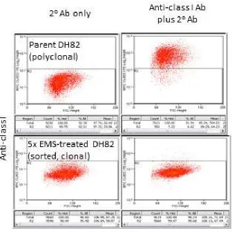
Figure 2. Example of a mutated DH82-derived cell clone displaying reduced DLAI expression ... 44
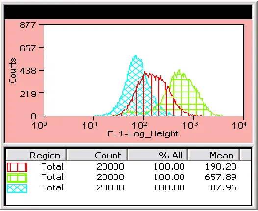
Figure 3. Overlay of flow cytometry histograms indicate RMAS cells display
increased MHCI expression following bulk peptide pulse protocols ... 45
Figure 4. The transfection of K562 cells with pCCR5 and both TALEN plasmids results in GFP conversion, indicating TALEN activity ... 52
Figure 5. Surveyor assay indicates that the K562 TALEN-recipients (TAL) possess insertion/deletion events that result in cleave by the endonuclease enzyme following
PCR product hybridization, while control product hybrids are not cleaved ... 52
Chapter 4.
Figure 1. TALEN and GFP reporter co-transfection into CRFK cells increases GFP+ MHCI- population ... 64
Figure 2. Cell clones of GFP+ MHCI- TALEN transfectants have modified genomic TAP2 sequences... 71
Figure 3. Exogenous TAP2 plasmid transfection into TAP2-KO clones restores surface FLAI expression ... 74
Figure 4. The 1A3-derived PoBoy2 clone retains the FLAG-tagged FLAI allele ... 75
Figure 5. Cold incubation increases surface FLAI expression on TAP2-KO clones ... 78
xiii
Chapter 5.
Figure 1. Immune cell interactions in T1D ... 92
Chapter 6.
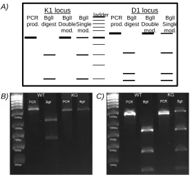
Figure 1. TALEN design for the NIT-1, classical MHCI genes ... 126
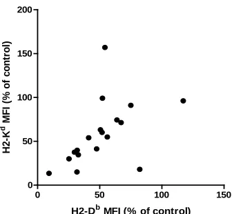
Figure 2. The co-transfection of Bgl left and right TALEN plasmids with the GFP reporter plasmid into NIT-1 cells increases GFP+ cells, genomic modifications, and
the percentage of cells that display modified levels of MHCI surface protein ... 128
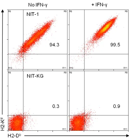
Figure 3. GFP-sorted, TALEN-transfected NIT-1 cell clones express altered surface protein values compared to an MHCIhigh clone... 138 Figure 4. The GFP+, TALEN-recipient cell clone, NIT-KG, lacks surface MHCI protein ... 139
Figure 5. The loss of restriction site assay indicates the NIT-KG clone contains modified genomic MHCI loci ... 140
Figure 6. The NIT-KG clone possesses modified genomic MHCI loci that result in frame-shift mutations and early stop codons ... 142
Figure 7. CD8+ T cells specific for IGRP-Kd molecules do not respond to the KG
cell line pulsed with the high affinity cognate peptide (NRP-V7) ... 144
xiv
Supplemental figure 2. Nucleotide sequence of the BglI TALEN left insert that was ligated into the JDS74 expression construct prior to co-transfection... 152
Supplemental figure 3. Nucleotide sequence of the BglI TALEN right insert that was ligated into the JDS70 expression construct prior to co-transfection ... 153
1
CHAPTER 1: Role of classical class I MHC in regulating cellular immune responses
Introduction
This dissertation will be focused upon two broad topics, as depicted in Figure
1, relating to the intentional downregulation of classical class Ia major
histocompatibility complex (MHCI) protein expression: 1) manipulating surface
expression in order to elucidate cluster of differentiation (CD)8+ T-cell responses in
the Canis lupus familiaris and Felis catus species, as this will contribute to the
sparse data available in these companion animals; and 2) preventing surface
expression in order to promote immune system evasion by graft tissues, as a
potential therapy for type 1 diabetes (T1D) patients. In this chapter, I will provide a
brief review of the current understanding of the mechanisms of MHCI expression as
these molecules restrict CD8+ T-cell and inhibit natural killer (NK)-cell responses
across species. Overall, we aim to 1) characterize and knock out the transporters
associated with antigen processing (TAP) genes so as to create an invaluable tool
kit for studying canine and feline T-cell responses; and to 2) use transcription
activator-like effector nucleases (TALENs) to knock out (KO) the MHCI heavy chains
2
Figure 1. Schematic of dissertation organization depicting aims, species included in studies, and proposed
outcomes. Chapters will be proceed as shown from left to right.
Overall Aim
Investigating and
manipulating MHC class I
restricted peptide epitopes
and cognate T cells
responses
Sub-aim
Downregulation of MHCI for studying CD8+ T-cell
responses Species Canis lupus familiaris Characterization of TAP1/2 genes Mutagenesis and selection of MHCIlow
cell lines
Species
Felis catus
Confirm TAP2 locus
TAP2 gene KO via TALEN technology
Sub-aim
Downregulation of MHCI for evasion from autoreactive
CD8+ T-cell recognition
Species
Mus musculus
TALEN-mediated disruption of MHCI in insulinoma cells
3
REGULATION OF IMMUNE CELLS BY MHCI EXPRESSION
The genomic region encoding a plethora of genes primarily involved in
immune responses is called the major histocompatibility complex (MHC), and is
thought to have co-evolved by way of duplication events and exon shuffling, while
being conserved across Bilateria (Danchin et al., 2003, 2004). Genes important for
immune function and antigen presentation, such as the classical and nonclassical
class I MHC loci, the class II MHC genes, the TAP1/2 genes, and proteasome
genes, are located within the MHC class 1 and 2 regions. The genes relevant to this
dissertation are those that encode the heterodimeric TAP1 and 2 subunits and also
the heavy chain units of the trimolecular peptide:MHCI complexes. For a detailed
discussion of the organization and function of the TAP1/2 genes, see Chapter 2. The
rest of this chapter will be dedicated to the function of the highly polymorphic MHCI
genes as they restrict immune responses.
The classical heterotrimeric MHCI complex is comprised of the MHCI heavy
chain, the beta-2-microglobulin (β2M) light chain, and a short peptide fragment that
is derived from proteins within the cytosol, after degradation by the proteasome, and
shuttled by the TAP heterodimer into the endoplasmic reticulum (ER), where these
peptides can then interact with the multitude of molecules that create the peptide
loading complex (Cresswell et al., 2006). The peptide-loaded MHCI (pMHCI)
complex is transported to the extracellular surface via the golgi apparatus where
receptors on NK- and CD8+ T-cells can interact with the complexes. Thus, MHCI
4
the surface membrane, where it may then mediate multifaceted interactions with the
immune system.
Numerous points along the above biochemical processes limit the breadth of
peptides presented within the groove of the MHCI molecule, thereby restricting the
ability of the T-cell receptor on the surface of CD8+ T cells to “see” all possible
peptides presented by MHCI complexes [reviewed in (Yewdell, 2006)], resulting in
certain antigen-specific clonotypes of CD8+ T cells to be more prevalent during
infections. Peptides that have been fragmented from intracellular proteins by the
proteasome and which are of sufficient affinity for MHCI peptide-binding residues,
found within the highly variable regions within the alpha 1 and 2 domains of the MHC
heavy chain (Bjorkman et al., 1987), may stabilize the MHCI complex to create a
peptide-loaded mature state (Mage et al., 2013; Neefjes et al., 2011; Trombetta and
Mellman, 2005). The constitutive proteasome and its interferon gamma
(IFNγ)-induced altered state, known as the immunoproteasome, have been shown to result
in differential peptide presentation (Toes et al., 2001) and influence CD8+ T-cell
responses (Chen et al., 2001), though the overall reduction of possible epitopes
presented following these antigen-editing pathways does not contribute significantly
to the restriction of T-cell responses (Yewdell, 2006). Instead, it has been shown that
allelic polymorphisms at the MHCI residues responsible for peptide-binding are what
ultimately influence the repertoire of peptides displayed at the cell surface (Bleek
and Nathenson, 1991), and thereby result in immunodominant CD8+ T-cell
5
allomorphs to bind peptides (often of variable lengths) is a result of conformational
differences created during folding of the MHCI peptide-binding groove (Madden et
al., 1991; Collins et al., 1994). MHCI folding results in the formation of pockets within
the alpha 1 and 2 domains that allow for interactions with residues at specific
positions of the peptide, thereby resulting in so-called peptide-binding motifs, which
are useful for predicting the ability of any peptide to bind to an MHCI allele
(Lundegaard et al., 2008). An example of such a peptide-binding motif is the
preferential binding of the canine MHCI allele, DLA-88*50801, with peptide
fragments possessing hydrophobic residues at positions 2 and 3, and either an
arginine or lysine at position 9 [(Ross et al., 2012); manuscript in submission].
Furthermore, some allomorphs of MHCI also possess the characteristic of binding
peptides with submotif-matching sequences (Fremont et al., 1995), thereby making
peptide binding predictions more complicated. Other steps that also restrict the
ability of pMHCI complexes from being recognized by CD8+ T cells include the
inability of TAP to transport all degraded protein fragments into the ER and the
availability of T-cell receptors (TCRs) capable of interacting with presented peptides
in the MHCI complex, but these are purported to have minimal influence (Yewdell,
2006). Thus, the steps that reduce the pool of presented pMHCI complexes, antigen
editing and MHCI binding primarily, result in a hierarchical distribution of CD8+ T
cells capable of responding to specific intracellular infections or self peptides.
As mentioned above, the ability to predict peptides derived from pathogens
6
useful tool and numerous databases are available to assist in these predictions
(Nielsen et al., 2007; Rammensee et al., 1999; Meydan et al., 2013). By applying
“reverse immunology” strategies, researchers are able to derive peptides from the
proteome of a pathogen, which can include some 135,000 proteins as in the
poxvirus (Yewdell, 2006), that have a high likelihood of binding the MHCI allomorph
of interest and stimulate cognate T-cell responses, which may ultimately provide
vaccination strategies or tools for disease diagnoses (van Endert et al., 2006). The
confirmation of a T-cell response to a putative MHCI-binding peptide can be
achieved by detecting T-cell effector responses, which will be discussed later,
including measurement of cytokine release, calcium flux, surface protein up- or
downregulation, or proliferation, to name a few (Tobery et al., 2006; Abu-Khader and
Krause, 2013). Yet, while pathogen-specific CD8+ T-cell responses have been
described in the canine and feline species (Hartley et al., 2014; Miller et al., 2013),
data regarding the precise roles of the MHCI loci responsible for restricting these
responses is lacking for companion animals.
In order to derive such peptide prediction databases for canid and felid
responses, the motifs of peptides bound by these species classical MHCI
allomorphs must first be empirically determined; one of our primary. Such MHCI
peptide motif characterization requires proper tool kits for 1) the collection of
naturally processed and presented peptides by MHCI allomorphs for the purposes of
creating binding motif hypotheses, and 2) the empirical testing of peptide-binding
7
a tool kit for analyzing the peptide motif of the classical canine DLAI-88*50801
allomorph (Ross et al., 2012), and in this dissertation I will discuss efforts to create a
similar tool for analyzing feline MHCI peptide-binding. The next subsections will
briefly describe the functions of MHCI complexes in restricting immune responses of
specific cellular subsets.
MHCI and T cells
Following the loading of edited peptides into the groove of the MHCI heavy
chain associated with β2M, pMHCI is displayed at the cellular membrane, where the
complex may interact with the TCRs on CD8+ T cells to create an immunological
synapse. The CD8 co-receptor on αβ TCR-possessing T cells is comprised of a
heterodimeric αβ glycoprotein complex that interacts with an invariant region of the
alpha 3 domain of MHCI (Salter et al., 1990) to stabilize the pMHCI-TCR interaction
and to enhance antigen sensitivity [reviewed in (Cole et al., 2012)]. Furthermore, the
co-localization of CD8 co-receptor and TCR at the cell surface has been shown to
relate to antigen sensitivity of T cells, possibly explaining the mechanism of
functional avidity maturation that occurs during ongoing infection (Borger et al.,
2014). The multidimensional pMHCI-TCR synapse [reviewed in (Zhu et al., 2013)] is
often considered signal one of the processes that orchestrate a T-cell response. This
synapse formation, which is not a binary type of interaction (Krummel et al., 2000),
induces protein-tyrosine kinase signaling cascades by the TCR/CD3 complexes to
8
and gene transcription depending on the maturation state of the T cell. Data are
available, however, to indicate that only this signal may be required for T-cell
responses to occur (Wang et al., 2000). Yet, it is widely accepted that further
signaling cascades are associated with the initial synapse formation to enhance or
prevent T-cell responses. Signal two is governed by co-receptors, such as activating
CD28 or inhibitory PD-1 complexes (Okazaki et al., 2002), on the CD8+ T cell that
provide further feedback signals to mediate responses by the T cell. Finally, the local
cytokine milieu, produced by antigen-presenting cells and/or CD4+ T cells in close
proximity to synapse formation, further enhances the regulatory feedback within
CD8+ T cells following TCR-MHCI ligation (Leung et al., 2010). Thus, the production
of an effective T-cell response is a complexly regulated pathway that starts with the
interaction of pMHCI-TCR complexes.
The accumulation of stimulatory and inhibitory signals in T cells ultimately
results in the activation of transcriptional regulatory factors that alter gene
expression to allow for effector function acquisition [reviewed in (Taniuchi, 2009)].
The relatively new understanding of the plasticity in the spectrum of T-cell lineage
differentiation has further highlighted the importance of the presence and qualitative
differences of secondary and tertiary signals (Arens and Schoenberger, 2010;
Murphy and Stockinger, 2010; Crispín and Tsokos, 2009). Thus, recent data reaffirm
the long-held conception that CD8+ T cells are not only capable of cytotoxic activity,
but also suppressor functions (Shafer-Weaver et al., 2009), though this concept will
9
cytotoxic effector functions of CD8+ T cells in the realm of diabetes, but here I will
briefly highlight general functions. Specifically, interferon-gamma (IFNγ) is one of the
primary cytokines produced by CD8+ T cells and functions to amplify T-cell
responses early after synapse formation (Teixeira et al., 2005), and to enhance
CD8+ T-cell cytotoxicity against malignant or infected cells, and also specifically
against the insulin producing beta (β) cells in type 1 diabetes (Chong et al., 2011).
The release of tumor necrosis factor-alpha (TNFα) cytokine, the release of cytotoxic
granules containing perforin and granzyme B onto target cells, and the presence of
apoptosis-inducing Fas ligands at the cell surface are also well-described effector
mechanisms utilized by CD8+ T cells (Janeway et al., 2001). Thus, the differentiation
of CD8+ T-cell effector functions is a plastic spectrum involving transcriptional
differences regulated epigenetically, which may result in the capacity to acquire
cytotoxic capability, one of the primary means by which these cells induce immunity
or tolerance.
The primary role of CD8+ T cells is to utilize effector functions to contain the
spread of the intracellular pathogen and to create an inhospitable environment for
pathogen replication [reviewed in (Demers et al., 2013)]. This is achieved by using
cytotoxic functions (e.g. Fas ligand and granzyme) to induce apoptosis in cells
displaying cognate peptides derived from intracellular pathogens by MHCI
complexes, in order to directly destroy cells infected by pathogen. During
differentiation, cytotoxic cells accumulate lytic molecules within granules
10
of target cells by way of polarization of granules towards the area of pMHCI
synapses [reviewed in (Galandrini et al., 2013)], thereby limiting the directional
release of cytotoxic molecules towards the infected cells. Furthermore, CD8+ T cells
can release non-cytotoxic beta-chemokines or lymphokines that may inhibit viral
entrance into the cell or viral replication within the cell (Demers et al., 2013).
Cytotoxic effectors are therefore primed by activation signals and respond rapidly to
intracellular pathogen to prevent spread of the infection.
Before circulating in the periphery where CD8+ T cells may perform the above
functions, they are first “educated” in the thymus. Here, thymocytes are exposed to
two primary types of processes, called positive and negative selection, mediated by
cortical and medullary antigen-presenting thymic epithelial cells, respectively
(Holländer and Peterson, 2009). These processes are responsible for producing
CD8 single positive T cells that have TCR with affinity for self MHCI complexes, but
have only low affinity for self peptides presented by MHCI complexes. Such TCR
selection is thought to occur based on the zipper model of pMHCI-TCR interaction
that correlates dwell time with activation thresholds that result in cell survival or
apoptosis [reviewed in (Palmer and Naeher, 2009)]. A recent study quantified thymic
selection processes in the mouse and determined that six times more thymocytes
undergo apoptosis in negative selection than survive positive selection, and that the
TCR signaling that induces cell death is only slightly stronger than signals that
induce survival (Stritesky et al., 2013). The thymic education, and routine peripheral
11
described to have a profound effect on the magnitude and type of response elicited
by these cells during infection (Persaud et al., 2014). The signaling cascades that
influence activation thresholds involve TCR-proximal adaptor proteins, such as
ZAP-70. Autoreactive T cells may possess mutations in ZAP-70 (Hsu et al., 2009) and
therefore do not become anergic. Furthermore, inducible expression of a negative
regulator of TCR signaling, CD5, on T cells during the negative selection process
helps to fine tune TCR signaling, and CD5 expression correlates with the affinity of
TCR for antigen (Azzam et al., 2001). When transgenic TCR with relatively high
affinity for self-MHC are expressed in the absence of CD5 expression, these
clonotypes undergo negative selection, likely due to strong TCR-mediated signal
transduction levels being too high. As a counter-balancing regulatory mechanism,
thymic processes can trigger survival, also a result of TCR affinity-based survival
(Lee et al., 2012), of regulatory T cells with higher affinity for self pMHCI that assist
in preventing autoreactivity by secreting immunosuppressive cytokines or expressing
membrane-bound effector molecules [reviewed in (Josefowicz et al., 2012)]. Thus,
the thymus produces CD8+ T cells displaying TCR with sufficient affinity for self
MHCI complexes to allow for effector responses, but can also produce regulatory T
cells that assist in preventing autoreactivity in the periphery.
As further discussed in chapter 5, T1D (and other autoimmune) patients
possess CD8+ T cells in the periphery capable of recognizing self peptides at
frequencies that overpower the regulatory T-cell effects (Tsai et al., 2010). While
12
extrathymic autoimmune regulator (Aire)-expressing cells (Bluestone and
Bour-Jordan, 2012; Metzger and Anderson, 2011), autoreactive T cells in these patients
somehow persist. There are numerous hypotheses regarding how such cells are not
anergized during tolerization processes and how these cells become activated in
peripheral circulation. Some hypotheses involve the peptide-centric model of TCR
binding by autoreactive CD8+ T cells (Bulek et al., 2012) and the ability of a single
TCR to recognize more than one peptide (Wooldridge et al., 2012). Furthermore, the
concept of T cells displaying more than one rearranged TCR gene product at the cell
membrane is also a possibility (Simpson et al., 1995; Fossati et al., 1999). The
exact mechanisms by which these autoreactive cells are activated in the periphery to
respond to self are still highly debatable. Two such hypotheses involve infection of
islets by enteroviruses and the concept of viral mimicry (Laitinen et al., 2014; Enouz
et al., 2012), resulting in release of cognate peptides to levels sufficient for CD8+
T-cell activation or viral peptides being displayed by MHCI on β T-cells that appear
similar to those recognized by autoreactive TCR, respectively. Therefore, peripheral
tolerance mechanisms are not always able to prevent autoreactivity to self peptides
mediated by CD8+ T cells that escape central tolerance mechanisms.
CD4+ T cells interact with peptide:MHCII complexes and are also under the
control of central and peripheral tolerance mechanisms, but will not be a focal point
of this dissertation. Yet, CD4+ T-cell responses do play a large role in susceptibility
to autoimmune diseases, as highlighted by the strong correlation between MHCII
13
allomorphs are believed to contribute heavily to disease progression in individuals
possessing risk-associated alleles (Ettinger et al., 2006), and inadequate suppressor
functions by CD4+ T cells are also believed to contribute to diabetes pathogenesis
(Long et al., 2010). Normally, CD4+ T cells help to provide the necessary cues for
mediating activation of CD8+ T cells against intracellular pathogens. Such cues
including the cytokines interleukin (IL)-4, IL-10, or IFNγ (and many others), are
involved in producing the milieu that results in an inflammatory or anti-inflammatory
environment, and direct humoral- or cell-mediated responses. This environment can
alter the activation statuses of antigen-presenting cells, e.g. upregulation of CD80
and CD86 on macrophages, and thereby enhance CD8+ T-cell activation by
providing co-stimulation. CD4+ T-cells, therefore, help to propagate CD8+ T
cell-responses, which are, normally, responsible for preventing the spread of intracellular
infections.
In summary, this subsection highlights the function of pMHCI complexes to
restrict CD8+ T-cell responses, which have been educated in the thymus to
recognize primarily non-self antigens presented by self MHCI, and are assisted by
CD4+ T cells [and other subsets (Lehuen et al., 2010)] that promote a cytokine
milieu conducive to cell-mediated immunity. Furthermore, peripheral regulatory
mechanisms, such as regulatory CD4+ and suppressor CD8+ T cells, assist in
preventing autoreactive responses, but may be overwhelmed by genetic variations
resulting in defective signaling within T cells of predisposed individuals. Thus,
14
standard CD8+ T-cell cytotoxic capabilities, but are directed against self pMHCI
complexes due to incomplete anergy induction.
MHCI and NK cells
Another immune cell subset that interacts directly with pMHCI complexes are
natural killer (NK) cells, which are considered an intermediate between innate and
adaptive immunity (Long et al., 2013). NK cells lack receptor diversity created by
rearrangement of DNA, but do possess a wide array of receptors encoded in the
germline, which use stimulatory (such as immunoreceptor tyrosine-based activation
motifs; ITAM) and inhibitory (immunoreceptor tyrosine-based inhibitory motifs; ITIM)
signaling cascades also present in other immune cell subsets. This section will only
briefly describe the function of this cell type and provide insights into the importance
of this cell type to our second major topic of this dissertation.
There are a plethora of NK cell receptors, but data regarding the function of
each are not currently available. The broadest of categories can be considered the
killer activation receptors (KAR) and killer inhibitory receptors (KIR) as based upon
the motifs found in the cytoplasmic tails; these groups paradoxically contain some
receptors that function antithetically to the group name (Orr and Lanier, 2011).
Furthermore, the naming of identical, orthologous receptors across species are not
consistent. However, the receptors primarily thought to be important for mediating
NK-cell responses (Long et al., 2013) are CD16 (a receptor for the Fc portion of
MHCI-15
specific ITIM-bearing receptors (a broad group of inhibitory receptors that interact
with classical and nonclassical MHCI complexes), which are of interest to our
discussion. While NK cells are known to display a multitude of receptors and
co-receptors that can propagate activation in synergistic and redundant manners, the
receptors that interact with MHCI license NK cell activity.
The specific MHCI environment of the individual organism results in licensing
of NK cells by interaction of inhibitory receptors on the NK cell with the individually
expressed MHCI complexes (Kim et al., 2005). The precise mechanisms involved in
NK cell licensing are debatable, as they may occur through a stimulatory or inhibitory
model [reviewed in (Yokoyama and Kim, 2006)] but ligation of receptors on the NK
cell with MHCI complexes is necessary for either. Such licensing corroborates the
“missing self” hypothesis of NK cell function (Kärre et al., 1986; Ljunggren and Kirre,
1990), and indicates the importance of MHCI expression as a negative feedback
mechanism that prevents NK cell cytotoxicity. The precise inhibitory signaling
induced by the ligation of MHCI to KIR appears to be propagated by ITIM domains
recruiting the Src homology region 2 domain-containing phosphatase-1 (SHP-1),
thereby preventing signal propagation of ITAM-bearing receptors (Long et al., 2013)
through dephosphorylation of a component in the activation pathway [possibly Vav1;
(Long, 2008)]. The licensing of NK cells has been described to be revocable, in that
NK cell hyporesponsiveness is found within hosts lacking MHCI expression, but may
be re-initiated following certain infections (Sun and Lanier, 2008; Joncker et al.,
hematopoietic-16
derived cell subsets (Kawamoto and Katsura, 2009). NK cells, therefore, display a
wide array of NK cell receptors, but the inhibitory receptors that recognize MHCI
complexes as ligands are crucial for preventing NK cell-mediated killing of normal
host cells and appear to be the dominant determinant of NK activation status.
Thus, MHCI complexes are critical for both NK- and CD8+ T-cell responses.
NK and CD8+ T cells share cytotoxic capabilities [specifically, lytic granules
(Galandrini et al., 2013)] and are very similar in their transcriptional profiles (Bezman
et al., 2012). However, unlike CD8+ T cells, new data indicate that NK cells may be
able to recognize viral infection due to the possession of pattern recognition
receptors, a poorly studied realm of NK-cell function (Muntasell et al., 2013).
Furthermore, NK cells require lower priming signals than CD8+ T cells in order to
respond to virally infected, malignantly transformed, or allogeneic cells, which are
the primary functions of NK cells. The capability of resting NK cells to rapidly
respond also makes these cells dangerous to transplant survival (Liu et al., 2012),
but also possibly helpful (Kroemer et al., 2008). Specifically, NK cells are not able to
recognize allogeneic MHCI allomorphs, and thus destroy allographs due to “missing
self”; but data also exist indicating that NK cells assist in destroying donor
antigen-presenting cells (APC), thereby reducing recipient alloreactive CD4+ T-cell
responses that are primed by donor APC. NK-cell activity detected against
bioengineered graft tissues that display reduced levels of MHCI indicate destruction
of these grafts as a likelihood (de la Garza-Rodea et al., 2011), but the precise
17
reactivity are ill defined. NK cells therefore function as cytotoxic mediators in the
early stages of cancer protection, viral infection, and graft rejection by using similar
effector functions as CD8+ T cells, and these responses are MHCI-dependent,
although NK-cell restrictions involve both classical and nonclassical MHCI.
NK cells are regarded as a primary line of defense against viral infection and
cancer, but situations do arise in which NK cell surveillance is evaded. Viruses may
possess specifically adapted protein mimicry, such as the murine cytomegalovirus
m157 protein (Orange et al., 2002), and cancers that have undergone the
elimination phase of cancer cell immunoediting are able to escape attack by NK cells
(Dunn et al., 2002). One example of viral immunevasin gene application in tissues
for transplant was described in the paper by de la Garza-Rodea cited above, in
which the cytomegalovirus US11 protein was used to reduce MHCI expression in
xenogeneic stem cells. While this application required the depletion of recipient NK
cells for graft survival, other virally derived proteins, such as MHCI homologs, may
further assist in graft survival (Orange et al., 2002). These naturally occurring
mechanisms for immune system evasion, therefore, offer insights into strategies by
which insulin producing grafts may be engineered to escape recognition by both
autoreactive CD8+ T cells and licensed NK cells in diabetic recipients. Chapter 6
contains a discussion of methods to address these issues with the assistance of
18
Summary of objectives
This chapter contains a brief review of the class I antigen processing pathway
and how MHCI presentation restricts CD8+ T-cell and NK-cell responses Results
presented in this dissertation will add to the understanding of the companion animal
MHCI-restricted T-cell responses by characterizing the canine TAP genes, and by
confirming the feline TAP2 locus while also creating KO cell lines. Additional results
demonstrate the use of biotechnological tools to create insulin producing tissues
19
References
Arens, R., and S.P. Schoenberger. 2010. Plasticity in programming of effector and memory CD8 T-cell formation. Immunol. Rev. 235:190–205.
doi:10.1111/j.0105-2896.2010.00899.x.
Azzam, H.S., J.B. DeJarnette, K. Huang, R. Emmons, C.-S. Park, C.L. Sommers, D. El-Khoury, E.W. Shores, and P.E. Love. 2001. Fine Tuning of TCR Signaling by CD5. J. Immunol. 166:5464–5472.
Baker, P.R., and A.K. Steck. 2011. The Past, Present, and Future of Genetic Associations in Type 1 Diabetes. Curr. Diab. Rep. 11:445–453.
doi:10.1007/s11892-011-0212-0.
Bezman, N.A., C.C. Kim, J.C. Sun, G. Min-Oo, D.W. Hendricks, Y. Kamimura, J.A. Best, A.W. Goldrath, L.L. Lanier, and T.I.G.P. Consortium. 2012. Molecular definition of the identity and activation of natural killer cells. Nat. Immunol.
13:1000–1009. doi:10.1038/ni.2395.
Bjorkman, P.J., M.A. Saper, B. Samraoui, W.S. Bennett, J.L. Strominger, and D.C. Wiley. 1987. Structure of the human class I histocompatibility antigen, HLA-A2. Nature. 329:506–512. doi:10.1038/329506a0.
Bleek, G.M. van, and S.G. Nathenson. 1991. The structure of the antigen-binding groove of major histocompatibility complex class I molecules determines specific selection of self-peptides. Proc. Natl. Acad. Sci. 88:11032–11036.
Bluestone, J.A., and H. Bour-Jordan. 2012. Current and future immunomodulation strategies to restore tolerance in autoimmune diseases. Cold Spring Harb. Perspect. Biol. 4. doi:10.1101/cshperspect.a007542.
Borger, J.G., R. Zamoyska, and D.M. Gakamsky. 2014. Proximity of TCR and its CD8 coreceptor controls sensitivity of T cells. Immunol. Lett. 157:16–22. doi:10.1016/j.imlet.2013.11.005.
Bulek, A.M., D.K. Cole, A. Skowera, G. Dolton, S. Gras, F. Madura, A. Fuller, J.J. Miles, E. Gostick, D.A. Price, J.W. Drijfhout, R.R. Knight, G.C. Huang, N. Lissin, P.E. Molloy, L. Wooldridge, B.K. Jakobsen, J. Rossjohn, M. Peakman, P.J. Rizkallah, and A.K. Sewell. 2012. Structural basis for the killing of human beta cells by CD8(+) T cells in type 1 diabetes. Nat. Immunol. 13:283–289. doi:10.1038/ni.2206.
Chen, W., C.C. Norbury, Y. Cho, J.W. Yewdell, and J.R. Bennink. 2001.
20
T Cells at the Levels of T Cell Repertoire and Presentation of Viral Antigens.
J. Exp. Med. 193:1319–1326. doi:10.1084/jem.193.11.1319.
Chong, M.M.W., H.E. Thomas, and T.W.H. Kay. 2011. y-Interferon Signaling in Pancreatic beta-Cells Is Persistent but Can Be Terminated by
Overexpression of Suppressor of Cytokine Signaling-1. Diabetes. 50.
Cole, D.K., B. Laugel, M. Clement, D.A. Price, L. Wooldridge, and A.K. Sewell. 2012. The molecular determinants of CD8 co-receptor function. Immunology.
137:139–148. doi:10.1111/j.1365-2567.2012.03625.x.
Collins, E.J., D.N. Garboczi, and D.C. Wiley. 1994. Three-dimensional structure of a peptide extending from one end of a class I MHC binding site. Nature.
371:626–629. doi:10.1038/371626a0.
Cresswell, P., N. Bangia, T. Dick, and and G. Diedrich. 2006. The nature of the MHC class I peptide loading complex. Immunol. Rev. 172:21–8.
Crispín, J.C., and G.C. Tsokos. 2009. Transcriptional regulation of IL-2 in health and autoimmunity. Autoimmun. Rev. 8:190–195.
doi:10.1016/j.autrev.2008.07.042.
Danchin, E., V. Vitiello, A. Vienne, O. Richard, P. Gouret, M.F. McDermott, and P. Pontarotti. 2004. The major histocompatibility complex origin. Immunol. Rev.
198:216–232. doi:10.1111/j.0105-2896.2004.00132.x.
Danchin, E.G.J., L. Abi-Rached, A. Gilles, and P. Pontarotti. 2003. Conservation of the MHC-like region throughout evolution. Immunogenetics. 55:141–148. doi:10.1007/s00251-003-0562-0.
Demers, K.R., M.A. Reuter, and M.R. Betts. 2013. CD8+ T-cell effector function and transcriptional regulation during HIV pathogenesis. Immunol. Rev. 254:190– 206. doi:10.1111/imr.12069.
Dunn, G.P., A.T. Bruce, H. Ikeda, L.J. Old, and R.D. Schreiber. 2002. Cancer immunoediting: from immunosurveillance to tumor escape. Nat. Immunol.
3:991–998. doi:10.1038/ni1102-991.
Van Endert, P., Y. Hassainya, V. Lindo, J.-M. Bach, P. Blancou, F. Lemonnier, and R. Mallone. 2006. HLA class I epitope discovery in type 1 diabetes. Ann. N. Y. Acad. Sci. 1079:190–197. doi:10.1196/annals.1375.030.
21
Ettinger, R.A., G.K. Papadopoulos, A.K. Moustakas, G.T. Nepom, and W.W. Kwok. 2006. Allelic variation in key peptide-binding pockets discriminates between closely related diabetes-protective and diabetes-susceptible HLA-DQB1*06 alleles. J. Immunol. Baltim. Md 1950. 176:1988–1998.
Fossati, G., A. Cooke, R.Q. Papafio, K. Haskins, and B. Stockinger. 1999. Triggering a second T cell receptor on diabetogenic T cells can prevent induction of diabetes. J. Exp. Med. 190:577–583.
Fremont, D.H., E.A. Stura, M. Matsumura, P.A. Peterson, and I.A. Wilson. 1995. Crystal structure of an H-2Kb-ovalbumin peptide complex reveals the interplay of primary and secondary anchor positions in the major
histocompatibility complex binding groove. Proc. Natl. Acad. Sci. U. S. A.
92:2479–2483.
Galandrini, R., C. Capuano, and A. Santoni. 2013. Activation of lymphocyte cytolytic machinery: where are we? Front. NK Cell Biol. 4:390.
doi:10.3389/fimmu.2013.00390.
De la Garza-Rodea, A., M. Verweij, H. Boersma, I. van der Velde-van Dijke, A. de Vries, R. Hoeben, D. van Bekkum, E.J. Wiertz, S. Knaän-Shanzer, and D. Unutmaz. 2011. Exploitation of Herpesvirus Immune Evasion Strategies to Modify the Immunogenicity of Human Mesenchymal Stem Cell Transplants.
PLoS ONE. 6:e14493. doi:10.1371/journal.pone.0014493.
Hartley, A.N., G. Cooley, S. Gwyn, M.M. Orozco, and R.L. Tarleton. 2014. Frequency of IFNγ-producing T cells correlates with seroreactivity and activated T cells during canine Trypanosoma cruzi infection. Vet. Res. 45:6. doi:10.1186/1297-9716-45-6.
Holländer, G.A., and P. Peterson. 2009. Learning to be tolerant: how T cells keep out of trouble. J. Intern. Med. 265:541–561.
doi:10.1111/j.1365-2796.2009.02093.x.
Hsu, L.-Y., Y.X. Tan, Z. Xiao, M. Malissen, and A. Weiss. 2009. A hypomorphic allele of ZAP-70 reveals a distinct thymic threshold for autoimmune disease versus autoimmune reactivity. J. Exp. Med. 206:2527–2541.
doi:10.1084/jem.20082902.
Janeway, J.C., P.J. Travers, and M. walport. 2001. T cell-mediated cytotoxicity. In
22
Joncker, N., N. Shifrin, F. Delebecque, and D. Raulet. 2010. Mature natural killer cells reset their responsiveness when exposed to an altered MHC
environment. J. Exp. Med. 207:2065–2072. doi:10.1084/jem.20100570.
Josefowicz, S.Z., L.-F. Lu, and A.Y. Rudensky. 2012. Regulatory T Cells:
Mechanisms of Differentiation and Function. Annu. Rev. Immunol. 30:531– 564. doi:10.1146/annurev.immunol.25.022106.141623.
Kärre, K., H.G. Ljunggren, G. Piontek, and R. Kiessling. 1986. Selective rejection of H-2-deficient lymphoma variants suggests alternative immune defence strategy. Nature. 319:675–678. doi:10.1038/319675a0.
Kawamoto, H., and Y. Katsura. 2009. A new paradigm for hematopoietic cell lineages: revision of the classical concept of the myeloid–lymphoid dichotomy. Trends Immunol. 30:193–200. doi:10.1016/j.it.2009.03.001.
Abu-Khader, A., and S. Krause. 2013. Rapid monitoring of immune reconstitution after allogeneic stem cell transplantation--a comparison of different assays for the detection of cytomegalovirus-specific T cells. Eur. J. Haematol. 91:534– 545. doi:10.1111/ejh.12187.
Kim, S., J. Poursine-Laurent, S.M. Truscott, L. Lybarger, Y.-J. Song, L. Yang, A.R. French, J.B. Sunwoo, S. Lemieux, T.H. Hansen, and W.M. Yokoyama. 2005. Licensing of natural killer cells by host major histocompatibility complex class I molecules. Nature. 436:709–713. doi:10.1038/nature03847.
Kroemer, A., K. Edtinger, and X.C. Li. 2008. The innate natural killer cells in transplant rejection and tolerance induction: Curr. Opin. Organ Transplant.
13:339–343. doi:10.1097/MOT.0b013e3283061115.
Krummel, M., C. Wülfing, C. Sumen, and M.M. Davis. 2000. Thirty–six views of T– cell recognition. Philos. Trans. R. Soc. Lond. B. Biol. Sci. 355:1071–1076. doi:10.1098/rstb.2000.0644.
Laitinen, O.H., H. Honkanen, O. Pakkanen, S. Oikarinen, M.M. Hankaniemi, H. Huhtala, T. Ruokoranta, V. Lecouturier, P. André, R. Harju, S.M. Virtanen, J. Lehtonen, J.W. Almond, T. Simell, O. Simell, J. Ilonen, R. Veijola, M. Knip, and H. Hyöty. 2014. Coxsackievirus B1 Is Associated With Induction of β-Cell Autoimmunity That Portends Type 1 Diabetes. Diabetes. 63:446–455.
doi:10.2337/db13-0619.
23
Lehuen, A., J. Diana, P. Zaccone, and A. Cooke. 2010. Immune cell crosstalk in type 1 diabetes. Nat. Rev. Immunol. 10:501–513. doi:10.1038/nri2787.
Leung, S., X. Liu, L. Fang, X. Chen, T. Guo, and J. Zhang. 2010. The cytokine milieu in the interplay of pathogenic Th1/Th17 cells and regulatory T cells in
autoimmune disease. Cell. Mol. Immunol. 7:182–189. doi:10.1038/cmi.2010.22.
Liu, W., X. Xiao, G. Demirci, J. Madsen, and X.C. Li. 2012. Innate NK cells and macrophages recognize and reject allogeneic nonself in vivo via different mechanisms. J. Immunol. Baltim. Md 1950. 188:2703–2711.
doi:10.4049/jimmunol.1102997.
Ljunggren, H., and K. Kirre. 1990. In search of the “missing self”: MHC molecules and NK cell recognition. Immunol. Today. 11:237–44.
Long, E.O. 2008. Negative signalling by inhibitory receptors: the NK cell paradigm.
Immunol. Rev. 224:70–84. doi:10.1111/j.1600-065X.2008.00660.x.
Long, E.O., H. Sik Kim, D. Liu, M.E. Peterson, and S. Rajagopalan. 2013.
Controlling Natural Killer Cell Responses: Integration of Signals for Activation and Inhibition. Annu. Rev. Immunol. 31:227–258.
doi:10.1146/annurev-immunol-020711-075005.
Long, S.A., K. Cerosaletti, P.L. Bollyky, M. Tatum, H. Shilling, S. Zhang, Z.-Y. Zhang, C. Pihoker, S. Sanda, C. Greenbaum, and J.H. Buckner. 2010. Defects in IL-2R signaling contribute to diminished maintenance of FOXP3 expression in CD4(+)CD25(+) regulatory T-cells of type 1 diabetic subjects.
Diabetes. 59:407–415. doi:10.2337/db09-0694.
Lundegaard, C., K. Lamberth, M. Harndahl, S. Buus, O. Lund, and M. Nielsen. 2008. NetMHC-3.0: accurate web accessible predictions of human, mouse and monkey MHC class I affinities for peptides of length 8-11. Nucleic Acids Res.
36:W509–512. doi:10.1093/nar/gkn202.
Madden, D.R., J.C. Gorga, J.L. Strominger, and D.C. Wiley. 1991. The structure of HLA-B27 reveals nonamer self-peptides bound in an extended conformation.
Nature. 353:321–325. doi:10.1038/353321a0.
Mage, M.G., M.A. Dolan, R. Wang, L.F. Boyd, M.J. Revilleza, H. Robinson, K. Natarajan, N.B. Myers, T.H. Hansen, and D.H. Margulies. 2013. A structural and molecular dynamics approach to understanding the peptide-receptive transition state of MHC-I molecules. Mol. Immunol. 55:123–125.
24
Messaoudi, I., J.A. Guevara Patiño, R. Dyall, J. LeMaoult, and J. Nikolich-Zugich. 2002. Direct link between mhc polymorphism, T cell avidity, and diversity in immune defense. Science. 298:1797–1800. doi:10.1126/science.1076064.
Metzger, T.C., and M.S. Anderson. 2011. Control of central and peripheral tolerance by Aire. Immunol. Rev. 241:89–103. doi:10.1111/j.1600-065X.2011.01008.x.
Meydan, C., H.H. Otu, and O.U. Sezerman. 2013. Prediction of peptides binding to MHC class I and II alleles by temporal motif mining. BMC Bioinformatics. 14 Suppl 2:S13. doi:10.1186/1471-2105-14-S2-S13.
Miller, M.M., E.M. Thompson, S.E. Suter, and J.E. Fogle. 2013. CD8+ clonality is associated with prolonged acute plasma viremia and altered mRNA cytokine profiles during the course of feline immunodeficiency virus infection. Vet. Immunol. Immunopathol. 152:200–208. doi:10.1016/j.vetimm.2012.12.005.
Muntasell, A., M. Costa-Garcia, A. Vera, N. Marina-Garcia, C.J. Kirschning, and M. López-Botet. 2013. Priming of NK cell anti-viral effector mechanisms by direct recognition of human cytomegalovirus. Front. Immunol. 4:40.
doi:10.3389/fimmu.2013.00040.
Murphy, K.M., and B. Stockinger. 2010. Effector T cell plasticity: flexibility in the face of changing circumstances. Nat. Immunol. 11:674–680. doi:10.1038/ni.1899.
Neefjes, J., M.L.M. Jongsma, P. Paul, and O. Bakke. 2011. Towards a systems understanding of MHC class I and MHC class II antigen presentation. Nat. Rev. Immunol. 11:823–836. doi:10.1038/nri3084.
Nielsen, M., C. Lundegaard, T. Blicher, K. Lamberth, M. Harndahl, S. Justesen, G. Røder, B. Peters, A. Sette, O. Lund, and S. Buus. 2007. NetMHCpan, a method for quantitative predictions of peptide binding to any HLA-A and -B locus protein of known sequence. PloS One. 2:e796.
doi:10.1371/journal.pone.0000796.
Okazaki, T., Y. Iwai, and T. Honjo. 2002. New regulatory receptors: inducible co-stimulator and PD-1. Curr. Opin. Immunol. 14:779–782. doi:10.1016/S0952-7915(02)00398-9.
Orange, J., M. Fassett, L. Koopman, J. Boyson, and and J. Strominger. 2002. Viral evasion of natural killer cells. Nat. Immunol. 3:1006–12.
25
Palmer, E., and D. Naeher. 2009. Affinity threshold for thymic selection through a T-cell receptor–co-receptor zipper. Nat. Rev. Immunol. 9:207–213.
Persaud, S.P., C.R. Parker, W.-L. Lo, K.S. Weber, and P.M. Allen. 2014. Intrinsic CD4+ T cell sensitivity and response to a pathogen are set and sustained by avidity for thymic and peripheral complexes of self peptide and MHC. Nat. Immunol. 15:266–274. doi:10.1038/ni.2822.
Rammensee, H.-G., J. Bachmann, N.P.N. Emmerich, O.A. Bachor, and S.
Stevanović. 1999. SYFPEITHI: database for MHC ligands and peptide motifs.
Immunogenetics. 50:213–219. doi:10.1007/s002510050595.
Ross, P., J.C. Holmes, G.S. Gojanovich, and P.R. Hess. 2012. A cell-based MHC stabilization assay for the detection of peptide binding to the canine classical class I molecule, DLA-88. Vet. Immunol. Immunopathol. 150:206–212. doi:10.1016/j.vetimm.2012.08.012.
Salter, R.D., R.J. Benjamin, P.K. Wesley, S.E. Buxton, T.P.J. Garrett, C. Clayberger, A.M. Krensky, A.M. Norment, D.R. Littman, and P. Parham. 1990. A binding site for the T-cell co-receptor CD8 on the α3 domain of HLA-A2. Nature. 345:41–46. doi:10.1038/345041a0.
Sanchez-Ruiz, Y., S. Valitutti, and L. Dupre. 2011. Stepwise Maturation of Lytic Granules during Differentiation and Activation of Human CD8+ T
Lymphocytes. PLoS ONE. 6:e27057. doi:10.1371/journal.pone.0027057.
Shafer-Weaver, K.A., M.J. Anderson, K. Stagliano, A. Malyguine, N.M. Greenberg, and A.A. Hurwitz. 2009. Cutting Edge: Tumor-Specific CD8+ T Cells
Infiltrating Prostatic Tumors Are Induced to Become Suppressor Cells. J. Immunol. 183:4848–4852. doi:10.4049/jimmunol.0900848.
Simpson, E., P. Chandler, A. Sponaas, M. Millrain, and P.J. Dyson. 1995. T cells with dual antigen specificity in T cell receptor transgenic mice rejecting allografts. Eur. J. Immunol. 25:2813–2817. doi:10.1002/eji.1830251015.
Stritesky, G.L., Y. Xing, J.R. Erickson, L.A. Kalekar, X. Wang, D.L. Mueller, S.C. Jameson, and K.A. Hogquist. 2013. Murine thymic selection quantified using a unique method to capture deleted T cells. Proc. Natl. Acad. Sci. 110:4679– 4684. doi:10.1073/pnas.1217532110.
Sun, J., and L. Lanier. 2008. Viral Infection Breaks NK Cell Tolerance to “Missing Self.” J. Immunol. 181:7453–7.
26
Teixeira, L.K., B.P.F. Fonseca, A. Vieira-de-Abreu, B.A. Barboza, B.K. Robbs, P.T. Bozza, and J.P.B. Viola. 2005. IFN-gamma production by CD8+ T cells depends on NFAT1 transcription factor and regulates Th differentiation. J. Immunol. Baltim. Md 1950. 175:5931–5939.
Tobery, T.W., S.A. Dubey, K. Anderson, D.C. Freed, K.S. Cox, J. Lin, M.T. Prokop, K.J. Sykes, R. Mogg, D.V. Mehrotra, T.-M. Fu, D.R. Casimiro, and J.W. Shiver. 2006. A comparison of standard immunogenicity assays for
monitoring HIV type 1 gag-specific T cell responses in Ad5 HIV Type 1 gag vaccinated human subjects. AIDS Res. Hum. Retroviruses. 22:1081–1090. doi:10.1089/aid.2006.22.1081.
Toes, R.E.M., A.K. Nussbaum, S. Degermann, M. Schirle, N.P.N. Emmerich, M. Kraft, C. Laplace, A. Zwinderman, T.P. Dick, J. Müller, B. Schönfisch, C. Schmid, H.-J. Fehling, S. Stevanovic, H.G. Rammensee, and H. Schild. 2001. Discrete Cleavage Motifs of Constitutive and Immunoproteasomes Revealed by Quantitative Analysis of Cleavage Products. J. Exp. Med. 194:1–12. doi:10.1084/jem.194.1.1.
Trombetta, E.S., and I. Mellman. 2005. Cell Biology of Antigen Processing in Vitro and in Vivo. Annu. Rev. Immunol. 23:975–1028.
doi:10.1146/annurev.immunol.22.012703.104538.
Tsai, S., A. Shameli, J. Yamanouchi, X. Clemente-Casares, J. Wang, P. Serra, Y. Yang, Z. Medarova, A. Moore, and P. Santamaria. 2010. Reversal of Autoimmunity by Boosting Memory-like Autoregulatory T Cells. Immunity. 32:568–580. doi:10.1016/j.immuni.2010.03.015.
Wang, B., R. Maile, R. Greenwood, E. Collins, and J. Frelinger. 2000. Naive CD8+ T cells do not require costimulation for proliferation and differentiation into cytotoxic effector cells. J. Immunol. 164.
Weiss, A. 1993. T cell antigen receptor signal transduction: A tale of tails and cytoplasmic protein-tyrosine kinases. Cell. 73:209–212. doi:10.1016/0092-8674(93)90221-B.
27
Yewdell, J.W. 2006. Confronting complexity: real-world immunodominance in antiviral CD8+ T cell responses. Immunity. 25:533–543.
doi:10.1016/j.immuni.2006.09.005.
Yokoyama, W.M., and S. Kim. 2006. Licensing of natural killer cells by self-major histocompatibility complex class I. Immunol. Rev. 214:143–154.
doi:10.1111/j.1600-065X.2006.00458.x.
Zhu, C., N. Jiang, J. Huang, V.I. Zarnitsyna, and B.D. Evavold. 2013. Insights from in situ analysis of TCR-pMHC recognition: response of an interaction network.
28
CHAPTER 2: TAP1/2 genes in the dog – sequence and function
Summary
This chapter contains our published article, highlighting the coding sequences and
the promoter regions of the canine TAP1 and 2 genes, and also showing that the
putative canine TAP2 gene is able to restore murine MHCI expression on the murine
TAP2 negative cell line RMAS. As discussed in Chapter 3, the purpose for this
survey of canine TAP gene polymorphism was to produce data that would be used
to identify mutations in TAP genes of canine cells that express low levels of dog
leukocyte antigen class I (DLAI) protein. These data can be used to design
expression constructs used for transfection of mutated cell clones to confirm that
supplementation with a functional TAP sequence results in rescue of DLAI
expression. The results herein provide useful information regarding the canine class
38
Supplemental Data
a For performing RLM-RACE, 5’ outer and inner primers supplied by the manufacturer that
annealed to the 5’ RACE adapter sequence were used.
b Lower case letters designate restriction enzyme recognition sites.
Supplemental Table 1
Primers used in this study.
Gene Sequence (5'- 3')
TAP1 PCR Forward GCTGCTGCTTCTCGCCGACT
Reverse AGCCGATCTTCCCTTCTCTGTACC
5' RACEa 3' Outer CCCGAGACTGGAGAGAACCA
3' Inner ACTGAGGGCGAACACATCCA
TAP2 PCR Forward TCCAGGCCTGTGGCTGGAGG
Reverse ACCTGCTCTACCCCTCCATACCTTAG
5' RACEa 3' Outer AAGATGGCGGTGGCAAAGGCGT
3' Inner GAAGAAGAAGGCTGCGACGA
pEGFP-TAP2 Forward TAATaagcttAGATAACACCATGCGGTTCCCTGA
b
39
Supplemental Table 2
Transporter sequences used in comparative analyses.
Gene Species Scientific name Database accession numbera
ABCB9 Cow Bos taurus DAA20670
Dog Canis lupus familiaris XP_858668
Human Homo sapiens Q9NP78
Mouse Mus musculus Q9JJ59
Rat Rattus norvegicus Q9QYJ4
TAP1 Cow Bos taurus DAA16696
Dog Canis lupus familiaris AJ630364
Gorilla Gorilla gorilla AAA91200
Human Homo sapiens Q03518
Monkey Macaca mulatta XP_001115506
Hamster Mesocricetus auratus AAB58721
Mouse Mus musculus P21958
Chimpanzee Pan troglodytes XP_001166911
Rat Rattus norvegicus NP_114444
TAP2 Cow Bos taurus DAA16704
Dog Canis lupus familiaris AJ630364
Gorilla Gorilla gorilla AAA36588
Human Homo sapiens NP_000535; NP061313 (isoform 2)
Monkey Macaca mulatta XP_002808427
Hamster Mesocricetus auratus AAB58724
Mouse Mus musculus NP_035660
Chimpanzee Pan troglodytes XP_003311251
Rat Rattus norvegicus P36372 (2a);NP_114445 (2b)
40
CHAPTER 3: Knocking out genes in the classical MHCI pathway to disrupt surface protein expression
Introduction
The primary objectives in this dissertation involve the disruption of surface
MHCI protein expression; numerous strategies could be utilized to achieve this
outcome, but here we will discuss the rational for use of genetic engineering
techniques targeting the TAP2 and the class I MHC heavy chain loci (Chapters 4
and 6, respectively). In this chapter, we will also briefly cover our early efforts at
knocking out TAP genes in canine cells, and describe the genome editing technique,
transcription activator-like effector nucleases (TALENs), that we ultimately used to
produce TAP2- and MHCI-KO cell lines.
Alternative target genes for prevention of surface MHCI expression would be
any of those involved in the complex antigen processing pathway (see Chapter 1),
such as chaperones involved in the peptide loading complex or the β2M gene. For
the purpose of detecting stabilization of MHCI complexes following exposure to
media containing putative MHCI-binding peptides, targeting the β2M gene would be
a poor choice as it is also required for optimal MHCI stabilization (Elliott et al., 1991).
Furthermore, this protein may be present in the cell culture medium (Pollack et al.,
1988), which would result in false identification of MHCI protein on feline cells,
though it is unknown whether these proteins would assist in stabilizing canine MHCI