Understanding and predicting storage stability of
UHT milk
A thesis submitted in fulfillment of the requirements of the degree of
Doctor of Philosophy
By
Manpreet Kaur Grewal
B.Tech. (Agril. Engg.), M.Tech. (Food Process Engg.)
College of Health and Biomedicine
Institute of Sustainable Industries and Liveable Cities
Dedicated to my beloved husband Sukhjiwan, parents S. Balwinder Singh and
Smt. Sukhmeet Kaur, parents-in-law S. Inderjeet Singh and Smt. Bhupinder
iii
I would like to express my sincere gratitude to my principal supervisor, Todor
Vasiljevic, Professor of Food Science and Leader, Advanced Food Systems Research Unit,
College of Health and Biomedicine, Victoria University for his professional guidance,
scientific contribution, vision and encouragement. I am grateful to him for listening with
patience to my ideas and steering me into the right direction with his ability to think clearly in
all sort of situations. His ‘question for a question’ style has honed my thinking skills and helped
developing capability to handle research projects independently. Because of him, I am now
confident to write a research paper on my own. His supervision fulfilled a lot of expectations,
I had from my PhD degree.
I also acknowledge scientific contribution, technical support and constructive feedback
of my co-supervisors by Dr. Osaana Donkor, Prof Vasso Appostopoulos and Prof. Lily
Stojanovska. I sincerely acknowledge timely academic and moral support of Dr. Jayani
Chandrapala in my PhD degree. She has always been supportive and willing to discuss different
challenges I faced in experimentation, analysis and interpretation. I cannot thank you enough
for making a positive impact on my personal and professional development.
I gratefully acknowledge the contribution and technical assistance of the laboratory
manager, Mrs. Stacey Lloyd and the laboratory technicians Joseph Pelle and Charmaine
DiQuattro. Special thanks to Joseph Pelle for his technical training for different instruments
and prompt support with their troubleshooting. Special thanks to colleagues at Victoria
University, Manjula Nishanthi, Nuwan Vithanage, Narges Dargahi, Winston Liyanaarachchi
for healthy discussions. Special thanks to Muditha for training me with different scientific
iv
Robin and Derrick Dsouza. Mere thanks will not be enough for my dear friends Anthony and
Robin for moral support and healthy arguments and discussions over these years. Your
invaluable time and efforts has helped me enormously to overcome difficult emotions, focus
on my study and succeed. Another valuable friend I gained during this PhD journey is Zeinab
Ahmed, whose kindness is matchless.
I will like to express gratitude to my supervisor Dr. SN Jha during my role as Food
Scientist at CIPHET, ICAR, India who introduced me to FTIR spectroscopy and chemometrics.
Without his mentorship and guidance, I would not have been able to acquire expertise and skill
in the area of spectroscopy, which helped me to sail smoothly throughout my PhD.
Finally, I would like to express my heartiest and sincerest gratitude to my husband,
Sukhjiwan, for always been positive, supportive, strongly believing in me and standing by my
side in every situation. Your love, enthusiasm, encouragement and moral support is the main
reason I survived the PhD course. No words would describe how grateful I am for the patience
exhibited by my kids Harjas and Tejal. I wish to thank my mother-in-law Bhupinder Kaur for
her continuous love and support over the last few years. I have to thank my dad Balwinder
Singh for constant reminder that I cannot quit. I acknowledge love and encouragement
provided by my mother Sukhmit Kaur.
I acknowledge the Australian India Institute for bestowing me with the Victoria India
Doctoral scholarship and state government of Victoria for providing me child care rebate and
vi
Grewal M.K., C. J., Donkor O., Apostolopoulos V., Vasiljevic T. (2016). Predicting storage
stability of UHT milk proteins using FTIR spectroscopy. Paper presented at the NIZO Dairy
vii
CERTIFICATE ... i
DECLARATION... ii
ACKNOWLEDGEMENT ... iii
DETAILS OF INCLUDED PAPERS... v
ORAL PRESENTATION ... vi
TABLE OF CONTENTS ... vii
LIST OF FIGURES ... xi
LIST OF TABLES ... xii
ABSTRACT ... xiii
CHAPTER 1 ... 1
1.1 Introduction ... 2
1.1 Thesis outline ... 6
CHAPTER 2 ... 8
2.1 Literature review ... 9
2.2 Milk as a ‘complete’ food ... 9
2.3 Milk composition and structure ... 9
2.4 Major milk constituents ... 10
viii
2.4.2.1 αs1-Casein... 13
2.4.2.2 αs2-Casein... 14
2.4.2.3 β-Casein ... 14
2.4.2.4 κ-Casein ... 14
2.4.2.5 Casein micelle... 15
2.4.2.6 Casein micelle structure ... 16
2.4.3 Whey proteins... 18
2.4.3.1 β-Lactoglobulin ... 18
2.4.3.2 α-Lactalbumin ... 19
2.4.3.3 Other whey proteins... 20
2.4.4 Minerals ... 20
2.4.5 Sugars ... 21
2.4.6 Vitamins and enzymes ... 22
2.5 Secondary structure of milk proteins ... 22
2.6 Milk processing ... 23
2.6.1 Thermisation ... 23
2.6.2 Low Pasteurisation... 25
2.6.3 High pasteurisation ... 25
ix
2.8 Changes to milk during UHT treatment ... 26
2.8.1 Chemical changes ... 26
2.8.1.1 Changes in pH and mineral balance ... 26
2.8.1.2 Whey protein denaturation and disulphide crosslinking ... 27
2.8.1.3 Structural modifications of proteins and non-disulphide protein crosslinking 29 2.8.1.4 Dissociation of proteins from the casein micelle ... 33
2.8.1.5 Other changes ... 33
2.8.2 Physical changes ... 36
2.8.2.1 Fouling ... 36
2.8.2.2 Sediment formation ... 38
2.9 Storage changes ... 39
2.9.1 Chemical changes ... 39
2.9.1.1 Protein crosslinking ... 40
2.9.1.2 Maillard reactions (glycosylation/ lactosylation) ... 41
2.9.1.3 Deamidation... 41
2.9.1.4 Proteolysis ... 42
2.9.2 Physical changes (visible storage instabilities/defects) ... 43
x
2.10 Shelf life testing ... 47
2.11 Fourier-Transform Infrared (FTIR) Spectroscopy ... 49
2.11.1 Spectral regions attributed to different milk components ... 51
2.11.2 Major applications in dairy... 53
2.11.3 Chemometrics/multivariate analysis (MVA) ... 54
2.11.3.1 Principal component analysis (PCA) ... 55
2.11.3.2 Partial least squares regression (PLSR) ... 59
CHAPTER 3 ... 62
CHAPTER 4 ... 78
CHAPTER 5 ... 90
CHAPTER 6 ... 103
CHAPTER 7 ... 114
7.1 Conclusions ... 115
7.2 Future research directions ... 118
CHAPTER 8 ... 120
xi
Figure 2.1 Structure of bovine β-Lg indicating disulphide bonds and thiol group ... 19
Figure 2.2. Formation of lanthionine, lysinoalanine and histidinoalanine from dehydroalanine
... 31
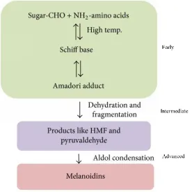
Figure 2.3. Overview of the early intermediated and advanced stages of Maillard reaction .. 32
Figure 2.4 Typical FTIR spectra of milk ... 52
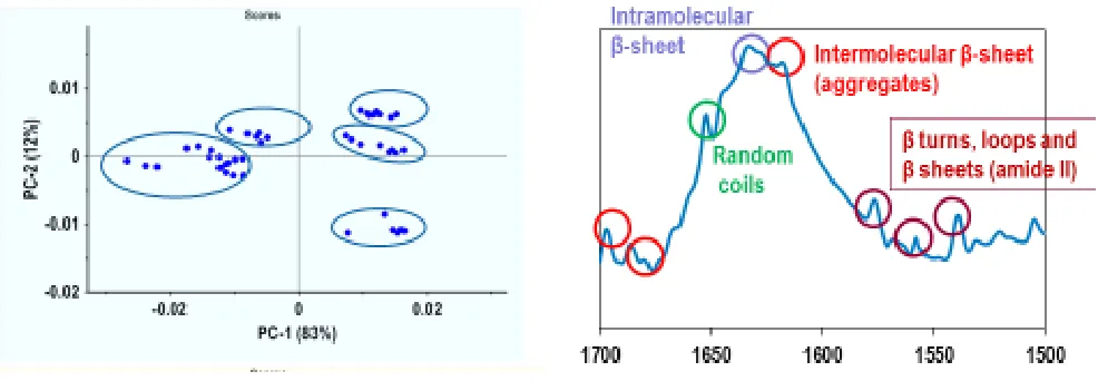
Figure 2.5 Typical score and loading plot of PCA ... 58
xii
Table 2.1 Composition of bovine milk... 11
Table 2.2 Key characteristics of four caseins ... 13
Table 2.3 Different models proposed for casein micelle structure ... 17
Table 2.4 Calcium partition in cow milk by Holt and Jenness (1984). ... 21
Table 2.5 Secondary Structure of milk proteins ... 24
xiii
UHT milk is gaining market share worldwide for being a healthy, safe and convenient food
product with a long shelf life at room temperature. However, the UHT treatment (usually
140-150 °C for 2-10 s) contributing to its long shelf life may induce some changes, which may
make it unstable during storage. Considerable levels of sedimentation (up to 1 % dry weight
basis) of proteinaceous material at the bottom of the storage container, or formation of a gel
(age-gelation), or both, can mark the end of shelf life of UHT milk, and hence affect its market
potential and incur financial losses to the UHT dairy processors. Irrespective of a mechanism,
gelation or sedimentation appears to be governed and preceded by changes in the extent and
nature of interactions among milk proteins leading to their aggregation. Though recent
investigations have added significantly to the understanding of the different protein
interactions, the exact mechanism involving changes in these molecular interactions leading to
sedimentation and gelation, is not clear yet. Moreover, less is known about the role of
protein-lipid interactions in the development of a storage instability. In addition, the need of a rapid
technique which can detect the shelf life of UHT milk has been emphasised in recent years.
This is mainly because applying full-length shelf life tests in a product such as UHT milk would
be time- and resource-intensive. Thus, the overall objective of this project was to understand
and rapidly predict the storage stability of UHT milk.
Accelerated shelf life testing is one such rapid alternative applied to a variety of food
products to save time. The industry has not been successful in applying this approach to
ultra-high temperature (UHT) milk because of the chemical and physical changes in the milk
proteins that take place during its processing and storage. Thus, the first objective of the study
was to investigate the feasibility of applying accelerated shelf life principles to UHT milk
xiv
polyacrylamide gel electrophoresis). Samples of UHT skim (SM) and whole milk (WM) were
stored at 20, 30, 40, and 50 °C for 28 days. Irrespective of fat content, UHT treatment had a
similar effect on the electrophoretic patterns of milk proteins. At the start of testing, proteins
were bonded mainly through disulphide and noncovalent interactions. However, storage at and
above 30 °C enhanced protein aggregation via covalent interactions. The extent of aggregation
appeared to be influenced by fat content, implying aggregation via melted or oxidized fat, or
both. Based on the reduction in loss in the absolute quantity of individual proteins, covalent
crosslinking in WM was facilitated mainly by products of lipid oxidation. Whereas, Maillard
and dehydroalanine products appeared to be the main contributors to protein changes in SM.
Protein crosslinking appeared to follow a different pathway at higher temperatures (≥40 °C),
making it difficult to extrapolate these changes to lower temperatures. The changes identified
under the accelerated shelf life conditions using electrophoretic analysis assisted in evaluating
the potential of using another rapid technique, Fourier transform infrared spectroscopy (FTIR)
in detecting changes in structure and interactions of milk proteins (the second objective).
The feasibility of using FTIR to detect changes in conformational rearrangements,
protein-protein and protein-lipid interactions was studied with accelerated shelf life protocols.
WM and SM were stored at 20, 30, 40 and 50 °C for 28 days. The changes in FTIR spectra
were observed concomitant with increased sedimentation in SM (by 50 %) and WM (by 20 %)
at higher temperatures (40 °C) after 14 days of the storage period. Milk samples stored at 40
and 50 °C showed marked changes in the bands corresponding to the conformations of milk
lipids and formation of intermolecular β-sheets, indicating protein-lipid interactions and
aggregation. Dried sediment contained fat confirming protein-lipid participation in the
sedimentation. FTIR was also able to detect changes that led to increased sedimentation in SM
xv
observed correlation between spectral changes and sediment formation at accelerated
temperatures has to align with that at normal storage temperature (third objective of the study).
SM and WM were stored at 20 °C for 9 months to investigate the feasibility of using
FTIR to predict sedimentation in UHT milk. Identified spectral marker variables corresponding
to changes in the structure and interactions of lipids, proteins and carbohydrates successfully
predicted sedimentation in SM (R2 -0.92) and WM (R2 - 0.60). Low predictability in WM may
be due to the influence of fat. These markers were similar to those observed during accelerated
shelf life testing, hence implying that the accelerated shelf life testing could be used in UHT
milk.
Among several changes in milk protein interactions during its heating and storage,
conformational changes specific to an interaction remain largely unknown. Hence, the fourth
objective of the project evaluated the possibility of fingerprinting two selected changes, i.e.,
deamidation and dephosphorylation, using FTIR. Enzymatic deamidation and
dephosphorylation were carried out prior to heat treatment. Principal component analysis
revealed that the heat treatment induced different changes in the secondary structure of control,
deamidated and dephosphorylated milk samples. In contrast to a significant (P<0.05) decrease
in β-sheet (1624 cm-1) and a rise in β-turns (1674 cm-1) in heated control samples, both
deamidation and dephosphorylation of SM before heat treatment created more ordered
secondary structure (significant (P<0.05) increase in α-helix (1650-52 cm-1) and β-sheet at the
expense of 310-helix (1661 cm-1), random (1645-46 cm-1) and β-turn (1674 cm-1). The only
difference between heated deamidated and dephosphorylated samples was decrease in large
xvi
with FTIR spectroscopy has a potential as a rapid tool to forecast sedimentation and other
instabilities in UHT milk, and hence the shelf life of UHT milk. Further, a complete
understanding of the conformational changes affecting the storage stability of UHT milk could
also be attained by studying the structural changes specific to different known interactions
1
CHAPTER 1
2 1.1 Introduction
UHT milk has gained acceptance worldwide for being a healthy, safe and convenient food
product with a long shelf life at room temperature (Newswire, 2014; Malmgren et al., 2017). It
owes its long shelf life to ultra-high temperature (UHT) treatment involving heating of milk at
high temperature (usually 140-150 °C) for 2-10 s followed by aseptic packaging (Deeth & Lewis,
2016). The treatment and packaging renders UHT milk as a ‘commercially sterile’ product with
minimal changes to its quality. More importantly elimination of the requirement of cold chain and
refrigeration facilities for transportation and storage contributes in reducing energy loads and the
greenhouse gas emissions. However, UHT milk might get unstable during storage and experience
physical defects like sedimentation of proteinaceous material at the bottom of the storage container
or formation of a gel (age-gelation), or both (Andrews & Cheeseman, 1972; McMahon, 1996;
Deeth & Lewis, 2016). Occurrence of considerable levels of sedimentation (up to 1 % dry weight
basis (Deeth & Lewis, 2016)) or gelation can mark the end of the shelf life of UHT milk. Hence,
the appearance of these defects in UHT milk could affect its market potential and incur financial
losses to the UHT dairy processors.
Sedimentation and gelation in UHT milk can be a result of non-enzymatic changes induced by
thermal (UHT) treatment, mainly whey protein denaturation, Maillard reaction and mineral
imbalance. Likewise, indigenous or exogenous enzyme induced proteolysis, another main
underlining mechanism of gelation, also induces protein network formation (i.e., interaction or
linking between proteins or their fragments) (Deeth & Lewis, 2016). Irrespective of mechanism,
gelation or sedimentation appears to be governed and preceded by changes in the extent and nature
3
Changes in protein interactions primarily involve noncovalent interactions, via either weak
bonding (such as hydrophobic, van der Waals, or electrostatic interactions) or covalent
crosslinking with other proteins through formation of disulphide bonds, advanced Maillard
products (AMP), and dehydroalanine (Singh, 1991; Friedman, 1999; Wang, Nema, & Teagarden,
2010; Holland, Gupta, Deeth, & Alewood, 2011). The most assessed pathway of protein
aggregation is mediated by denaturation of whey proteins as a result of UHT treatment (Singh,
1991). Denaturation exposes a free sulphydryl group of β-lactoglobulin (β-Lg), which is involved
in intramolecular and intermolecular thiol-disulphide exchange reactions with other whey proteins
and caseins [κ-Casein (k-CN) and αs2-Casein (αs2-CN)] present on the surface and the interior of
the casein micelles, respectively, and homogenised fat globules (Sharma & Dalgleish, 1993).
Though recent investigations have expanded the understanding of the above-mentioned
interactions, the exact mechanism leading to sedimentation and gelation in UHT milk, is not clear
yet. Moreover, the focus has been mainly on the protein-protein linking, less is known about the
role of protein-lipid interactions in the initiation and buildup of sediment or a gel. All that is known
is that there is lower extent of proteolysis (López-Fandiño, Olano, Corzo, & Ramos, 1993;
Garcia-Risco, Ramos, & Lopez-Fandino, 1999), and the formation of Maillard reaction products (Valero,
Villamiel, Miralles, Sanz, & Martı́nez-Castro, 2001) in stored UHT milk with higher fat content.
These reports however give an indication that the milk fat plays some role in the extent of protein
interactions during storage, and hence needs to be further explored, which would be undertaken in
this project.
In addition to exploring the progression and extent of changes in different protein interactions
4
these changes and hence predict their development in advance has been emphasised in recent years.
This is mainly because applying full-length shelf life tests for the purpose in a product such as
UHT milk would be time- and resource-intensive. Additionally, it is estimated that 40 % of the
cost of developing a new food emulsion is incurred in shelf life testing (Robins, 2000). Accelerated
shelf life tests could be one of the suitable alternative for predicting shelf life of UHT milk in
comparably short time. By accelerating the rate of deterioration of sensory attributes with the use
of elevated storage temperatures, Richards, De Kock, and Buys (2014) successfully developed a
model to predict shelf life of UHT milk. Likewise, changes in protein-protein interactions linked
to shelf life ending defects such as sediment or gelation can be enhanced using higher storage
temperatures (Al-Saadi & Deeth, 2008). Proteomic analysis in combination with accelerated shelf
life testing have added information about different types of prospective crosslinking during storage
of UHT milk at elevated temperatures, but these techniques are still limited in their approach
(Holland et al., 2011). Main reason being the fairly laborious sample preparation accompanied
with a complex analysis, all of which require highly trained experts. In recent years, application of
vibrational spectroscopy techniques has increased in the food industry. This has been due to these
techniques being rapid, non-invasive, non-destructive, requiring very less sample preparation and
relatively low cost as compared to the conventional techniques. These techniques are based on the
unique spectral analysis of stretching and bending of molecular bonds, and thus collectively
producing fingerprint spectra for a particular molecule (Wang, He, Labuza, & Ismail, 2013).
Among these techniques, FTIR (Fourier Transform Infrared Spectroscopy) is most accepted and
is sensitive to changes in covalent bonding and non-covalent electrostatic and hydrophobic
5
has already been successfully used to study ageing of pharmaceuticals (Masmoudi, Dréau,
Piccerelle, & Kister, 2005) and oil in water emulsions (Whittinghill, Norton, & Proctor, 1999) by
determination of changes in molecular structure and interactions, but has not been used in UHT
milk.
Hence, the project for the first time envisaged studying feasibility of using FTIR technique and
accelerated shelf life testing to detect changes in protein structure and interactions, which lead to
storage instability mainly sedimentation in UHT milk. Besides tracking the development of storage
instabilities in milk using FTIR, the project would also focus on developing a rapid method for
prediction of sedimentation in advance. To achieve this, a new approach correlating changes in
FTIR spectral profiles and sedimentation using multivariate analysis tools (chemometrics) would
be developed. The appropriateness of the developed approach using FTIR and accelerated shelf
life testing would be established by comparing the findings to those obtained during full length
shelf life testing.
The focus was only on build-up of sediment and not on gelation because higher storage
temperatures (>40 °C) inhibit age-gelation (Kocak & Zadow, 1985; McMahon, 1996; Deeth &
Lewis, 2016) and result in increased non-dispersible sediment (Nieuwenhuijse & van Boekel,
2003). Thus, accelerated shelf life testing in UHT milk could only be used to predict sedimentation.
The results could be extrapolated to predict aggregation mechanisms at lower storage
temperatures, avoiding lengthy real-time analysis. The developed approach can also be used to
determine and predict storage instabilities in other dairy and non-dairy emulsions.
Besides having an idea of overall changes in the secondary structure and related protein
6
to individual protein interactions needs to be known in order to improve the predictive ability of
model developed to predict sedimentation in UHT milk and identify the main interactions
responsible for this storage instability.
Thus, the overall objective of this project was to understand and predict the storage stability of
UHT milk using FTIR spectroscopy and accelerated storage temperatures. The main objectives of
this research were:
i) To investigate feasibility of using elevated temperatures during storage of UHT milk
containing different fat content to elucidate changes in interactions of milk proteins at
ambient temperature using electrophoretic analysis
ii) To determine the feasibility of FTIR technique to detect changes in protein structure
and interactions leading to sedimentation under accelerated storage temperatures in
skim and whole UHT milk.
iii) To develop a rapid method for detection and prediction of sedimentation in UHT milk
using FTIR and multivariate analysis (chemometrics) by comparing findings obtained
under accelerated storage temperatures with a long storage study at room temperature.
iv) To detect specific changes in milk protein structure and interactions held responsible
for sedimentation using FTIR spectroscopy
1.1 Thesis outline
This thesis comprises of seven chapters. Chapter 1 presents introduction of the project and
its aims. A thorough literature review of the current scientific knowledge on the scope of the
7
addressing the four objectives in the same sequence mentioned. An overall conclusion from
8
CHAPTER 2
9 2.1 Literature review
2.2 Milk as a ‘complete’ food
Milk, a fluid secreted by females of all mammals, is designed to meet the complete
nutritional requirement of their neonates. Mainly, milk contains ~90 % water, life’s vital
requirement, lactose and lipids for energy, proteins and peptides (immunoglobulins, enzymes,
enzyme inhibitors, growth factors, hormones, antibacterial agents) for growth, immunity and other
physiological functions (O’Mahony & Fox, 2013). Furthermore, milk is free from toxins and
antinutritional factors and has a pleasant and attractive flavour and mouthfeel. Such a nutritional
and sensory profile has given milk the label of ‘complete’ food and is not restricted only to meet
the nutritional requirements of neonates. Milk from domesticated cows (mainly) has become an
integral part of human diet in many parts of the world. In addition, the ease with which it can be
converted to a wide range (several thousands) of different and attractive products has made it a
favourite ingredient for food industry. As a result, in the USA, Canada, Australia and New Zealand
and many European countries, about 30 % of dietary protein is supplied by milk and dairy products
(O’Mahony & Fox, 2013). In this thesis, unless otherwise stated, the word milk refers to bovine
milk.
2.3 Milk composition and structure
Milk, a white or yellowish white complex fluid, contains on average mainly water (91 %),
sugar (lactose) (4.6 %), lipids (3.9 %), proteins (3.25 %) and mineral substances (0.65 %) (Walstra,
Jenness, & Badings, 1984; Walstra, Wouters, & Geurts, 2005). Milk also contains organic acids
such as citrate, enzymes and a range of vitamins that do not fit into the previously mentioned major
10
stages of lactation and animal’s health. However, this biological variability will not be discussed
in detail here.
Milk structure is mainly due to colloidal existence of its lipids as fat globules (fat droplets
stabilized by mixture of lipoproteins and bilayer structures), protein casein as casein micelles
sterically stabilized by hairy layer of κ-CN and whey proteins as oligomers. These structural
components are mainly responsible for optical properties, viscosity and physical stability of milk.
These components will be discussed in more detail in section 2.3.1. Typical constituents of milk
and their distribution between the colloidal and serum, defined as milk depleted of caseins and fat
globules, phases is summarised in Table 2.1.
2.4 Major milk constituents
2.4.1 Lipids
Almost 99 % of milk lipid exists as milk fat globules (MFG) (Heid & Keenan, 2005), which
are lipid spheres with diameter ranging from 0.1 to 15 µm (average 4 µm) and surrounded by a
multilayered structure called milk fat globule membrane (MFGM) (Leser & Michel, 2008). The
membrane comprises about 2 % of the fat globules and is composed of a phospholipid monolayer,
an inner protein coat, a bilayered phospholipid membrane, and a glycosylated surface (Mather &
Keenan, 1998). MFGM acts as a natural emulsifying agent preventing flocculation and
coalescence of fat globules. The membrane also prevents hydrolysis of fat by enzymes.
Approximately 95-98 % of the total lipids in these globules consist of triacylglycerides, with lesser
amounts of partial glycerides, sterols, phospholipids, glycolipids, and hydrocarbons (Table 2.1)
membrane-11
fragments in the skim milk phase. A detailed review has been published recently about MFG and
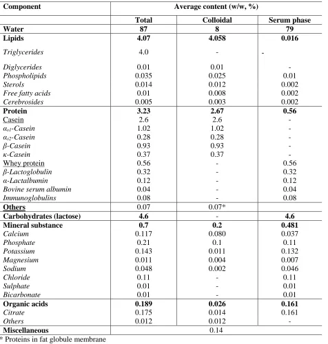
MFGM (Singh & Gallier, 2017).
Table 2.1 Composition of bovine milk (adapted from Walstra et al., (1984) and Walstra et al. (2005))
Component Average content (w/w, %)
Total Colloidal Serum phase
Water 87 8 79
Lipids 4.07 4.058 0.016
Triglycerides 4.0 - -
Diglycerides 0.01 0.01 -
Phospholipids 0.035 0.025 0.01
Sterols 0.014 0.012 0.002
Free fatty acids 0.01 0.008 0.002
Cerebrosides 0.005 0.003 0.002
Protein 3.23 2.67 0.56
Casein 2.6 2.6 -
αs1-Casein 1.02 1.02 -
αs2-Casein 0.28 0.28 -
β-Casein 0.93 0.93 -
κ-Casein 0.37 0.37 -
Whey protein 0.56 - 0.56
β-Lactoglobulin 0.32 - 0.32
α-Lactalbumin 0.12 - 0.12
Bovine serum albumin 0.04 - 0.04
Immunoglobulins 0.08 - 0.08
Others 0.07 0.07*
Carbohydrates (lactose) 4.6 - 4.6
Mineralsubstance 0.7 0.2 0.481
Calcium 0.117 0.080 0.037
Phosphate 0.21 0.1 0.11
Potassium 0.143 0.011 0.132
Magnesium 0.011 0.004 0.007
Sodium 0.048 0.002 0.046
Chloride 0.11 - 0.11
Sulphate 0.01 - 0.01
Bicarbonate 0.01 - 0.01
Organic acids 0.189 0.026 0.161
Citrate 0.175 0.014 0.161
Others 0.012 0.012 -
Miscellaneous 0.14
* Proteins in fat globule membrane
** The values are average of the biological variability between individual animals, breeds, seasons, feed,
12 2.4.2 Protein-Caseins
Casein is the main milk protein constituting about 80 % of the total protein in milk. It is a
milk specific group of proteins that precipitates upon acidification to pH 4.6. These proteins are
also coagulated by action of rennet (chymosin and other proteinases). Caseins are phosphorylated
proteins, containing an average of 0.85 % phosphorous, and thus having an ability to bind relatively
large amounts of calcium (Fox, Uniacke-Lowe, McSweeney, & O'Mahony, 2015b). Caseins exist
in the milk in a form of large colloidal aggregates called casein micelles (106-109 Da) which include
all four types of caseins namely αs1, αs2, β and κ-caseins, along with the colloidal calcium
phosphate and water (Table 2.1). The key properties of these four types of caseins are listed in
Table 2.2.
Caseins are relatively heat stable. However, heating above 120 °C induces some chemical
changes which render them insoluble (Walstra et al., 2005). Heat stability of caseins can be
attributed to their less order structural organization including secondary and tertiary structures,
which is further owed to high proline content. However, the full secondary and tertiary structure
13
Table 2.2 Key characteristics of four caseins (adapted from Huppertz (2013) and Fox, Uniacke-Lowe, McSweeney, and O'Mahony (2015a))
αs1-Casein αs1-CN B-8P
αs2-Casein αs2-CN A-11P
β-Casein β-CN A2-5P
κ-Casein κ-CN A-1P
Molecular weight (Da) 23599 25206 23973 19052
Number of amino acid residues 199 207 209 169
Serine 16 17 16 13
Proline 17 10 35 20
Cysteine 0 2 0 2
Lysine 14 24 11 9
Positively charged residues 25 33 20 17
Negativley charged residues 40 39 28 28
Aromatic residues 20 20 14 14
Grand average of hydropathicity (GRAVY)
-0.704 -0.918 -0.355 -0.557
2.4.2.1 αs1-Casein
αs1-Casein (αs1-CN) is the most abundant of all caseins contributing about 40 % to the total
casein in milk (Huppertz, 2013). It is moderately hydrophobic protein with two predominantly
hydrophobic regions and one highly charged polar zone (Fox et al., 2015a). Around eight genetic
variants have been detected until now with 8-9 phosphorylated amino acid residues (mostly
serine). More details about these variants can be found elsewhere (Huppertz, 2013). αs1-CN is
characterized by a lack of cysteine residue and is a calcium sensitive casein precipitating at 3-8
mM CaCl2. At pH 6.6 and ionic strength of 0.003, αs1-CN exists as a monomer. With increase in
ionic strength to 0.01 at same pH, a monomer-dimer equilibrium exists. Further increase in ionic
strength to 0.2 at the same pH favours formation of dimers and tetramers. However, increase in
14 2.4.2.2 αs2-Casein
αs2-Casein constitutes upto 10 % of the total casein in milk. It exhibits varying levels of
phosphorylation with 10-13 phosphates (Thompson, Boland, & Singh, 2009) and intermolecular
disulphide bonding due to the presence of two cysteine residues per mole. It is the most hydrophilic
of the caseins with a dipolar structure where the N terminus is negatively charged and the C
terminus is positively charged (Fox et al., 2015a). At neutral pH, αs2-CN self-associates with
increase in ionic strength with maximum at 0.2. αs2-CN has also the highest number of
phosphorylated residues and is also most sensitive to calcium-induced precipitation. It is insoluble
in calcium solution over 2 mM.
2.4.2.3 β-Casein
β-Casein (β-CN) constitutes about 35 % to the total caseins in milk (Huppertz, 2013). It
has amphipathic properties due to a strong negatively charged N-terminal portion with all the
phosphorylated residues (4-5) and apolar C terminal characterized by little charge and high
hydrophobicity. It is most hydrophobic casein and thus its self-association is strongly dependent
on temperature, only a monomeric form is present at 4 °C but large polymers are assembled at
room temperature. Compared to αs1- and αs2-CN, β-CN is less sensitive to calcium-induced
precipitation. At 37 °C, it precipitates in the range of 8-15 mM calcium ions. However, at 1 °C, it
does not precipitate up to 400 mM of CaCl2.
2.4.2.4 κ-Casein
κ-Casein is the smallest of the caseins with low levels of phosphorylation (1-3P) and low
sensitivity to calcium. Of all the caseins, only κ-CN is glycosylated (Huppertz, 2013). κ-CN has
15
without any aromatic and a few apolar residues and a strongly hydrophobic N-terminus. This is an
important characteristic of κ-CN, which explains its presence on the surface of the casein micelle
and its micelle size determinations and stabilizing properties (Swaisgood, 2003). κ-CN is rapidly
hydrolyzed by an enzyme chymosin and other proteases at Phe (105)-Met (106) yielding
N-terminal fragment (para κ-CN) with two cysteine residues and C-N-terminal fragment (macropeptide)
containing all carbohydrate and phosphate groups. Hydrolysis of κ-CN is the first step in the
manufacturing of most cheese varieties. In a naturally occurring mixture, κ-CN polymerizes into
subunits containing 3-8 monomers through S-S linkages, which further polymerize into complexes
with 1180 kDa. Self-association of κ-CN has a lower sensitivity to temperature when compared to
β-CN (Huppertz, 2013).
2.4.2.5 Casein micelle
Above mentioned four caseins, αs1, αs2, β and κ casein associate with each other in an
approximate ratio of 4: 1: 3.5: 1.5, respectively, in presence of Ca2+ and produce a dominant feature
of milk, the casein micelle, a macromolecular assembly with an average size of 200 nm (Dalgleish,
2011). In addition to caseins (approx. 94 %), micelles also contain inorganic minerals (~6 %) (Fox
et al., 2015a). Inorganic minerals mainly include calcium bound to phosphorus in calcium
phosphate nanoclusters referred to as colloidal calcium phosphate (CCP) and lower levels of
magnesium and citrate. This ability of integrating CCP into the micelle makes them a primary
transport vehicle for calcium and phosphorus, essential minerals (Singh, 2014). In addition, the
presence of CCP also avoids the risk of significant increase in milk viscosity due to open structure
16
colour of the milk. The micelles are very stable and can withstand heat treatment, commercial
homogenization and high calcium concentration (Singh, 2014).
2.4.2.6 Casein micelle structure
Casein micelle structure is a topic of continuing research over the last thirty years. Different
models have been proposed to explain the structure of casein micelle. The main idea, supporting
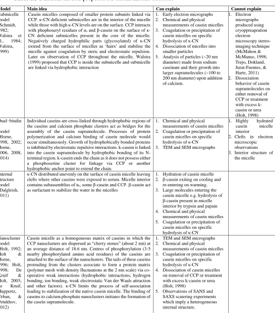
experimental observations, strengths and limitations of different models are discussed in Table
17
Table 2.3 Different models proposed for casein micelle structure
Model Main idea Can explain Cannot explain
Submicelle model (Schmidt, 1982; Walstra et al., 1984; Walstra, 1999)
Casein micelles composed of smaller protein subunits linked via CCP. κ-CN deficient submicelles are in the interior of the micelle while those with high κ-CN levels are on the surface. CCP interacts with phophoseryl residues of αs and β-casein on the surface of
κ-CN deficient submicelles present in the core of the micelle. Negatively charged hydrophilic parts (glycosylated) of κ-CN extend from the surface of micelles as ‘hairs’ and stabilise the micelle against coagulation by steric and electrostatic repulsion. Later on observation of CCP throughout the micelle, Walstra (1999) proposed that CCP is inside the submicelle and submicelle are linked via hydrophobic interaction
1. Early electron micrographs 2. Chemical and physical
measurements of casein micelles 3. Coagulation or precipitation of
casein micelles on specific hydrolysis of κ-CN 4. Dissociation of micelles into
smaller particles
5. Analysis of particles (~20 nm diameter) made from sodium caseinate and their growth into larger supramolecules (~100 to 200 nm diameter) upon addition of calcium. 1. Electron micrographs produced using cryoppreapration electron microscopy stereo- imaging technique (McMahon & McManus, 1998; Trejo, Dokland, Jurat-Fuentes, & Harte, 2011) 2. Dissociation
behavior of casein supramolecules on either removal of CCP or treatment with excess k-casein or urea (Holt, 1998) Dual−bindin
g model (Horne, 1998, 2002; Horne, 2006, 2008, 2014)
Individual caseins are cross-linked through hydrophobic regions of the caseins and calcium phosphate clusters act as bridges for the assembly of the casein supramolecule. Processes of protein polymerization and calcium binding of casein molecule would occur simultaneously. Growth of hydrophobically bonded proteins is inhibited by electrostatic repulsive interactions. k-casein is linked into the casein supramolecule by hydrophobic bonding of its N-terminal region. k-casein ends the chain as it does not possess either a phosphoserine cluster for linkage via CCP or another hydrophobic anchor point to extend the chain.
1. Chemical and physical
measurements of casein micelles 2. Coagulation or precipitation of
casein micelles on specific hydrolysis of κ-CN
3. TEM and SEM micrographs
1. Highly hydrated
casein micelle
interior
2. Clefts in electron microscopic observations 3. Interior structure of
the micelle Internal structure model (Dalgleish, 2011)
κ-CN distributed unevenly on the surface of casein micelle leaving clefts where other caseins were exposed to serum. Micelle interior contains subassemblies of αs, some β-casein and CCP. β-casein act
as surfactant to stabilize the water in the micelles
1. Hydration of casein micelle 2. β-casein exiting on cooling and
re-entering on warming. 3. Large molecules entering the
casein micelle e.g. hydrolysis of β-casein present in micelle interior by trypsin and papain 4. Chemical and physical
measurements of casein micelles 5. Coagulation or precipitation of
casein micelles on specific hydrolysis of κ-CN Nanocluster
model (Holt, 1992; Holt & Horne, 1996; Holt,
1998; De
Kruif & Holt, 2003;
de Kruif,
Huppertz, Urban, & Petukhov, 2012)
Casein micelle as a homogeneous matrix of caseins in which the CCP nanoclusters are dispersed as “cherry stones” (about 2 nm) at an average distance of 18.6 nm. Centres of phosphorylation (3-5 nearby phosphorylated amino acid residues) of the caseins are attached to the surface of the nanoclusters. The tails of these caseins protruding from the clusters associate to form a protein matrix (polymer mesh with density fluctuations at the 2 nm scale) via co-operative weak interactions (hydrophobic interactions, hydrogen bonding, ion bonding, weak electrostatic Van der Waals attraction and other factors). κ-CN limits the process of self-association leading to stabilization of the native casein micelle. The binding of caseins to calcium phosphate nanoclusters initiates the formation of the casein supramolecule.
1. TEM and SEM micrographs
2. Chemical and physical
measurements of casein micelles 3. Coagulation or precipitation of
casein micelles on specific hydrolysis of κ-CN
4. Dissociation of casein micelles on removal of CCP or treatment with excess k-casein or urea (Holt, 1998)
18 2.4.3 Whey proteins
Whey proteins constitute 20 % of the total protein content in bovine milk. The term ‘whey proteins’
encompasses five different groups of proteins, namely, β-lactoglobulin (β-Lg), α-lactalbumin
(α-La), lesser amounts of bovine serum albumin (BSA), immunoglobulins (Ig) and proteose peptones
(pp). These proteins have a globular to ellipsoid structure and are relatively soluble. These proteins
are more heat sensitive and less sensitive to calcium than caseins.
2.4.3.1 β-Lactoglobulin
β-Lg is the most abundant (~ 60 %) whey protein in milk (Table 2.1). It is a globular protein with
162 amino acid residues and a molecular weight of approximately18 kDa. Ten genetic variants of
Lg have been known with most common being named A and B (Edwards & Jameson, 2014).
β-Lg can self-associate to varying degrees depending on temperature, pH, protein concentration and
ionic strength. At ambient temperature and natural pH of milk, β-Lg exists as dimer. A β-Lg
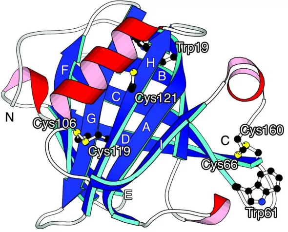
monomer contains two intra-molecular disulphide bonds and a free sulfhydryl group (-SH, Cys121).
β-Lg (Figure. 2.1) denatures at temperatures above 65 °C at pH 6.7 exposing its free thiol group,
which can undergo thiol-disulphide exchange reactions with other denatured β-Lg and/or milk
proteins such as κ-CN and α-La. Denaturation and following crosslinking reactions of β-Lg affect
the rennet coagulation and heat stability of the milk. Denaturation temperature of β-Lg depends on
pH. It is most heat sensitive near pH 4.0 and most stable at pH 6.0 (Sawyer, 2003). The
physiological role of β-Lg is not known. However, due to the presence of a hydrophobic cavity, it
is considered as transporter for small hydrophobic molecules like vitamin A (Fox et al., 2015a). In
addition, β-Lg is one of the milk proteins responsible for milk protein intolerance or allergy in
19
Figure 2.1 Structure of bovine β-Lg indicating disulphide bonds and thiol group (Yagi, Sakurai, Kalidas, Batt, & Goto, 2003)
2.4.3.2 α-Lactalbumin
α-Lactalbumin (α-La) constitutes 20 % of whey proteins and has two genetic variants A (most
common in bovine milk) and B with a molecular weight of 14.147 kDa and 14.175 kDa,
respectively (Fox et al., 2015a). α-La is composed of 123 amino acid residues and has an oblate
ellipsoid tertiary structure with a deep cleft dividing the protein in two lobes (α and β). The cleft
is binding site for Ca2+, an integral part of its molecular structure,which plays a role in its stability
and minimized susceptibility to denaturation. The α-lobe has three α-helices and three short 310-
helices whereas the β-lobe contains three stranded β sheets and a short 310- helix. α-La contains
four disulphide bonds and lacks free thiol group, which further adds to its heat stability. α-La is a
component of enzyme lactose synthetase which defines its physiological role being transferring
20 2.4.3.3 Other whey proteins
Bovine serum albumin (BSA), immunoglobulins (Ig) and proteose peptone (pp) are minor
proteins present in the serum at low levels (Table 2.1). BSA accounts for 6 % of whey proteins
and has a molecular weight of ~65 kDa with 582 amino acid residues. Ig are antibodies synthesised
in response to foreign antigens. These proteins provide immunity to the young and thus their
concentration is high in colostrum but decreases rapidly after postpartum. Proteose peptones is a
complex mixture of peptides, most of which produced by the action of indigenous enzyme plasmin
(Fox et al., 2015a).
The contributions of these whey proteins to the physio-chemical changes in milk during
UHT processing and subsequent storage are considered relatively minor, compared to α-La and
β-Lg and therefore not discussed in detail here. Detailed information about these proteins can be
found elsewhere (Hurley & Theil, 2013; O’mahony & Fox, 2013; Wynn & Sheehy, 2013; Fox et
al., 2015a).
2.4.4 Minerals
Milk minerals occur in one or more chemical forms, including ions and salts (organic and
inorganic), complexes or constituents of organic molecules like proteins. They constitute only a
small fraction (8-9 g/L) of milk and includes cations (mainly calcium, magnesium, sodium and
potassium) and anions (mainly inorganic phosphate, citrate and chloride) (Gaucheron, 2005).
These cations and anions are partitioned between the colloidal and aqueous phase of milk. About
one third of calcium, two thirds of magnesium, half of the inorganic phosphate and over 90 %
citrate is in aqueous phase whereas the rest is associated with casein molecules and are an integral
21
CCP, as discussed in previous sections 2.4.2.5 and 2.4.2.6, plays a pivotal role in the casein
micelle structure and stability. There is an equilibrium between calcium in the colloidal and
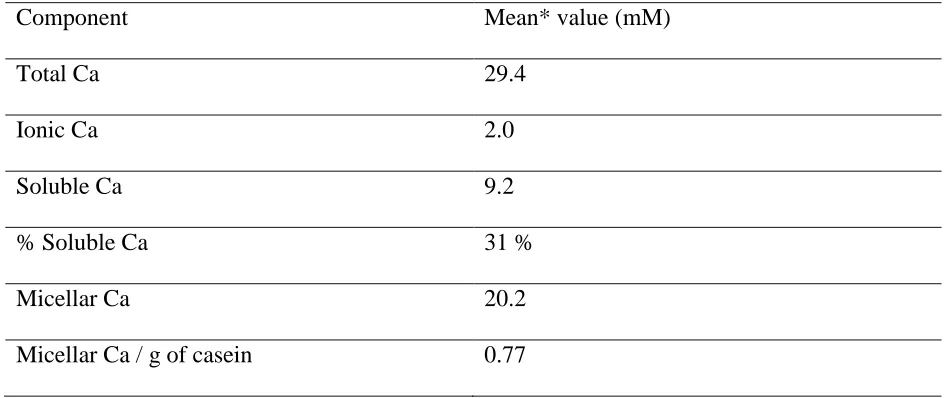
aqueous phase. Partition of calcium in milk between different phases has been tabulated in Table
2.4. The formation of CCP is an effective way of “burying” a considerable amount of Ca and
phosphate within casein micelles, therefore determines to a large extent the physicochemical
properties of casein micelles (Lucey, 2003). About 70 % of total Ca and 30 % of total phosphate
as principle colloidal salt are in the colloidal phase, which combines with four major caseins
(αs1-, αs2-(αs1-, β- and k-CN) in the casein micelle (Gaucheron(αs1-, 2005).
Table 2.4 Calcium partition in cow milk by Holt and Jenness (1984).
*Mean values include the biological variability over different breeds of cows
2.4.5 Sugars
Main sugar of milk is lactose, a reducing disaccharide composed of glucose and galactose.
Lactose is responsible for slight sweet taste of milk. Being the principal carbon source for
microorganisms in milk, it is indispensable for manufacture of fermented dairy products like
cheese and yogurt. In addition to lactose, a large number of free saccharides mainly
Component Mean* value (mM)
Total Ca 29.4
Ionic Ca 2.0
Soluble Ca 9.2
% Soluble Ca 31 %
Micellar Ca 20.2
22
oligosaccharides and small amount of monosaccharides and glycosylated proteins κ-CN, mucins
and glycoproteins in fat globule membrane are also present (O’Mahony & Fox, 2014)
2.4.6 Vitamins and enzymes
Milk is a source of fat soluble (A, D and E) and the water-soluble vitamins (C, B1, B2, B6,
B12, pantothenic acid, niacin, biotin and folic acid). Milk also contains a large number of enzymes
including proteases, lipases and oxidoreductases. Plasmin is the major serine protease in milk and
its concentration increases during very early or very late lactation or in animal with mastitis.
Plasmin is heat stable at pH of milk and can retain substantial activity after pasteurisation and thus
affect protein stability of milk. Lipoprotein lipase in the major lipase in milk. The enzyme is heat
labile and thus of no significance in heat treated milk. Lactoperoxidase is one of the main
oxidoreductases present in milk and is able to catalyze oxidation of unsaturated fatty acids
resulting in oxidized taste. Lactoperoxidase is also a component of potentially important
lactoperoxidase/thiocyanate/hydrogen peroxide system. Alkaline phosphatase does not affect milk
stability but is important due to its widespread use as index of adequate heat treatment (Varnam &
Sutherland, 2001; Walstra et al., 2005).
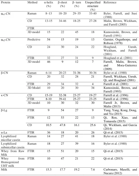
2.5 Secondary structure of milk proteins
In last 30 years, spectral studies and predictive modelling has thrown some light on
secondary structure of different caseins individually and in micellar state (Huppertz, 2013) (Table
2.5). This has uplifted their status from ‘random coil proteins’ to ‘rheomorpic proteins’ with an
open, flexible and mobile conformation (Holt & Sawyer, 1993). Contents of secondary structures
of different caseins individually and in the micellar state determined using different methods in
23
to be the one with the highest amount of secondary structure among caseins. The C-terminal half
of αs2-CN is suggested to be having a globular conformation with α-helix and a β-sheet whereas
the N-terminal region forms a randomly structured hydrophilic tail. αs1-CN contains little α-helix
but considerable amount of β-sheets and β-turns (Fox et al., 2015a). β-Lg has a lower proportion
of α-helix (~ 12 %) compared to α-La (36 %) (Figure 2.1). However, β-sheet content in β-Lg (50
%) is far greater than that in α-La. β-Lg is a typical lipocalin (family of proteins found in gram
negative bacteria, vertebrate cells, and invertebrate cells, and in plants which transport small
hydrophobic molecules such as steroids, bilins, retinoids and lipids) with a structure that contains
a β-barrel with eight antiparallel β-strands, labelled A–H and a three-turn α-helix that lies parallel
to three of the β strands. Strands A–D form one surface of the barrel while strands E–H form the
other (Kontopidis et al., 2004).
2.6 Milk processing
Raw milk is highly perishable and may contain pathogenic bacteria. Therefore, in order to
make it safe for drinking and extending its shelf life, milk is generally heat-treated. Heat treatment
may vary in its intensity, which is a combination of temperature and the duration of heating.
Heating processes are classified into different categories on the basis of their intensity.
2.6.1 Thermisation
Thermisation is a heat treatment (20 s, 60-69 °C) of intensity lower than pasteurisation
aimed to kill bacteria, especially psychrotrophs. Many psychrotrophs produce heat resistant lipases
and proteinases that may eventually deteriorate quality parameters of milk. Thermisation kills
many vegetative microorganisnms and partially inactivates some enzymes with negligible changes
24 Table 2.5Secondary Structure of milk proteins
Protein Method α-helix (%) β-sheet (%) β- turn (%) Unspecified structure (%) Reference
αs1-CN Raman 8–13 18–20 29–35 33-40 Byler, Farrell, and Susi (1988)
CD 13-15 34-46 18-25 27-28 Malin, Brown, Wickham, and Farrell (2005)
FTIR
3D model 15 22 45 18 Kumosinski, Brown, and Farrell (1991)
αs2-CN Predictive 54 15 19 13 Garnier, Osguthorpe, and Robson (1978)
CD 24 30 24 22 Hoagland, Unruh,
Wickham, and Farrell (2001)
FTIR 32 27 31 9 Hoagland et al. (2001) 3D model 46 9 12 7 Farrell, Malin, Brown,
and Mora-Gutierrez (2009)
β-CN Raman 6-14 20-23 31-36 30-36 Byler et al. (1988)
CD 20 32 28 21 Farrell, Wickham, Unruh,
Qi, and Hoagland (2001)
FTIR 29 34 32 4 Farrell et al. (2001)
3D Model 10 20 30 34 Kumosinski, Brown, and Farrell (1993)
κ-CN CD 15-19 32-38 23-27 19-27 Farrell et al. (1996)
FTIR 8-10 39-41 25-27 23-25 Farrell et al. (1996)
3D model 10 30 32 30 Farrell Jr, Brown, and Malin (2013)
β-Lg FTIR 9 54 27 9 Yang, Yang, Kong, Dong,
and Yu (2015)
FTIR 12 53 22 13 Qi, Ren, Xiao, and
Tomasula (2015)
CD 10.5 47.8 16.1 25.6 Qi, Wickham, and Garcia
(2014)
α-La FTIR 36 18 20 26 Qi et al. (2015)
Lyophilized micellar casein
Raman 14 27 41 18 Byler et al. (1988)
Lyophilized submicellar casein
Raman 18 27 39 16 Byler et al. (1988)
Whey from Raw Milk
FTIR 15 51 20 15 Qi et al. (2015)
Whey from Homogenized UHT milk
FTIR 10 47 21 22 Qi et al. (2015)
25 2.6.2 Low Pasteurisation
Low pasteurisation is a heat treatment with intensity (30 min, 63 °C or 15 s, 72 °C)
optimized to inactivate enzyme alkaline phosphastase. The treatment kills all yeasts and moulds,
almost all pathogens specifically Mycobacterium tuberculosis and some vegetative bacteria. In
addition, only some enzymes are inactivated and there are no alterations in flavour, cold
agglutination and bacteriostatic properties of milk. Whey proteins are also not denatured with low
pasteurisation treatment, thus no significant effect on their secondary structure.
2.6.3 High pasteurisation
High pasteurisation is a heat treatment (20 s, 85 °C) sufficient to destroy activity of enzyme
lactoperoxidase. The treatment kills virtually all vegetative microorganisms but no spores and
inactivates most of the enzymes except plasmin and some bacterial proteinases and lipases. In
addition to a cooked flavour, the treatment denatures partly the whey proteins and destroys the
bacteriostatic properties of milk.
2.6.4 Sterilisation
Sterilisation is a heat treatment designed to eliminate most of the microorganisms and
bacterial spores present in milk. This can be sufficed by either in-container sterilisation (30 min,
110 °C) (Walstra et al., 2005) or Ultra high temperature (UHT) short time treatment (1-10 s,
135-145 °C) (Deeth & Lewis, 2016). In-container sterilisation inactivates all milk enzymes except
bacterial lipases and proteinases. However, it results in browning due to extensive Maillard
reactions, a sterilized milk flavour, loss of some available lysine, denaturation of whey proteins
26
treatment results in less chemical changes and a weak cooked flavour with no inactivation of
plasmin, proteinases and bacterial lipases.
2.7 UHT processing
UHT heat treatment in combination with aseptic packaging can extend shelf life of milk up
to 12 months with less chemical changes compared to in-container sterilization. Time-temperature
combination or intensity of UHT process are decided to achieve inactivation of heat resistant
spores of Bacillus stearothermophilus with minimum chemical changes so that sensory and
nutritional qualities of the ‘commercially sterile’ UHT milk are not lowered.
UHT heating can be achieved using either ‘direct’ or ‘indirect’ heating methods. Direct
heating results in rapid heating due to mixing with superheated steam and rapid cooling using
vacuum flash cooling process. Indirect heating process involves slower heating and cooling as the
heat transfer occurs across wall of stainless steel plate or tube exchanger. The comparison between
direct heating and indirect heating systems in terms of their heating profiles, economics and
changes in UHT milk quality have been discussed in detail elsewhere (Datta, Elliott, Perkins, &
Deeth, 2002; Tran, Datta, Lewis, & Deeth, 2008; Lewis & Deeth, 2009).
2.8 Changes to milk during UHT treatment
2.8.1 Chemical changes
2.8.1.1 Changes in pH and mineral balance
Both pH and ionic calcium (Ca2+) concentration decrease with increase in the temperature
of milk. Milk pH has been reported to decrease to 6.0 at 100 °C (Walstra et al., 1984) and may be
27
ionic calcium, there has been studies in the past and recently that reported the exact opposite
(Tessier & Rose, 1958; Chandrapala, McKinnon, Augustin, & Udabage, 2010; On-Nom,
Grandison, & Lewis, 2010). Deeth and Lewis (2017a) suggested that both the pH and Ca2+ at 140
°C affect the heat stability at this temperature.
UHT processing has less effect on the pH of white milk and is rarely reported in the
literature (Deeth & Lewis, 2017a). According to Gaucher et al. (2008), pasteurisation increased
the pH of raw milk from 6.69-6.76 to 6.70-6.78 and after subsequent UHT treatment, it was
decreased to 6.66-6.76. However, the pH of milk is reduced substantially (0.2-0.3 units) after
in-container sterilisation (Deeth & Lewis, 2017a). In contrast to little effect on pH of milk, UHT
treatment substantially reduces Ca2+ concentration, which recovers on further storage but not to its
original value (Geerts, Bekhof, & Scherjon, 1983). The decrease in the solubility of calcium
phosphate during UHT treatment might lead to its precipitate formation (fouling deposit) on the
heat exchanger walls or association with casein micelle. Overall, UHT treatment has a minor effect
on mineral partitioning (Deeth & Lewis, 2017a).
2.8.1.2 Whey protein denaturation and disulphide crosslinking
The most discussed change in milk proteins induced by UHT treatment (or any heat
treatment) is the denaturation of whey proteins. The degree of denaturation of whey proteins is
less in UHT milk as compared to 100 % in in-container sterilisation and depends on the type of
processing (i.e. direct or indirect) and the temperature-time profile of the heating process. Among
the whey proteins, Ig and BSA are completely denatured by sterilisation processes followed by
β-Lg and α-La. Proteose peptones are not denatured by heat (Mulvihill & Donovan, 1987). Indirect
28
processing (Datta et al., 2002; Tran et al., 2008). β-Lg is almost completely denatured by
commercial indirect heating process compared to only 74-92 % denaturation in the direct one.
However, the percentage denaturation of α-La was 25-90 % and 27-58 % for indirect and direct
plants, respectively (Tran et al., 2008; Deeth & Lewis, 2016). Due to sufficient (non-zero) residual
levels of α-La in UHT milk, its concentration has been suggested as an index of heat treatment
(Dupont, Rolet-Repecaud, & Muller-Renaud, 2004; Tran et al., 2008).
Denaturation of whey proteins involves their unfolding and hence exposing reactive
sulfhydryl groups and hydrophobic regions, which subsequently result in sulfhydryl-disulphide
and hydrophobic protein-protein interactions. The most significant interaction involves
crosslinking between β-Lg and κ-CN. Denaturation of β-Lg involves unfolding of the globular
monomer exposing hydrophobic amino acids and a free sulfhydryl group, at Cys121, which is
normally buried inside the native globular structure. The native monomer undergoes
intramolecular sulfhydryl-disulphide reactions with other exposed sulfhydryl groups resulting in
the formation of reactive non-native monomers. These reactive monomers primarily react with
κ-CN located on the surface of the micelle but may do so after reacting with α-La via both
hydrophobic interactions and intermolecular sulfhydryl-disulphide interchange reactions. Thus,
UHT treatment at normal pH 6.7 results in formation of the casein micelles with whey proteins
attached. However, due to dissociation of some κ-CN from the micelle on heating, it also appears
in the serum in associations with denatured whey proteins (Anema, 2008). The amount of these
complexes in the serum phase depends on several factors such as the nature of the heat treatment
and pH (Corredig & Dalgleish, 1996a; Corredig & Dalgleish, 1996b). Most of the denatured whey
proteins are associated with the casein micelle in indirect heated UHT milk compared to only 50
29
also more α-La attached to the casein micelle compared to β-Lg. This difference has significant
practical implications because the micelle with increased amount of α-La attached becomes more
hydrophobic, thus, increasing the water-holding capacity, an important consideration for some
products. Other than type of heating process, the pH of heating also significantly affects the
distribution of denatured whey proteins. At pH ~6.5, most of the denatured whey proteins attach
to the casein micelle while at pH >6.8, most of them are in the serum with κ-CN (Deeth & Lewis,
2016).
2.8.1.3 Structural modifications of proteins and non-disulphide protein crosslinking
Dephosphorylation of the phosphoserine residues present in milk caseins occurs during
sterilisation by either hydrolysis with a release of a phosphate group and serine (Eq. 1) or by
β-elimination with formation of dehydroalanine and phosphate (Eq. 2) (Belec & Jenness).
R − CH2− O − PO32−+ H
2O → R − CH2− OH + HPO42− (1) (phosphoserine) (serine)
R − CH2− O − PO32−→ R = CH2− OH + HPO42− (2)
(phosphoserine) (dehydroalanine)
More phosphate and serine were reportedly formed during heating of caseinate solutions
than dehydroalanine (van Boekel, 1999). Dehydroalanine is also formed from O -glycosylserine
or cysteine residues by elimination of a sugar or H2S, respectively. Once formed dehydroalanine
reacts with the amino group of lysine residues, the imidazole group of histidine, or the thiol group
of cysteine, resulting in intra- or intermolecular lysinoalanine, histidinoalanine, or lanthionine
crosslinks, respectively (O’Connell & Fox, 2003). Proteins cross-linked via dehydroalanine unlike
30
mercaptoethanol and on hydrolysis yield isodipeptides such as lysinoalanine, histidinoalanine and
lanthionine (Deeth & Lewis, 2016). UHT milk contains very little lysinoalanine (0 to 50 μg/g
protein) compared to autoclaved milk (110 to 710 μg/g protein) (Fritsch, Hoffmann, &
Klostermeyer, 1983). Higher temperatures, higher pH and longer heating times increased
31
Figure 2.2. Formation of lanthionine, lysinoalanine and histidinoalanine from dehydroalanine β-elimination
Dehydroalanine Cysteine
Lysine
32
Lysine residues of milk proteins link with reducing sugar lactose to initiate Maillard
reaction (Figure 2.2), the products of which such as glyoxal and methylglyoxal serve as another
source of non-disulphide crosslinks in UHT milk (Andrews & Cheeseman, 1971; Andrews &
Cheeseman, 1972; Andrews, 1975). Recently it has been shown that on incubating milk protein
concentrate with methylglyoxal, polymerised proteins produced were similar to those formed in
this product during storage (Le, Holland, Bhandari, Alewood, & Deeth, 2013). Protein
crosslinking, whether via dehydroalanine or Maillard reaction products, reduces the nutritional
availability of amino acids such as lysine and reduces the digestibility of the proteins (Deeth &
Lewis, 2016). However, on a positive note, the non-disulphide crosslinking especially via Maillard
reaction products may inhibit age-gelation (storage instability) of UHT milk during storage
(McMahon, 1996).
33 2.8.1.4 Dissociation of proteins from the casein micelle
Considerable dissociation of individual caseins from the micelle takes place during heating
of milk. Extent and nature of this dissociation depend on the heat intensity, pH and milk solids
concentration. Dissociation of caseins is higher in in-container sterilised and concentrated milk
products than unconcentrated UHT milk (Deeth & Lewis, 2017a). Most investigations have
focused on the dissociation of κ-CN from the micelle before or after its interactions with whey
proteins, especially β-Lg (Corredig & Dalgleish, 1996b; Anema, 2008; Anema, 2014).
Dissociation of κ-CN from the surface of the micelle can make it more susceptible to calcium
ion-mediated heat- induced aggregation, so this has a marked effect on the heat stability of the milk.
Singh and Latham (1993) studied the aggregation and dissociation of protein in milk heated at
UHT temperatures and found that initial heating gave rise to the formation of high molecular
weight complexes composed of whey proteins and κ-CN. On extended heating, most of the whey
proteins/κ-CN complexes remained associated with the micelles at the natural pH of milk (6.67)
with little dissociation of monomeric proteins from the casein micelle. Increasing the pH under the
same heating conditions resulted in increased quantities of whey proteins/κ-CN complexes and
monomeric proteins in the serum phase. However recently, Anema (2017) proposed that
dissociation of κ-CN occurs before interaction with the denatured whey protein and the complexes
are formed in the serum phase.
2.8.1.5 Other changes
In addition to changes discussed above, another main UHT heat treatment induced changes
are formation of lactulose, loss of thiamine, heat induced flavours and the effect on different
enzymes. Lactulose, a disaccharide of glucose and galactose, is produced by epimerisation of