DOI: 10.1534/genetics.105.045286
The Drosophila Dosage Compensation Complex Binds to Polytene
Chromosomes Independently of Developmental
Changes in Transcription
I. V. Kotlikova,*
,1O. V. Demakova,*
,1V. F. Semeshin,* V. V. Shloma,* L. V. Boldyreva,*
M. I. Kuroda
†and I. F. Zhimulev*
,2*Institute of Cytology and Genetics, Siberian Branch of Russian Academy of Sciences, Novosibirsk 630090, Russia and†Howard Hughes Medical Institute, Harvard-Partners Center for Genetics and Genomics, Harvard Medical School, Boston, Massachusetts 02115
Manuscript received May 10, 2005 Accepted for publication July 22, 2005
ABSTRACT
In Drosophila, thedosagecompensationcomplex (DCC) mediates upregulation of transcription from the single male X chromosome. Despite coating the polytene male X, the DCC pattern looks discon-tinuous and probably reflects DCC dynamic associations with genes active at a given moment of devel-opment in a salivary gland. To test this hypothesis, we compared binding patterns of the DCC and of the elongating form of RNA polymerase II (PolIIo). We found that, unlike PolIIo, the DCC demonstrates a stable banded pattern throughout larval development and escapes binding to a subset of transcriptionally active areas, including developmental puffs. Moreover, these proteins are not completely colocalized at the electron microscopy level. These data combined imply that simple recognition of PolII machinery or of general features of active chromatin is either insufficient or not involved in DCC recruitment to its targets. We propose that DCC-mediated site-specific upregulation of transcription is not the fate of all active X-linked genes in males. Additionally, we found that DCC subunit MLE associates dynamically with develop-mental and heat-shock-induced puffs and, surprisingly, with those developing within DCC-devoid regions of the male X, thus resembling the PolIIo pattern. These data imply that, independently of other MSL proteins, the RNA-helicase MLE might participate in general transcriptional regulation or RNA processing.
D
OSAGE compensation in Drosophila represents aunique example of chromosome-specific upregu-lation of genetic activity, which results in the equaliza-tion of levels of X chromosome products in homo- and hemizygous sexes. This process is shown to be regulated by an RNA-protein complex, called thedosagec ompen-sationcomplex (DCC). The DCC comprises five protein subunits, MSL1, MSL2, MSL3, MLE, and MOF (products ofmale-specific lethal-1,-2,-3,maleless, andmales absent on the firstgenes, respectively), and at least two noncoding
RNAs, roX1 and roX2 (for review, see Meller and
Kuroda2002; Gilfillanet al.2004). Also there is evi-dence that a histone phosphokinase JIL-1 participates in the DCC as well as having essential functions in both sexes ( Jinet al.1999; Wanget al.2001). The dosage compensation in Drosophila is achieved by a twofold increase in transcription from the single male X chromo-some. The long-standing model predicts that the acety-lase and phosphokinase activities of the DCC specifically modify histones; this renders the chromatin on the male X open, which directly enhances transcription of the X-linked genes (Kurodaet al.1991; Boneet al.1994; Jin
et al.1999; Akhtarand Becker2000; Smithet al.2000). An alternative model assumes that dosage compensation is caused by an ‘‘inverse dosage effect’’ (Birchler1996), where DCC functions mainly to sequester the histone modifications and transcriptional factors to the male X, thereby preventing the imbalance of these factors on the X vs. the autosomes that would otherwise lead to an increase in transcription throughout the genome (Bhadraet al. 1999; PalBhadraet al. 2005).
On the polytene male X, the DCC is localized in
300 discrete regions of decompacted chromatin, re-ferred to as interbands (Kelley et al. 1999). Still, the DCC-binding pattern is far from uniform, since many chromosomal regions were mapped and shown to be reproducibly devoid of DCC binding (DCC gaps) (Bakeret al. 1994; Demakovaet al. 2003). In general, one of the fundamental questions of the dosage com-pensation mechanism is what are the factors that deter-mine association of the functional complex with the whole set of its targets, on the one hand, and arrest it in distinct regions on the other hand. Of hundreds of DCC-binding sites along the male X chromosome70 associ-ate with an incomplete complex (Lymanet al.1997; Gu et al.1998; Demakovaet al.2003). These were proposed to be DNA targets serving aschromatinentrysites (CESs) and playing a key role both in recruitment of DCC to the X chromosome and in subsequent complex spreading
1These authors contributed equally to this article.
2Corresponding author: Institute of Cytology and Genetics, Siberian
Branch of Russian Academy of Sciences, Lavrentjeva 10, Novosibirsk 630090, Russia. E-mail: [email protected]
to all additional sites (Lymanet al.1997; Kelleyet al. 1999; Parket al.2002). Further studies on DCC binding to the X under varying levels of MSL2 have led to the idea that this process is directed by a hierarchy of target sites displaying different affinities for the complex (Demakova et al. 2003). Nevertheless, this model ac-cepts that, finally, upon achievement of high DCC titers, the CES might provoke complex spreading into adja-cent low-affinity sites. Unfortunately, it still remains un-known what CESs are in molecular terms. Moreover, some authors suggest that CESs do not possess any spe-cial molecular properties except their high affinity to the complex (Fagegaltierand Baker2004).
It was shown earlier thatroXRNAs, as well as histone acetyltransferase and ATPase activities of MOF and MLE, respectively, are needed for recruiting the DCC to mul-tiple non-CES binding sites (Guet al.2000; Parket al. 2002). Recently, it was reported that the DCC-binding pattern on the male X reflects the distribution of genes, which are active in the tissue at this moment of develop-ment (Sasset al.2003). Thus, the transcriptional activity of a locus could be determinative for DCC binding to most of its targets. The same idea has also been used to explain discontinuous DCC spreading patterns from auto-somalroXtransgenes (Kelleyet al.1999; Sasset al.2003). To test this hypothesis and to further understand the requirements of additional non-CESs for DCC target-ing, we compared the binding patterns of DCC and of PolIIo, the form of RNA polymerase II engaged in efficient transcription (Weekset al.1993; Komarnitsky et al.2000; Chengand Sharp 2003). Productive tran-scription is characterized by the phosphorylation of the carboxy-terminal domain (CTD) at serine 5 and 2 in the largest subunit of PolII and the recruitment of a num-ber of elongation and RNA-processing factors (Hampsey and Reinberg2003; Palancadeand Bensaude2003). In this study, we make use of antibody H14, which recognizes PolII molecules carrying the phosphoserine 5 epitope. Since phosphorylation of serine 5 is required to initiate elongation complex assembly (Komarnitskyet al.2000; Palancadeand Bensaude2003) and is foundin vivo dis-tributed across the entire transcription unit (Schwartz et al.2003), the H14 staining patterns mark entire active areas along the polytene male X.
We tracked visible changes of PolIIo and DCC distri-bution through larval development both on the male X and on an autosomal region with DCC spreading from a roX1transgene. Temporal changes in the expression of distinct genes active in the salivary gland are accompa-nied by puffing of distinct chromosome regions in well-described patterns that mark the steps of development through larval and pupal stages (Ashburneret al. 1974; reviewed in Zhimulev1999). Therefore, we were able to compare binding patterns of the proteins with the main visible developmental changes of gene expression.
We found that transcriptionally active areas can be revealed within all the DCC gaps, and in some cases these
corresponded to the ecdysone-induced puffs. While the PolIIo appeared dynamic and predominantly associated with puffs, DCC was reproducibly absent from them and generally demonstrated surprising stability of binding patterns on the male X during larval developmental stages. Moreover, in the regions where both proteins are abundant, complete colocalization at the electron microscopy (EM) level appears not to be a rule. We also found that in the ectopic DCC spreading system, similarly to the wild-type male X, a number of transcrip-tionally active regions are reproducibly devoid of DCC binding. These data combined indicate that many active genes escape DCC binding, suggesting that the tran-scriptional activity could be necessary but not sufficient for DCC recruitment and stabilization at all its targets.
In this study, we also tested the expectation that, in addition to its proposed function in assembling roX RNAs into DCC (Melleret al.2000), the RNA-helicase MLE might direct the complex to active genes by associ-ating with nascent transcripts (Richter et al. 1996; Stuckenholzet al. 1999). Since both the data on its localization on polytene chromosomes and the inter-pretations of these data are still contradictory (Kuroda et al.1991; Leeet al.1997; Bhadraet al.1999; Ruizet al. 2000), we set out to map all MLE targets in the nucleus. To this end, we separately investigated the distribution through larval development of MLE in both sexes in a range of wild-type and mutant backgrounds as well as upon heat-shock treatment. Our data indicate that, sep-arately from the DCC, MLE associates dynamically with all puffs, thus resembling the PolIIo distribution. This type of puff stage-dependent binding is demonstrated by MLE on female X chromosomes, on autosomes of both sexes, and, surprisingly, in puffs developing within some DCC gaps on the male X chromosome—that is to say, whenever it functions independently of the other MSL proteins. Thus, it is tempting to assume that MLE might contribute to the general mechanisms of transcription regulation and RNA processing in addition to its still-unknown functions in dosage compensation.
MATERIALS AND METHODS
Fly strains and genetic crosses:Flies were raised on standard cornmeal-yeast-agar-molasses medium. Descriptions of all mu-tants and rearrangements not specifically mentioned can be found in Lindsleyand Zimm(1992). To analyze MLE binding to the X chromosome, the stockw; msl3 [w1
; H83M2-61]/TM6, Tbcarrying the MSL2-expressing transgene was used (Kelley et al.1995). The stocksw; msl1L60/CyOandmsl3Pred/TM6, Tb
bear null alleles formsl1andmsl3, respectively (Gormanet al. 1995; Changand Kuroda1998).
To generate males with extensive DCC spreading from the autosomalroXtransgene, we utilized two stocks:y w roX1ÿroX2ÿ
second stock. The sons of the genotypey w roX1ÿ
roX2ÿ
cos4D; GM roX1-DDHS-72Dwere used for cytological analysis.
All crosses to generate larvae for immunostaining were carried out at 18°. A laboratory stock ofDrosophila simulanswas used.
Staging of larvae:Each developmental stage of third instar larvae or prepupae displays a specific and exclusively constant puffing pattern. To classify a puff stage (PS) correctly, we used a detailed schedule of puff changes in ontogenesis, which was established earlier (reviewed in Zhimulev1999) and is rou-tinely used for this purpose.
Immunofluorescent staining: The immunostaining proce-dure was as in Kurodaet al. (1991) and as in Demakovaet al. 2003). Both primary affinity-purified rabbit anti-MSL2 and MLE antibodies were used at a dilution of 1:100, and anti-MSL1 was used at 1:50 and detected with a 1:150 dilution of fluorescein isothiocyanate (FITC)-conjugated goat anti-rabbit IgG secondary antibodies (Sigma, St. Louis). Primary affinity-purified goat anti-MLE antibodies were used at a 1:150 dilution and detected with a 1:400 dilution of FITC-conjugated rabbit anti-goat IgG secondary antibodies (Sigma). Primary affinity-purified goat anti-MSL3 antibodies were used at a 1:50 dilution and detected with a 1:500 dilution of Cy3-conjugated donkey anti-goat IgG secondary antibody (Rockland, Gilbertsville, PA). Primary monoclonal H14 mouse antibodies against the CTD of RNA polymerase II phosphorylated at Ser5 (Covance) were used at a dilution of 1:50 and detected with a 1:250 dilution of FITC-conjugated goat anti-mouse IgM secondary antibody (Sigma). For double-staining experiments, the antibodies raised in different hosts were incubated simultaneously overnight at 4°
in a humidified chamber. After primary antibody incubation, slides were thoroughly washed in PBT and subsequently incu-bated with the secondary antibodies, first specific for one anti-gene and then specific for the other (2 hr each). Chromosomes were viewed using epifluorescent optics with the Olympus micro-scope ( Japan) or with Axiomicro-scope 2 plus (Zeiss). Images were obtained and treated using the corresponding software: DPCon-troller 1.2.1.108 for Olympus and ISIS–CPD1E for Axioscope.
The descriptions of protein-binding patterns are based on the data obtained from at least five slides (100–120 nuclei in total) in each experiment.
Heat-shock treatment:To obtain heat-shocked salivary glands, third instar larvae were collected in a polypropylene tube and submerged in a 37°water bath for 30 min. Squashes were made and immunostained as described above.
Electron microscopy double immunostaining:For these ex-periments the transgenic w; msl3 [w1
; H83M2-61]/TM6, Tb females were used, since their two X chromosomes in most of the regions demonstrate the DCC pattern resembling that of the wild-type males (Demakova et al.2003) but display the morphology, which is more convenient for electron micros-copy analysis (Semeshin et al. 2002). The immunostaining procedure was as in Semeshinet al.(2002) with minor modi-fications. Primary affinity-purified rabbit anti-MSL2 antibodies and primary monoclonal H14 mouse IgM antibodies against the phosphorylated at Ser5 CTD of RNA polymerase II (Covance) were used at a dilution of 1:50. For EM colocalization experiments, the antibodies were incubated simultaneously overnight at 4° in a humidified chamber. Then slides were washed four times for 5 min in PBTand then incubated at room temperature for 3 hr with a mixture of secondary antibodies: anti-mouse IgM FITC-conjugated (developed in goat) (1:250) and anti-rabbit IgG Gold (6 nm) conjugated (developed in donkey) (1:30) ( Jackson ImmunoResearch, West Grove, PA) antibodies. After incubation with secondary antibodies, the slides were washed with double-distilled water not fewer than six times (3 min each), and then chromosomes were treated with Silver Enhancement reagent (Boehringer
Mann-heim, Indianapolis) for 20 min to increase the size of gold particles. Thereafter, slides were washed six times (3 min each) with double-distilled water and incubated with anti-goat IgG Gold (18 nm) conjugated antibodies (developed in donkey) (1:30) ( Jackson ImmunoResearch) for 3 hr at room tempera-ture, thoroughly washed in PBT and double-distilled water, dehydrated in a graded ethanol series (20, 35, 50, and 70%) for 5 min in each, and left overnight in a 1.5% solution of uranyl acetate in 70% ethanol for staining. Further procedures of de-hydration and embedding in epoxy resin have been described elsewhere (Semeshin et al. 1998). Ultra-thin sections were examined in a JEM-100C electron microscope at 80 kV.
To localize chromosome regions, we referred to the revised cytological maps of polytene chromosomes of C. B. Bridges (represented in Lindsleyand Zimm1992).
RESULTS
where the two adjacent bands split. PolIIo diffusely paints the puff material; however, no specific MSL bind-ing sites are observed in this region throughout the larval stages, and a typical DCC gap is formed (Figure 2, E–I). A number of other regions on the X chromosome that undergo puff formation during the third instar stage, such as 2C1-2 (Figure 3A), 9CD, and 16B (data not shown), also appear completely devoid of DCC.
However, there is an important exception to these observations, namely the largest X chromosomal puff in the region 2B. Its formation throughout the larval and prepupal stages is mainly due to the activity of the
ecdysone-dependent gene Broad-Complex, which maps
to region 2B3-5 (Zhimulevet al.1995). DCC localizes to this puff during all the larval developmental stages. However, in contrast to the diffuse PolIIo labeling of all the puffs, the DCC pattern in the region 2B appears as distinct differential binding sites lying across the
Figure1.—The distribution of DCC and PolIIo in the distal half of the male X chromosome. The wild-type male X chro-mosome is stained by MSL3 antibody (red) and anti-PolIIo (green). (A and C) From top to bottom: merged image, DCC, RNA polymerase II, and phase contrast. Asterisks indi-cate DCC gaps, where distinct RNA polymerase binding sites are seen (green arrows). Merged images demonstrate exam-ples in which MSL3 is detected at sites where RNA polymerase II is absent (separate red arrows), along with the cases in which the signals lie close to each other or partially overlap (adjacent arrows). (B) Fluorescence intensity profile for the two antibodies through the divisions 1–6 of the polytene chromosome map. Bar, 5mm. This image was obtained on Axioscope 2 plus (Zeiss) and processed with the ISIS–CPD1E software (http://www.metasystems.de).
puff material even during maximal puff development (Figure 3A). As this region has recently been demonstrated to harbor over five CESs inmsl3mutants (Demakovaet al. 2003), it is tempting to suggest the MSL binding in 2B is
mainly due to the molecular peculiarities of local CESs, rather than to the transcriptional activity of the un-derlying genes. In accordance with this idea, we found that PolIIo escapes from the region 2B3-5 at PS10-11 during the short period when the geneBroad-Complexis known to be inactive (Andres et al. 1993), whereas MSL3 binding remains unchanged (Figure 2J).
In general, in contrast to the PolIIo distribution, the DCC pattern looks stable over the developmental stages analyzed (region 3A-D in Figure 2, B and C). Some of the faint DCC-binding sites bordering some gaps, as exemplified by the regions 3C7-11 or 6EF, may appear undetectable in some nuclei of a single individual.
Discontinuous spreading of the DCC from a roX1
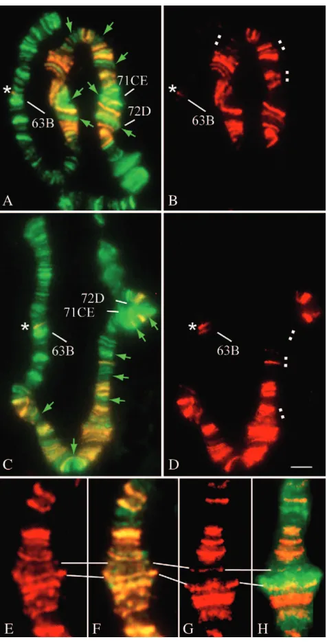
transgene does not fully reflect localization of active genes in the flanking autosomal region: We asked whether DCC chooses active genes when it spreads locally from autosomalroXtransgenes. To generate the most efficient DCC spreading in all nuclei, we tested male larvae, which carry the GM roX1-DDHS-72D trans-gene in aroX1ÿroX2ÿdouble-mutant background (Park
et al.2002; Baiet al.2004). We found that a large num-ber of good Po1IIo-binding sites, including some of ecdysone-induced puffs, do not associate with the DCC (Figure 4, A and C). The DCC spreading patterns are well known to be variable from nucleus to nucleus, al-though we failed to confirm this variability to be PS de-pendent (Figure 4, B and D). It should be noted that in the region 63BC the DCC binds one site, which was found to associate often with the complex in wild-type males (Demakovaet al.2003) and never binds to neigh-boring sites regardless of their active transcriptional status (Figure 4, A–D). Thus, similarly to the male X, the patterns of ectopic DCC binding do not directly reflect the localization of active transcriptional domains in au-tosomes carrying aGM roX1-DDHS-72Dtransgene.
EM double immunostaining reveals that DCC and Po1IIo can map to close but still distinct areas within a single interband:Since the majority of factors involved in transcription localize on polytene chromosomes to the regions of decondensed chromatin, namely to inter-bands and puffs (Champlin et al. 1991; Weeks et al. 1993; Stokeset al.1996; Kaplanet al.2000; Gerberet al. 2001; Gonzyet al.2002; Saunderset al.2003), it is no wonder that light-microscopy analysis of the DCC and PolIIo patterns on the male X revealed a significant degree of overlap in many regions (except puffs and cytologically extensive DCC gaps). However, we noted a number of regions where distinct MSL-binding sites could be observed, whereas the RNA polymerase was un-detectable (Figure 1). These might be classified as some of the CESs since unknown DNA features within CESs may serve as targets for the MSL proteins. Additionally, the recently characterized CES within the 18D region appears not to be transcribed (Ohet al.2004). Neverthe-less, many of such regions seem not to correspond to the known 70 CESs.
Figure3.—Localization of DCC and MLE looks different in the regions of prominent puffs developed in the distal part of the X chromosome. (A–D) Oregon-R (wild type) male. (E) w; msl3 [w1
Upon closer examination of the well-stretched mosomes, we managed to reveal also a number of chro-mosomal regions, in which the signals tend to show only partial overlap or to border each other (Figures 1 and 2, A–D). To determine precisely the relative localization of DCC and PolIIo within distinct small chromosomal regions, we carried out EM double immunostaining. Figure 5 shows data obtained for the 6A-D and the 8E-10A regions. Earlier it was shown that MSL2 tends to demonstrate unipolar labeling of narrow band/ interband borders along the whole male X (Semeshin et al.2002). Here we found that this specific localization of MSL2 looks different from PolIIo binding to decom-pacted interbands (Figure 5, C–F). The protein labels look partly overlapping in some regions (Figure 5, D–F) but localize apart in others (Figure 5C). Thus, EM data also indicate that relative localization of PolIIo and MSL2 within a distinct interband may be very close but still not identical.
MLE protein interacts with numerous transcription-ally active regions of polytene chromosomes indepen-dently of DCC: MLE possesses NTPase and both RNA- and DNA-helicase activities (Kuroda et al.1991; Leeet al. 1997). Furthermore, the physical binding of MLE to the X chromosome most likely occurs via RNA (Richteret al.1996). It is generally believed that MLE may act as a helicase to facilitate the targeting of the MSL proteins to male X or assembly of DCC (Pannuti and Lucchesi2000). Nevertheless, the possibility that the RNA-helicase activity of MLE could play a significant role in DCC stabilization in the regions of active genes via interaction with nascent transcripts has not been excluded to date (Richter et al. 1996; Stuckenholz et al.1999). In contrast to other MSLs, MLE is expressed in both sexes in comparable quantities (Kuroda et al. 1991) and can contribute to processes other than dos-age compensation (Kernan et al. 1991; Rastelli and Kuroda1998; Reenanet al.2000).
Although MLE is the first DCC subunit that has been immunolocalized on the polytene chromosomes, it is the only one of the MSLs whose detailed localization still remains unclear. MLE was first reported to bind pre-dominantly the male X chromosome and additionally to demonstrate numerous faint sites of association on male autosomes and all female chromosomes, these autoso-mal targets being uncharacterized (Kurodaet al.1991). Later, Bhadraet al.(1999), using the same MLE-specific antibody, restricted male MLE binding exclusively to the X chromosome. At the same time, in females, they
reported msl-dependent MLE binding to all
chromo-somes, and this finding catalyzed an idea about the existence of a reduced MSL complex in females that might contribute to an inverse dosage effect (Bhadra et al.1999). Finally, recently it was shown that in addition to labeling the X chromosome, MLE binds only a few autosomal sites in males, similar to other MSLs (Ruiz et al.2000).
To gain further insight into possible roles of MLE in transcription regulation, we performed precise map-ping of MLE-binding sites in both sexes at different points of larval development. First, we noted that MLE autosomal distribution looked identical in both sexes. As demonstrated in Figure 6, A and B, the RNA-helicase MLE binds essentially to all the developmental puffs typical for the PS analyzed and to many interbands. The observation that MLE binds puffs also is true for the female X chromosome (Figures 3F and 6B). Diffuse PS-dependent binding sites on female X look quite dif-ferent when compared with the reproducible banded pattern in males. Also, we found that several regions (18F, 19A, 22B, two sites in 29C, 30A, 50C, and 86C) re-producibly demonstrate intensive MLE staining, some of them appearing PS specific (Figure 6, A and B). To illustrate, the strong MLE-binding site in the region 30A can be detected only at PS1, whereas the one in the region 29C appears exclusively at the PS8-9. Thus, when associating with the DCC, the MLE distribution pattern on the male X is highly reproducible, whereas its MSL-independent chromosomal binding appears to be essen-tially a function of the transcriptional status of the region, resembling the PolIIo pattern. Additionally, we observed punctate MLE staining of the chromocenter with the intensity of staining varying from nucleus to nu-cleus (Figure 6, A, B, and D), as it was shown earlier for the species from the Obscura group (Boneand Kuroda 1996). Under the staining conditions used, no MLE-binding sites could be detected inmle1/mle1females (data not shown).
It is of special interest to understand whether MLE is able to function independently from DCC on the male X. As an intriguing exception, we detected MLE in puffs developed in the regions 2C, 3C, 16AB, that is, in the DCC gaps. (Figure 3, B and D). With theSgs4cluster
becoming inactive, the prominent MLE-binding site in the region 3C appears to regress, but at PS5-6 we still were able to detect one faint signal additionally to the DCC pattern typical for the region (Figure 3B). The MLE labeling of theSgs4puff is observed also inw; msl3 [w1
; H83M2-61]/TM6, Tb female larvae (Figure 3E). These animals carried the MSL2-expressing transgene and therefore exhibited ectopic dosage compensation (Kelleyet al.1995) but produced the DCC at a lower level compared to that of the wild-type male. We specu-late that reduced amounts of DCC might be the reason for much more intensive puff labeling by anti-MLE antibodies in the transgenic females. This proposal implies that there might be a competition for MLE between DCC and DCC-independent target sites on the male X (first of all, in the regions devoid of DCC). This might explain why MLE binding in the chromocenter appears to be more detectable in female cells than in male ones, where most of the protein is recruited to the X chromosome. Also, we observed, first, that unlike DCC, MLE demonstrates diffuse labeling in the 2B puff (Figure 3B) and, second, that the strongest MLE-binding sites sometimes can be seen in the 18F-19A region (Figure 6, C and D). We found these sites to be typical for females but never to serve as strong targets for the other MSL proteins. Thus, we believe that the RNA-helicase MLE has an additional function, separate from its participation as a DCC subunit on the male X.
MLE associates with heat-shock-induced puffs and apparently shows increased affinity to the ag-element, which produces the noncoding heat-shock RNA:Upon brief exposure to heat shock, the MLE protein can be found in many heat-shock-induced puffs. In particular, we observed a very robust permanent signal in the region 87C. Strong labeling could also be seen in the puff regions 93D and 95D, but never in the region 87A
Figure 5.—Detailed relative localization of MSL2 and PolIIo within distinct chromosomal re-gions at the electron microscopy level. The w; msl3 [w1;
(Figure 7, A and B). The two adjacent puffs 87A and 87C are known to develop due to the activity of a cluster of hsp70 genes (Ish-Horowicz et al. 1979). However, in contrast to the 87A region, the 87C region contains not only thehsp70 cluster, but also the ag-element, coding
for the hshRNA with unknown functions (Sharmaand
Lakhotia 1995). Since we never observed any MLE-binding site in region 87C in both sexes under non-heat-shock conditions, we speculate that this particular hshRNA, which belongs to the same class of RNA mole-cules as theroXRNAs, might be the target for MLE. To test the idea, we utilizedD. simulans, a sibling species for D. melanogaster, which lacks the sequences homologous to theag-elementin this region (Liset al. 1981). Con-sistent with this proposal, when staining the polytene chromosomes fromD. simulansheat-shocked larvae with MLE-specific antibodies, we failed to detect labeling of the 87C region (Figure 7C). It should be noted that
ag-element RNA sequence, when compared to that of theroX1or roX2, produced no significant homologies (http://www.ncbi.nlm.nih.gov/blast/).
Finally, by using the female larvae from the stocksw; msl1L60/CyO andmsl3P red/TM6, Tb, which were homo-zygous for themsl1andmsl3null alleles, respectively, we ascertained that neither of these mutations caused any detectable changes in MLE0binding patterns on the chromosomes (data not shown). This result is inconsis-tent with the idea that MLE association with female chromosomes is MSL dependent as described by Bhadraet al.(1999).
Thus, independently of other MSL proteins, MLE in-teracts with the most prominent transcriptionally active regions of chromosomes and likely shows increased affinity to the transcripts of certain genes.
DISCUSSION
The nature of targets for the DCC is a long-standing problem. Originally, it was proposed that specific enhancer-like sequences might reside close to individual X-linked genes, serving as targets for DCC (reviewed in Baker et al.1994). In contrast, the ‘‘spreading’’ model postulates
that the male X is marked by a quite limited number of
DNA sequences (35) that recruit the DCC and
accumulate locally at high levels, which in turn results in association of the complex with numerous sites of low affinity (Kelley et al. 1999; Kageyama et al. 2001). Nevertheless, the modern view of the problem assumes that there might be many more DNA sequences re-quired both for the initial recruitment/assembly of DCC (CES) (Demakovaet al.2003) and for the association of a functional complex with additional sites (non-CES) (Fagegaltierand Baker2004; Gilfillanet al.2004; Oh et al. 2004). In contrast to these postulated DNA sequences, most sites on the X, which are targets for functional DCC, are thought to mark genes actively transcribed in a given tissue and time of development (Kelleyet al.1999; Sasset al.2003). This idea implies that DCC mediates transcription enhancement via di-rect involvement in transcription regulation of each active gene. In this article, we made an effort to test further this model by precisely investigating the relative localization of DCC and PolIIo along the male X in the course of larval development.
Previously it was described that in vivo PolIIo and various elongation factors, as well as the H3.3 histone variant, dynamically associate with active genes, accom-pany their expression, and look colocalized in Drosoph-ila polytene chromosomes. This overlap is most obvious as diffuse labeling of developmental and heat-shock-induced puffs (Weeks et al.1993; Kaplanet al. 2000; Gerberet al.2001; Saunderset al.2003; Schwartzet al.
2003; Schwartz and Ahmad 2005). Intriguingly, we
found that DCC demonstrates striking stability in both the number and intensity of binding sites along the X throughout larval development. Moreover, despite being targets for MLE, the sites of the most intensive gene expression both on the male X and within an autosomal DCC-spreading area appear to not be targets for DCC at all. We also demonstrated all the DCC gaps that comprise active genes. Additionally, DCC skips over a number of transcriptionally active regions when it inappropriately spreads in cis from an autosomal roX1 transgene. We therefore suggest that active transcriptional status of the chromosomal region or association with MLE is not sufficient for DCC targeting and that, if DCC binds to actively transcribed regions, it does so very selectively.
bridge DCC with transcriptionally active regions, this might occur only if some additional requirements for DCC binding are realized.
Earlier, it was reported that a partial MSL complex lacking MSL2 protein is present in normal female nu-clei. Accordingly, mutations in variousmslgenes except
mof disassociate all MSLs from the chromosomes in
females (Bhadraet al. 1999). Nevertheless, our data on MLE distribution in polytene chromosomes of females homozygous formsl1or msl3null alleles indicate that even if MSLs form a partial complex in females, MLE binds the chromatin in an MSL-independent manner.
Our findings raise questions as to what are the reasons for exclusion of DCC from some active X-linked regions and whether dosage compensation does take place there. One can speculate that highly active chromatin in puffing regions turns into a poor substrate for the DCC due to drastic changes in packaging, possibly, up to nucleosome removal (Orphanidesand Reinberg2000; Kireevaet al.2002). However, a cluster of CESs bound by DCC is detected in the puffed 2B region throughout larval development. Moreover, strong transcription in-duced in EP transposons on the male X sometimes re-sults in ectopic DCC recruitment, suggesting that the complex is able to recognize very active chromatin (Sass et al.2003).
Revealing active genes within each of the cytologically extensive DCC gaps provides yet another puzzle. On the one hand, in the neo X chromosome ofD. miranda, the blocks of chromatin escaping dosage compensation do alternate with other blocks that are dosage compen-sated and therefore bind DCC (Boneand Kuroda1996; Marinand Baker1998). However, no data indicate that such clustering takes place inD. melanogaster (Ghosh et al.1992). If X-linked genes actually possess still un-known features needed for DCC targeting (Fagegaltier and Baker 2004), then DCC gaps might reflect evolu-tionary incompleteness of this process inD. melanogaster. It should be noted that, in contrast,70 autosomal re-gions are competent to recruit functional DCC in wild-type males (Demakova et al. 2003). Alternatively, the active genes located within the DCC gaps might serve as targets for the complex but cannot realize this ability probably due to the chromatin environment.
Whether the genes within puffs and DCC gaps un-dergo dosage compensation remains to be answered. It seems plausible to suggest that transcription upregula-tion could be not so essential for a subset of highly ex-pressed genes. Nevertheless, whatever the reasons for highly expressed loci to escape association with DCC, these genes are probably dosage compensated, which was shown at least forSgs4and theBroad-Complex(Breen and Lucchesi 1986; Kaiser et al. 1986; Chiang and Kurnit2003). If many active genes lack DCC-binding sites in the immediate vicinity, they might achieve dosage compensation by a yet unknown pathway. It is very possible that upregulation of active genes within
DCC gaps and puffing regions might be achieved, at least to some extent, via DCC-mediated establishment of a more open chromatin structure of the whole male X, suggesting that DCC affects transcription indirectly. Site-specific localization of H4Ac16 probably initiates a cascade of molecular remodeling events resulting in diffuse appearance of the whole male X chromosome (Bone et al. 1994). Generally, such a chromatin state would facilitate the access of various transcription and replication factors. Accordingly, in females having ec-topic dosage compensation induced, the DCC gap corre-sponding to the intercalary heterochromatin region on the polytene X demonstrated a greater extent of both polytenization and replication than in the wild type. This clearly correlated with the higher local concentrations of the DCC in neighboring areas (Alekseyenkoet al.2002). Thus, despite the fact that the DCC-mediated site-specific histone acetylation pattern correlates with an increase in transcription of the underlying sequences (Akhtar et al.2000; Henryet al.2001; Smithet al.2001), we believe it would be more accurate to suggest that there is no common scenario of dosage compensation for all the X-linked genes. Also, the DCC pattern appears essentially permanent and displays only negligible variations, both in the course of larval development (our data) and in different tissues (Sasset al.2003), which might point to the contribution of yet unidentified epigenetic factors in the establishment and maintenance of DCC binding. For example, the transcriptional activity of X-linked genes could govern DCC settling on the male X in early em-bryogenesis, and this pattern might be subsequently reproduced epigenetically. Hence, this scenario would imply high stability of the DCC pattern at least for the housekeeping genes, rather than dramatic changes in DCC distribution resulting from fine-tuned transcrip-tional programs further in development.
Future molecular studies of the dosage compensation status of active genes mapping to the DCC gaps could determine whether dosage compensation can also uti-lize some unknown mechanisms other than site-specific acetylation of H4 at lysine 16 leading to site-specific transcription enhancement. Alternatively, there may be many more X-linked genes whose expression does not require dosage compensation than was expected to date. Regardless, the important question remains how functional DCC recognizes its targets among the active genes on the male X chromosome.
We thank A. A. Gortchakov, Yu. B. Schwartz, and A. A. Alekseyenko for helpful discussions of our results. This work was supported by Fogarty International Research Collaboration Award (FIRCA) grant (International Cooperation Program) NI R03TW05669-01, Program for Scientific Schools RF 00-15-97984, and Program for Molecular and Cellular Biology NSH-918.2003.4.
LITERATURE CITED
essential for dosage compensation in Drosophila. Mol. Cell5:
367–375.
Akhtar, A., D. Zinkand P. B. Becker, 2000 Chromodomains are protein-RNA interaction modules. Nature407:405–409. Alekseyenko, A. A., O. V. Demakova, E. S. Belyaeva, G. F.
Makarevich, I. V. Kotlikovaet al., 2002 Dosage compensa-tion and intercalary heterochromatin in X chromosomes of Drosophila melanogaster. Chromosoma111:106–113. Andres, A. J., J. C. Fletcher, F. D. Karim and C. S. Thummel,
1993 Molecular analysis of the initiation of insect metamor-phosis: a comparative study of Drosophila ecdysteroid-regulated transcription. Dev. Biol.160:388–404.
Ashburner, M., C. Chihara, P. Meltzer and G. Richards, 1974 Temporal control of puffing activity in polytene chromo-somes. Cold Spring Harbor Symp. Quant. Biol.38:655–662. Bai, X., A. A. Alekseyenkoand M. I. Kuroda, 2004
Sequence-specific targeting of MSL complex regulates transcription of the roX RNA genes. EMBO J.23:2853–2861.
Baker, B. S., M. Gormanand I. Marin, 1994 Dosage compensation in Drosophila. Annu. Rev. Genet.28:491–521.
Belyaeva, E. S., L. S. Korochkina, I. F. Zhimulev and N. K. Nazarova, 1974 Puff characteristics in the X-chromosome of D. melanogaster females. Cytology16:440–445.
Bhadra, U., M. Pal-Bhadra and J. A. Birchler, 1999 Role of the male specific lethal (msl) genes in modifying the effects of sex chromosomal dosage in Drosophila. Genetics152:249– 268.
Birchler, J. A., 1996 X chromosome dosage compensation in Dro-sophila. Science272:1190–1191.
Bone, J. R., and M. I. Kuroda, 1996 Dosage compensation regula-tory proteins and the evolution of sex chromosomes in Drosoph-ila. Genetics144:705–713.
Bone, J. R., J. Lavender, R. Richman, M. J. Palmer, B. M. Turner
et al., 1994 Acetylated histone H4 on the male X chromosome is associated with dosage compensation in Drosophila. Genes Dev.8:96–104.
Breen, T. R., and J. C. Lucchesi, 1986 Analysis of the dosage com-pensation of a specific transcript in Drosophila melanogaster.
Genetics112:483–491.
Champlin, D. T., M. Frasch, H. Saumweber and J. T. Lis, 1991 Characterization of a Drosophila protein associated with boundaries of transcriptionally active chromatin. Genes Dev.5:
1611–1621.
Chang, K. A., and M. I. Kuroda, 1998 Modulation of MSL1 abun-dance in female Drosophila contributes to the sex specificity of dosage compensation. Genetics150:699–709.
Cheng, C., and P. A. Sharp, 2003 RNA polymerase II accumulation in the promoter-proximal region of the dihydrofolate reductase and gamma-actin genes. Mol. Cell. Biol.23:1961–1967. Chiang, P. W., and D. M. Kurnit, 2003 Study of dosage
compensa-tion in Drosophila. Genetics165:1167–1181.
Demakova, O. V., I. V. Kotlikova, P. R. Gordadze, A. A. Alekseyenko, M. I. Kurodaet al., 2003 The MSL complex levels are critical for its correct targeting to the chromosomes in Drosophila melanogaster. Chromosoma112:103–115.
Fagegaltier, D., and B. S. Baker, 2004 X chromosome sites auton-omously recruit the dosage compensation complex in Drosoph-ila males. PLoS Biol.2:e341.
Furia, M., P. P. D’Avino, S. Crispi, D. Artiaco and L. C. Polito, 1993 Dense cluster of genes is located at the ecdysone-regulated 3C puff of Drosophila melanogaster. J. Mol. Biol.231:531–538. Gerber, M., J. Ma, K. Dean, J. C. Eissenbergand A. Shilatifard,
2001 Drosophila ELL is associated with actively elongating RNA polymerase II on transcriptionally active sites in vivo. EMBO J.20:6104–6114.
Ghosh, S., J. C. Lucchesiand J. E. Manning, 1992 The non-dosage compensated LSP1-alpha gene of Drosophila melanogaster lies immediately downstream of the dosage compensated L12 gene. Mol. Gen. Genet.233:49–52.
Gilfillan, G. D., I. K. Dahlsveenand P. B. Becker, 2004 Lifting a chromosome: dosage compensation in Drosophila melanogaster. FEBS Lett.567:8–14.
Gonzy, G., G. V. Pokholkova, F. Peronnet, B. Mugat, O. V. Demakovaet al., 2002 Isolation and characterization of novel mutations of the Broad-Complex, a key regulatory gene of
ecdy-sone induction in Drosophila melanogaster. Insect Biochem. Mol. Biol.32:121–132.
Gorman, M., A. Frankeand B. S. Baker, 1995 Molecular character-ization of the male-specific lethal-3 gene and investigations of the regulation of dosage compensation in Drosophila. Development
121:463–475.
Gu, W., P. Szauterand J. C. Lucchesi, 1998 Targeting of MOF, a putative histone acetyl transferase, to the X chromosome of Drosophila melanogaster. Dev. Genet.22:56–64.
Gu, W., X. Wei, A. Pannutiand J. C. Lucchesi, 2000 Targeting the chromatin-remodeling MSL complex of Drosophila to its sites of action on the X chromosome requires both acetyl transferase and ATPase activities. EMBO J.19:5202–5211.
Hampsey, M., and D. Reinberg, 2003 Tails of intrigue: phosphory-lation of RNA polymerase II mediates histone methyphosphory-lation. Cell
113:429–432.
Henry, R. A., B. Tews, X. Liand M. J. Scott, 2001 Recruitment of the male-specific lethal (MSL) dosage compensation complex to an autosomally integrated roX chromatin entry site correlates with an increased expression of an adjacent reporter gene in male Drosophila. J. Biol. Chem.276:31953–31958.
Ish-Horowicz, D., S. M. Pinchin, J. Gausz, H. Gyurkovics, G. Benczeet al., 1979 Deletion mapping of two D. melano-gaster loci that code for the 70,000 dalton heat-induced protein. Cell17:565–571.
Jin, Y., Y. Wang, D. L. Walker, H. Dong, C. Conleyet al., 1999 JIL-1: a novel chromosomal tandem kinase implicated in transcrip-tional regulation in Drosophila. Mol. Cell4:129–135.
Kageyama, Y., G. Mengus, G. Gilfillan, H. G. Kennedy, C. Stuckenholz et al., 2001 Association and spreading of the Drosophila dosage compensation complex from a discrete roX1 chromatin entry site. EMBO J.20:2236–2245.
Kaiser, K., M. Furiaand D. M. Glover, 1986 Dosage compensation at the sgs4 locus of Drosophila melanogaster. J. Mol. Biol.187:
529–536.
Kaplan, C. D., J. R. Morris, C. Wuand F. Winston, 2000 Spt5 and spt6 are associated with active transcription and have character-istics of general elongation factors in D. melanogaster. Genes Dev.14:2623–2634.
Kelley, R. L., I. Solovyeva, L. M. Lyman, R. Richman, V. Solovyev
et al., 1995 Expression of msl-2 causes assembly of dosage com-pensation regulators on the X chromosomes and female lethality in Drosophila. Cell81:867–877.
Kelley, R. L., V. H. Meller, P. R. Gordadze, G. Roman, R. L. Davis
et al., 1999 Epigenetic spreading of the Drosophila dosage com-pensation complex from roX RNA genes into flanking chroma-tin. Cell98:513–522.
Kernan, M. J., M. I. Kuroda, R. Kreber, B. S. Baker and B. Ganetzky, 1991 napts, a mutation affecting sodium channel activity in Drosophila, is an allele of mle, a regulator of X chro-mosome transcription. Cell66:949–959.
Kireeva, M. L., W. Walter, V. Tchernajenko, V. Bondarenko, M. Kashlevet al., 2002 Nucleosome remodeling induced by RNA polymerase II: loss of the H2A/H2B dimer during transcrip-tion. Mol. Cell9:541–552.
Komarnitsky, P., E. J. Cho and S. Buratowski, 2000 Different phosphorylated forms of RNA polymerase II and associated mRNA processing factors during transcription. Genes Dev.14:
2452–2460.
Kuroda, M. I., M. J. Kernan, R. Kreber, B. Ganetzkyand B. S. Baker, 1991 The maleless protein associates with the X chro-mosome to regulate dosage compensation in Drosophila. Cell
66:935–947.
Lee, C. G., K. A. Chang, M. I. Kurodaand J. Hurwitz, 1997 The NTPase/helicase activities of Drosophila maleless, an essential factor in dosage compensation. EMBO J.16:2671–2681. Lindsley, D. L., and G. G. Zimm, 1992 The Genome of Drosophila
melanogaster.Academic Press, San Diego.
Lis, J. T., W. Neckameyer, R. Dubenskyand N. Costlow, 1981 Clon-ing and characterization of nine heat-shock-induced mRNAs of Drosophila melanogaster. Gene15:67–80.
Marin, I., and B. S. Baker, 1998 The evolutionary dynamics of sex determination. Science281:1990–1994.
Meller, V. H., and M. I. Kuroda, 2002 Sex and the single chromo-some. Adv. Genet.46:1–24.
Meller, V. H., P. R. Gordadze, Y. Park, X. Chu, C. Stuckenholz
et al., 2000 Ordered assembly of roX RNAs into MSL complexes on the dosage-compensated X chromosome in Drosophila. Curr. Biol.10:136–143.
Oh, H., J. R. Boneand M. I. Kuroda, 2004 Multiple classes of MSL binding sites target dosage compensation to the X chromosome of Drosophila. Curr. Biol.14:481–487.
Orphanides, G., and D. Reinberg, 2000 RNA polymerase II elon-gation through chromatin. Nature407:471–475.
Palancade, B., and O. Bensaude, 2003 Investigating RNA polymer-ase II carboxyl-terminal domain (CTD) phosphorylation. Eur. J. Biochem.270:3859–3870.
Pal Bhadra, M., U. Bhadra, J. Kundu and J. A. Birchler, 2005 Gene expression analysis of the function of the male-specific lethal complex in Drosophila. Genetics169:2061–2074. Pannuti, A., and J. C. Lucchesi, 2000 Recycling to remodel: evolu-tion of dosage-compensaevolu-tion complexes. Curr. Opin. Genet. Dev.
10:644–650.
Park, Y., R. L. Kelley, H. Oh, M. I. Kuroda and V. H. Meller, 2002 Extent of chromatin spreading determined by roX RNA recruitment of MSL proteins. Science298:1620–1623. Rastelli, L., and M. I. Kuroda, 1998 An analysis of maleless and
histone H4 acetylation in Drosophila melanogaster spermatogen-esis. Mech. Dev.71:107–117.
Reenan, R. A., C. J. Hanrahanand G. Barry, 2000 The mle(napts) RNA helicase mutation in Drosophila results in a splicing catas-trophe of the para Na1channel transcript in a region of RNA editing. Neuron25:139–149.
Richter, L., J. R. Boneand M. I. Kuroda, 1996 RNA-dependent association of the Drosophila maleless protein with the male X chromosome. Genes Cells1:325–336.
Ruiz, M. F., M. R. Esteban, C. Donoro, C. Godayand L. Sanchez, 2000 Evolution of dosage compensation in Diptera: the gene maleless implements dosage compensation in Drosophila (Brachy-cera suborder) but its homolog in Sciara (Nemato(Brachy-cera suborder) appears to play no role in dosage compensation. Genetics156:
1853–1865.
Sass, G. L., A. Pannutiand J. C. Lucchesi, 2003 Maspecific le-thal complex of Drosophila targets activated regions of the X chromosome for chromatin remodeling. Proc. Natl. Acad. Sci. USA100:8287–8291.
Saunders, A., J. Werner, E. D. Andrulis, T. Nakayama, S. Hirose
et al., 2003 Tracking FACT and the RNA polymerase II elonga-tion complex through chromatin in vivo. Science 301:1094– 1096.
Schwartz, B. E., and K. Ahmad, 2005 Transcriptional activation triggers deposition and removal of the histone variant H3.3. Genes Dev.19:804–814.
Schwartz, B. E., S. Larochelle, B. Suterand J. T. Lis, 2003 Cdk7 is required for full activation of Drosophila heat shock genes and RNA polymerase II phosphorylation in vivo. Mol. Cell. Biol.23:
6876–6886.
Semeshin, V. F., R. Artero, M. Perez-Alonsoand V. V. Shloma, 1998 Electron microscopic in situ hybridization of digoxigenin-dUTP-labelled DNA probes with Drosophila melanogaster poly-tene chromosomes. Chromosome Res.6:405–410.
Semeshin, V. F., E. N. Andreyeva, V. V. Shloma, H. Saumweber and I. F. Zhimulev, 2002 Immunogold electron microscope localization of proteins in Drosophila polytene chromosomes: applications and limitations of the method. Chromosome Res.
10:429–433.
Sharma, A., and S. C. Lakhotia, 1995 In situ quantification of hsp70 and alpha-beta transcripts at 87A and 87C loci in relation to hsr-omega gene activity in polytene cells of Drosophila mela-nogaster. Chromosome Res.3:386–393.
Smith, E. R., A. Pannuti, W. Gu, A. Steurnagel, R. G. Cooket al., 2000 The Drosophila MSL complex acetylates histone H4 at lysine 16, a chromatin modification linked to dosage compensa-tion. Mol. Cell. Biol.20:312–318.
Smith, E. R., C. D. Allisand J. C. Lucchesi, 2001 Linking global histone acetylation to the transcription enhancement of X-chromosomal genes in Drosophila males. J. Biol. Chem. 276:
31483–31486.
Stokes, D. G., K. D. Tartof and R. P. Perry, 1996 CHD1 is concentrated in interbands and puffed regions of Drosophila polytene chromosomes. Proc. Natl. Acad. Sci. USA 93:7137– 7142.
Stuckenholz, C., Y. Kageyama and M. I. Kuroda, 1999 Guilt by association: non-coding RNAs, chromosome-specific proteins and dosage compensation in Drosophila. Trends Genet.15:454– 458.
Wang, Y., W. Zhang, Y. Jin, J. Johansen and K. M. Johansen, 2001 The JIL-1 tandem kinase mediates histone H3 phosphor-ylation and is required for maintenance of chromatin structure in Drosophila. Cell105:433–443.
Weeks, J. R., S. E. Hardin, J. Shen, J. M. Leeand A. L. Greenleaf, 1993 Locus-specific variation in phosphorylation state of RNA polymerase II in vivo: correlations with gene activity and tran-script processing. Genes Dev.7:2329–2344.
Zhang, S., and F. Grosse, 2004 Multiple functions of nuclear DNA helicase II (RNA helicase A) in nucleic acid metabolism. Acta Biochim. Biophys. Sin. (Shanghai)36:177–183.
Zhimulev, I. F., 1999 Genetic organization of polytene chromo-somes. Adv. Genet.39:1–589.
Zhimulev, I. F., E. S. Belyaeva, O. M. Mazinaand M. L. Balasov, 1995 Structure and expression of the BR-C locus in Drosophila melanogaster, Diptera: Drosophilidae. Eur. J. Entomol.92:263– 270.