DOI: 10.1534/genetics.107.081869
A Gain-of-Function Suppressor Screen for Genes Involved in Dorsal–Ventral
Boundary Formation in the Drosophila Wing
Fernando Bejarano,
1Carlos M. Luque,
1,2He´ctor Herranz, Georgina Sorrosal,
Neus Rafel, Thu Thuy Pham and Marco Mila´n
3Institucio´ Catalana de Recerca i Estudis Avancxats and Institute for Research in Biomedicine, 08028 Barcelona, Spain Manuscript received September 13, 2007
Accepted for publication November 9, 2007
ABSTRACT
The Drosophila wing primordium is subdivided into a dorsal (D) and a ventral (V) compartment by the activity of the LIM-homeodomain protein Apterous in D cells. Cell interactions between D and V cells induce the activation of Notch at the DV boundary. Notch is required for the maintenance of the compartment boundary and the growth of the wing primordium. Beadex, a gain-of-function allele of dLMO, results in increased levels of dLMO protein, which interferes with the activity of Apterous and results in defects in DV axis formation. We performed a gain-of-function enhancer-promoter (EP) screen to search for suppressors ofBeadexwhen overexpressed in D cells. We identified 53 lines corresponding to 35 genes. Loci encoding for micro-RNAs and proteins involved in chromatin organization, transcriptional control, and vesicle trafficking were characterized in the context of dLMO activity and DV boundary formation. Our results indicate that a gain-of-function genetic screen in a sensitized background, as opposed to classical loss-of-function-based screenings, is a very efficient way to identify redundant genes involved in a developmental process.
I
N multicellular organisms, initially homogenoussheets of cells are often subdivided into adjacent cell populations by the activity of certain transcription
factors (reviewed in Irvine and Rauskolb 2001). In
many cases, cell interactions between these populations lead to the restricted expression of signaling molecules at their boundaries, which organize growth and/or the pattern of nearby cells. The stability of these bound-aries frequently relies on the acquisition of differential cell affinities between adjacent populations. When these boundaries behave as lineage restriction boundaries,
these populations are called compartments (Garcı´a
-Bellidoet al.1973). The Drosophila wing primordium,
a monolayered epithelium that gives rise to the adult wing and part of the thorax, is subdivided into an anterior and a posterior compartment by the activity of the homeodomain transcription factors Engrailed
and Invected in posterior cells (Garcı´a-Bellido and
Santamaria 1972; Lawrence and Morata 1976;
Tabataet al. 1995; Zecca et al. 1995). During larval
development, the wing primordium suffers a secondary compartment subdivision. The activity of the LIM-homeodomain transcription factor Apterous (Ap) is
res-ponsible for this later subdivision into a dorsal (D) and
a ventral (V) compartment (Diaz-Benjumeaand Cohen
1993).
Ap has three functions in wing development. It is re-sponsible for the establishment of the Notch-dependent signaling center, the generation of a lineage restriction at the DV boundary, and the acquisition of a dorsal iden-tity during cell differentiation. Ap exerts these functions through three classes of target genes. The complemen-tary expression of Serrate and Delta, two ligands of the receptor Notch, to D and V cells, respectively, initiates a cascade of short-range cell interactions that lead to the activation of Notch at the DV boundary (Figure 1B). Dorsally expressed Serrate and ventrally expressed Delta activate Notch symmetrically in cells on both sides of
the DV compartment boundary (Diaz-Benjumea and
Cohen1993;deCeliset al.1996b; Dohertyet al.1996).
Expression of the glycosyltransferase Fringe in D cells makes them more sensitive to Delta and less sensitive to
Serrate (Bruckneret al.2000; Moloney2000; Munro
and Freeman 2000), thus polarizing Notch activation
toward the DV boundary. Notch activation induces Wingless (Wg) expression in cells along this boundary. The combined activity of Notch and Wg organizes the growth and patterning of the whole wing primordium
(Giraldez and Cohen 2003). The transmembrane
proteins Capricious (Caps) and Tartan (Trn) belong to the second class of Ap target genes that contribute to the generation of an affinity difference between D and V cells (Mila´ net al. 2001a). Finally, the activity of the 1These authors contributed equally to this work.
2Present address:Centro Andaluz de Biologı´a del Desarrollo,
Universi-dad Pablo de Olavide-CSIC, 41013 Sevilla, Spain.
3Corresponding author:ICREA and Institute for Research in Biomedicine
(IRB), Josep Samitier, 1-5, 08028 Barcelona, Spain. E-mail: mmilan@pcb.ub.es
homeodomain protein Msh, another Ap target, confers D identity (Mila´ net al.2001b).
The activity of Ap must be tightly regulated during development to allow the dynamic change in the expression pattern of Serrate and Delta. Later in de-velopment, these proteins are restricted to the pre-sumptive vein tissue to help define the width of the adult longitudinal wing veins (deCeliset al.1997). Ap activity
depends on the formation of a higher order complex, in which two molecules of Ap are bridged by a dimer of its cofactor, the LIM-domain binding protein dLDB/Chip
(Fernandez-Funezet al.1998; Mila´ nand Cohen1999;
van Meyel et al. 1999). The level of Ap activity is
regulated during wing development by expression of
another LIM-domain protein, dLMO (Mila´ net al.1998;
Shoreshet al.1998; Zenget al.1998). dLMO competes
with Ap for binding to its cofactor Chip/dLDB, con-tributes to reducing the activity of Ap, and facilitates the transition in the expression pattern of Serrate and Delta, which, late in development, become symmetri-cally expressed along the wing veins in both D and V
compartments (Mila´ nand Cohen2000).
To further our understanding of the process of boundary formation in the Drosophila wing, we per-formed a gain-of-function suppressor screen. This screen is based on the capacity of genes to bypass the requirement of Ap protein activity in DV boundary formation when they are overexpressed in the domain
of ap. Here we identify, characterize, and discuss four
classes of genes in the context of DV boundary forma-tion or dLMO activity: chromatin organizaforma-tion genes, transcription factors, micro-RNAs, and proteins in-volved in vesicle trafficking and membrane fusion.
MATERIALS AND METHODS
Drosophila strains: Bx1,apgal4, andap-lacZare described in
Mila´ net al.(1998).UAS-Caps,UAS-Trn,caps65.2, andtrn25/4are
described in Mila´ net al.(2001a).AxM1is described in Perez
et al.(2005).lilli4u5andlilli632are described in Wittweret al.
(2001).UAS-p35is described in Hayet al.(1995).UAS-mtvand
mtv6are described in Funakoshiet al.(2001). Other stocks are
described in FlyBase. The following Drosophila genotypes were used to generate loss-of-function clones:
hs-FLP; lilli4u5FRT40A/Ubi-GFP FRT40A
hs-FLP; cbtE1FRT40A/Ubi-GFP FRT40A
hs-FLP; cbtE28FRT40A/Ubi-GFP FRT40A
hs-FLP; nmdK10909FRT40A/Ubi-GFP FRT40A
hs-FLP; FRT42D mtv6/FRT42D Ubi-GFP
hs-FLP; FRT42D l(3)04708/FRT42D Ubi-GFP hs-FLP; nufKG02305FRT80/Ubi-GFP FRT80
hs-FLP; draperd5FRT80/Ubi-GFP FRT80
hs-FLP; draperd19FRT80/Ubi-GFP FRT80
hs-FLP; FRT82 tara1/FRT82 Ubi-GFP.
Larvae were heat-shocked for 1 hr at 37°and dissected 60 hr later.
Crossing scheme:In a cross, four virginsof a Bx1/FM6; apgal4/
CyOstock were mated with 2–3 males of 4200 independentw; EP (white1)insertions (Rorthet al.1998), generated by the
groups of S. Cohen, A. Ephrussi, M. Mlodzik, and P. Rorth (EMBL, Heidelberg, Germany) and kindly maintained by Gu¨nter Bro¨nner in Go¨ttingen (Germany). At least 10Bx1/Y;
apgal4/EPmales per cross were scored for their wing phenotype.
Candidate enhancer-promoter (EP) lines were tested with the Bx1stock for their capacity to rescue the wing phenotype in a
Gal4-independent manner. Candidate EP lines were also tested for their capacity to cause a gain-of-function phenotype when overexpressed with theptc-gal4,en-gal4, andap-gal4wing drivers. Wings were mounted in Faure’s medium.
Molecular characterization of EP lines: To identify the
genes isolated by their gain-of-function capacity to suppress theBeadex1phenotype, flanking DNA was isolated by plasmid
rescue and the genomic region immediately downstream of the EP element promoter (at the 39end of thePelement) was sequenced. Flanking DNA was also isolated by inverse PCR to verify the presence of only one EP line per stock, and the genomic region immediately downstream of the EP element promoter was sequenced to confirm the plasmid rescue results. Details on the protocols followed can be found at the Figure1.—Design of the genetic screen for suppressors of
theBeadexwing phenotype. The wing primordium is subdi-vided into a dorsal (D) and a ventral (V) compartment by the restricted expression and activity of Apterous (Ap) in D cells. (A)ap-lacZexpression in a third instar wing disc visual-ized by histochemical staining forb-gal activity. (B) Early in development, Serrate (Ser) signals to V cells to activate Notch (N). Likewise, Delta (Dl) signals to D cells to activate Notch modified by Fringe (Fng) along the DV boundary. (C) Bea-dex1/FM6; apterousGal4/CyOflies, which have a strong loss of
Berkeley Drosophila Genome Project Web site (http:// www.fruitfly.org/).
Antibodies and constructs:Monoclonal antibodies against
Wingless (Wg) and Cut are described in the Developmental Hybridoma Bank. Antibody against dLMO is described in Mila´ net al.(1998) and was kindly provided by S. M. Cohen.
Other antibodies are commercially available.In situ hybrid-ization was carried out as in Mila´ net al.(1996). The dLMO 39
-UTR was amplified by PCR from genomic DNA and cloned into tubulin-enhanced green fluorescence protein (EGFP) as described in Brenneckeet al.(2003). ThemiR-14hairpin was
cloned downstream of dsRed2 in pUAST as described in Brenneckeet al.(2003).
RESULTS AND DISCUSSION
Several ways to rescue the Beadex1 wing phenotype:
Beadex1(
Bx1) is a gain-of-function allele of
dLMO that
results in higher levels of dLMO mRNA in the
de-veloping wing imaginal disc (Mila´ net al.1998; Shoresh et al.1998; Zenget al.1998). dLMO protein competes
with Ap for binding to its cofactor Chip. Consequently,
the activity of the Ap protein is reduced in a Bx1
background, its target genes are not expressed at appropriate levels, and the activation of Notch at the DV boundary and the formation of the adult wing margin are compromised (Figure 2B). Activation of the Notch signaling pathway by means of a gain-of-function allele ofNotch(Abruptex,deCelisand Garcı´a-Bellido
1994) or reduced levels of Hairless, an antagonist of
Notch signaling activity (Banget al.1995), rescued the
Bx1phenotype (Figure 2, I and J), indicating that the
wing-margin defects of Bx1 wings are a direct
conse-quence of reduced levels of Notch.
Overexpression ofapin its own expression domain
(using theapgal4driver) rescues theBx1phenotype (in
Bx1/Y; apgal4/UAS-apflies), and reduced levels ofapor
Chip enhance the Bx1 phenotype (Mila´ n et al. 1998,
2004). Two classes of Ap target genes contribute to the
formation of the DV boundary. fringe and Serrate are
directly involved in the activation of Notch at the DV
boundary (Irvineand Wieschaus1994; Diaz-Benjumea
and Cohen1995). As expected, overexpression of either
of these genes in theapexpression domain rescues the
Bx1
phenotype, and reduced levels of these two genes
enhance it (Mila´ net al. 1998, 2004). The leucine-rich
repeat (LRR) transmembrane proteins Caps and Trn are involved in the generation of an affinity difference
between D and V cells. Reduced levels ofcapsortrnhave
been shown to enhance theBx1phenotype (Mila´ net al.
2001a; compare also Figure 2D and 2F with 2B), in-dicating that these two genes contribute to the formation of the DV boundary. We then monitored the capacity of overexpressed Caps or Trn to rescue the wing-margin defects of Bx1
adult wings. Overexpression of either of these two proteins suppressed the wing-margin defects (Figure 2, C and E). Interestingly, they also rescued the Notch activation levels at the DV boundary, as shown by
the levels of Wg protein expression (compare Figure 2G and 2H and data not shown). Taken together, these re-sults indicate that Caps and Trn contribute to the acti-vation of Notch at the DV boundary, probably through
the generation of a stable DV affinity boundary. TheBx1
wing-margin phenotype therefore appears to be a highly sensitive background in which to search for genes in-volved in Ap activity and Notch activation, and as such we used it in a gain-of-function-based screen, described in the next section.
Figure 2.—Increased Notch activity or Caps/Tartan
ex-pression rescues theBeadexwing phenotype. (A, C, and E) Cu-ticle preparations ofBx1/Y; apgal4/1(A),Bx1/Y; apgal4/uas-caps
(C) andBx1/Y; apgal4/uas-trn(E) adult wings. Note rescue of
the wing-margin defects when Caps or Trn are expressed. Note also the blistered wing phenotype in C and E, probably due to defects in cell apposition between D and V wing surfa-ces in the presence of high levels of Caps or Trn expression. (B, D, and F) Cuticle preparations of Bx1/1 (B), Bx1/1,
caps65.2/1(D), andBx1/1, trn25/4/1(F) adult wings. Note
en-hancement of the wing-margin defects when eithercapsortrn are removed. (G and H)Bx1/Y; apgal4/1(G) andBx1/Y; apgal4/
uas-caps (H) wing discs labeled to visualize Gal4 (red) and Wingless (Wg, blue) protein expression. Dorsal (d) and ven-tral (v) compartments are marked. (I and J) Cuticle prepara-tions ofBx1/AxM1(I) andBx1/1; HE31
The suppression screen:A loss-of-function approach has been widely used in traditional genetic screenings but has several limitations. Redundancy between genes that have overlapping functions might partially or com-pletely mask gene function, and an early phenotype caused by a mutation might prevent the detection of later phenotypes. The gain-of-function approach by-passes these two limitations and allows the examination of the misexpression of phenotypes in the biological context of choice. This approach has been shown to be a powerful tool in identifying genes involved in
develop-ment (Rorthet al. 1998). Here we performed an
EP-mediated overexpression screening, in which 4200 randomly inserted and independently generated EP insertions, which allow the misexpression of genes that lie immediately downstream of the point of insertion, were driven in the developing wing by a dorsal-specific
Gal4 driver (apgal4) in a Bx1 sensitized background
(Figure 1C).Bx1/Y; apgal4/1male flies showed a strong scalloping phenotype (loss of wing-margin structures) as a result of reduced levels of Ap activity (note thatapgal4
is a loss-of-function allele ofap). Those EP insertions,
which have the capacity to rescue the wing-margin phenotypes of these flies, drive candidate genes in-volved in DV boundary formation, either as positive regulators of Ap activity or as downstream genes in-volved indirectly or directly in the activation of Notch at the boundary. Fifty-three EP lines partially or totally rescued the scalloping phenotype. Most of these lines (47/53) showed a complete rescue. Table 1 describes the relationship of the EP insertion with known and predicted genes. These EP insertions correspond to 35 genes. The finding of EP lines that drive the expression of fringe (EPs 3-511, 3-581, 3-612, and 3-934), ap(EP
3-1583) orosa(EPs3-473,3-619,3-900,3-941,3-1074,
3-1386, and3-1591), a member of the Brahma
chromatin-remodeling complex that binds Chip and modulates
the expression of Ap target genes (Collinset al.1999;
Heitzleret al. 2003; Mila´ net al.2004), confirms the
success of our screen.
Most EPs were located in the correct location (59end)
of the nearby genes that would generate Gal4-driven sense-strand full-length mRNAs. This result was also
observed in other published EP screens (Rorthet al.
1998; Tobaet al.1999). A low percentage of EP lines
were located in intronic or exonic sequences. Partial Gal4-driven sense-strand functional mRNAs might be generated in the former case. Note three EP lines were
inserted in the first intron of osa (Table 1) and the
rescue capacity of this gene has already been
demon-strated by means of an UAS-osa transgenic construct
(Mila´ net al.2004). When inserted in exonic sequences,
mutant alleles of the candidate genes might be pro-duced. Note those EP lines inserted inskuld,Draper, and
nonmitochondrial derivative(nmd) were located in exonic sequences (Table 1). The loss of function of these genes, and not its Gal4-mediated overexpression, was
respon-sible for the Bx1 rescue, as demonstrated by the
dominant genetic interaction betweenBx1and
loss-of-function alleles of these genes (see below). Surprisingly, some EPs (e.g.,3-1583driving the expression ofap;3-612
and3-934driving the expression offringe) were located in the opposite direction, suggesting that the Gal4 binding sites were duplicated during transposition, or
alternatively, that the EP drives the expression of 39
-located genes. Interestingly, a similar case was found in UAS transgenic constructs, in which a genomic frag-ment containing a micro-RNA in antisense orientation relative to the pUAST vector is transcribed in a Gal4-driven sense strand, probably as a result of the capacity of the Gal4-dependent transcription to increase the
activity of the endogenous promoter (Brenneckeet al.
2003).
Two different methods were used to validate the candidate genes whose overexpression was able to
rescue the Beadexwing-margin phenotype. First,
avail-able UAS transgenes, or alternatively, availavail-able EP
insertions located at the 59end of the candidate genes
were tested for the ability to rescue, in a Gal4-dependent
manner, theBeadexwing-margin phenotype. We found
that in the cases analyzed (32/53 EPs) this was the case (Table 1). In some cases, UAS transgenes or other EP
insertions were not available. We then carried outin situ
hybridization with RNA-labeled probes of the genes downstream of the EP insertions. We found that in the cases analyzed (4) the genes are overexpressed in a Gal4-dependent manner (Figure 3). Our experience with EP insertions somehow indicates, however, that this might not be the best way for the validation, since many genes in the neighborhood can be upregulated upon Gal4 transcriptional activation. This is the reason we
have not performedin situhybridization for more EP
insertions.
Since we performed a misexpression screening,
sev-eral of the candidate genes able to rescue theBx1
phe-notype may not be required for wing development and DV boundary formation in wild-type flies. To test this, we searched FlyBase for mutants in the candidate genes or deficiencies covering them, and checked whether there
were any dominant interactions withBx1
. Bx1
/1females
have a mild scalloping in the posterior compartment of the wing (Figure 2B). Males carrying a mutation in the candidate gene or a deficiency covering it were crossed
withBx1females, and the wing phenotype of the female
progeny was tested for enhancement. The vast majority of the candidate genes showed a dominant interaction with Beadex1 (Table 1; 28/34 genes tested; Figure 4).
Some of the genes, when removed, rescued theBx1
phe-notype (nmd,skuld/pap, andDraper,Figure 4R and data
not shown), indicating that the suppression is due to the loss of function of the candidate gene.
we used the following Gal4 drivers:ap-gal4(expressed in
dorsal cells),patched-gal4(expressed along the anterior–
posterior compartment boundary), and engrailed-gal4
(expressed in the posterior compartment). Ectopic
expression offringeorapin the V compartment, using
thepatched-gal4orengrailed-gal4drivers, induces ectopic
wing-margin structures (Mila´ n and Cohen 2003).
When using the same Gal4 drivers, none of the lines identified, with the exception of those driving the
expression offringeorap, caused this phenotype (data
not shown). This observation indicates that the number of genes with an instructive role likefringe,Serrate, orap
is very low, and that the newly identified genes encode for proteins that modulate the activity of the elements or pathways involved in DV boundary formation. Many of these genes caused the loss of macro- and micro-chaetae
in the adult notum (when expressed with theap-Gal4
driver), thereby resembling a Notch gain-of-function
phenotype (Table 1, Heitzlerand Simpson1991). This
suggests that the nature of the Bx1 rescue by
overex-pression of these genes is through an increase in Notch
activity levels, as occurred in an Abruptex or Hairless
mutant background (Figure 2, I and J). Taken together, these results indicate that the designed suppression screen is efficient in identifying modulators of the pathways and elements involved in DV boundary formation.
Classes of genes: Of the 35 genes identified in the
screen, 20 corresponded to genes previously character-ized. Five genes ½ap, osa, fng, skuld/pap, and E(spl-g)
participate in Notch signaling and/or DV boundary
formation in the Drosophila wing (Diaz-Benjumeaand
Cohen 1993; Irvine and Wieschaus 1994; de Celis
et al.1996a; Janodyet al.2003; Mila´ net al.2004). Other
genes are involved in other aspects of wing development (e.g.,capicuaandpointedin EGF receptor signaling,mtv
and schnurri in Dpp signaling) (Greider et al. 1995;
Funakoshiet al.2001; Rochet al.2002) and Hedgehog
signaling (Bejaranoet al.2007), suggesting that either
different signaling pathways are closely coordinated during DV boundary formation or distinct pathways share common elements. A large group of genes with essential roles in other developmental processes were identified (e.g.,PAR-5/14-3-3ein anterior–posterior axis
formation in the oocyte, cabut in JNK signaling, and
embryonic dorsal closure) (Bentonet al.2002; Mun˜ oz
-Descalzo et al. 2005). Many of these have not been
tested for their role in DV boundary formation; however, they might also be involved in this developmental pro-cess. These genes are described in Table 2. Here we will further discuss the function of several of these genes.
Chromatin organization genes: Eukaryotic
nucleo-some assembly and higher-order packaging produce a general repression of gene expression. Remodeling of chromatin structure is required for gene activation. ATP-dependent protein complexes with chromatin-remodeling activity can change nucleosomal pattern
and DNA packaging. In Drosophila, thePolycombgroup
of genes maintains repression of homeotic genes by inducing a repressive chromatin structure while some
members of the trithorax group of genes suppress
dominantPolycombphenotypes (Kennisonand Tamkun
1988). In our screen, we found two members of the
trithoraxgroup of genes:taranis(3-1575) andosa(3-473, 3-619, 3-900, 3-941, 3-1074, 3-1386, 3-1591), whose
misexpression rescues theBx1
mutant phenotype
(Fig-ure 4N and Mila´ net al.2004), and one member of the
Polycombgroup of genes:chameau(2-1069), whose, most
probably, loss of function dominantly rescues the Bx1
mutant phenotype (Figure 4E). osa, a trithorax gene,
associates with the Brahma chromatin remodeling
complex (Collins et al. 1999), binds and genetically
interacts with Chip, the Ap cofactor (Heitzler et al.
2003), and modulates the expression of Ap target genes (Mila´ n et al. 2004). taranis, another member of the trithoraxgroup of genes, appears to be involved in inte-grating chromatin structure with cell-cycle regulation
(Calgaroet al.2002). Althoughtaranisgenetically
inter-acts withosaandBeadex1(Figure 4T and Calgaroet al.
2002; Mila´ net al.2004), clones of cells mutant fortaranis
did not affect DV boundary formation (supplemental Figure S1 at http://www.genetics.org/supplemental/), thus suggesting that the role oftaranisin this process is
redundant with another gene.chameau, a member of the
Polycombgroup of genes, is involved in gene silencing.
The EP line2-1069 is inserted pointing reverse in the
fifth exon of chameau and it is thus supposed that it
drives transcription of antisense strandchameaumRNA.
Consequently, the rescued phenotype observed might
be the result of decreased expression ofchameau.
Genes involved in the control of transcription: The
percentage of genes involved in transcriptional regula-tion, as detected in the screen, was very high, corre-sponding to almost half of the lines. Three genes have already been well-characterized for their role in DV boundary formation and/or Notch signaling in the
Drosophila wing, including ap (2-1583, Figure 4H).
E(spl)-g(3-378, Figure 4K), a member of theEnhancer of splitgene complex, is a downstream effector of Notch, acts as a transcriptional repressor in controlling
neuro-nal cell fate decisions (Robey1997), and belongs to the
Hairy-related proteins with a proline basic HLH
do-main. skuld/pap/TRAP240(3-532,Figure 4L), a
homo-log of TRAP240, together with kohtalo, the TRAP230
homolog, are the largest subunits of the Drosophila mediator complex. Proteins of this complex act as transcriptional coactivators that link specific tion factors to RNA polymerase II and basal
transcrip-tional machinery. skuld and kohtalo are required to
maintain the difference in cell affinities between D
and V cells ( Janodyet al.2003). One might then expect
that mutations in these genes would enhance, likecaps
and tartan mutant alleles do, the Bx1
wing-margin
phenotype (Mila´ net al.2001a). However, this was not
intron, and loss-of-function mutations in skuld domi-nantly rescued theBx1
phenotype in a Gal4-independent manner (Figure 4R and data not shown), indicating that
skuldmight be involved in other aspects of wing devel-opment and not simply in controlling cell affinities.
Five genes have already been well-characterized for
their role in other aspects of wing development.capicua
(3-1005), a transcriptional repressor involved in
wing-vein patterning (Rochet al.2002), andpointed(3-637,
3-853,3-1638, and3-1859), a gene encoding two ETS-related proteins, are two transcriptional mediators of the EGF receptor pathway in Drosophila. The restricted expression of the EGF receptor ligand vein in the most dorsal part of the early wing primordium induces the activation of the EGF receptor pathway and the
expres-sion of ap, thus defining the dorsal compartment
(Wang et al. 2000; Zecca and Struhl 2002). Given
that transheterozygous combinations forpointed
loss-of-function alleles resemble the phenotypes caused by
early depletion of the EGF receptor pathway (Scholz
et al. 1993), the activity of the EGF receptor pathway
might be mediated bypointed. The role ofcapicuain this
process remains to be analyzed.
schnurri(2-1279and2-1743), a zinc finger-containing transcription factor,master of thickveins (mtv, 2-473), a
zinc finger-containing nuclear protein, and lilliputian
(lilli,2-27), the only member of the fragile X/Burkitt’s lymphoma family of transcription factors in Drosophila,
are involved in various aspects ofdpp-dependent
pattern-ing in embryonic and/or wpattern-ing development (Aurora
et al.1995; Greideret al.1995; Funakoshiet al.2001; Su et al.2001). Two distinct insertions driving the
expres-sion of schnurri were identified as suppressors of Bx1
(Figure 4G) and an independently generated EP
in-sertion driving schnurri expression (EP2359; Rorth
et al.1998) also suppressed theBx1wing-margin
pheno-type. Overexpression of lillior mtv in the D
compart-ment ofBx1males completely rescued the wing-margin
defects of the adult flies and Notch activity levels at the DV boundary, as shown by the expression of Wg protein (Figures 5, B and D, and 6A and data not shown). In these two cases, the rescue was verified with an UAS transgenic construct (Figure 5C and data not shown). Note the overexpression ofschnurri,lilli, ormtvcaused growth and wing-folding defects, indicating that these genes are involved in other developmental processes
(Greideret al. 1995; Funakoshi et al. 2001; Su et al.
2001; Wittweret al.2001; Bejaranoet al.2007). In all
cases, loss of one copy of the gene strongly enhanced
the Bx1heterozygous adult wing phenotype (compare
Figures 4O, 5A, and 6B with 2B).schnurrimutant cells
have been previously shown to cause loss of wing-margin
structures in the adult wing (Greideret al.1995). We
have analyzed the capacity oflilliormtvmutant cells to
activate the Notch pathway at the DV boundary. For this
purpose, we monitored the expression ofwgandcut, two
target genes of Notch at the wing margin. Notch activity
levels were strongly reduced inmtvmutant cells (Figure
5E). Consistent with this, mtv mutant clones induced
loss of wing-margin structures in the adult (Figure 5, F
and G). Loss of lilli caused a slight reduction in Wg
expression levels (Figure 6, D–F) and did not produce any overt adult wing phenotype. Altogether, these results indicate that these three genes are directly or indirectly required for proper Notch activation at the DV boundary. Mtv protein is known to work in the same
protein complex as Groucho (Bejaranoet al.2007), the
founding member of a superfamily of transcriptional corepressors that operate in many signaling pathways, including Notch in the Drosophila wing. We would then like to speculate that Mtv exerts its function together with Groucho in the Notch pathway.
Finally, overexpression of cabut (2-408), a gene
en-coding a zinc finger transcription factor and involved in embryonic dorsal closure (Mun˜ oz-Descalzoet al.2005),
rescued theBx1
phenotype (Figure 4B). It is interesting
to note that loss of function mutations of cabut
domi-nantly enhanced theBx1
phenotype and clones of cells
mutant forcabutdid not show any apparent wing
phe-notype (supplemental Figure S1 at http://www.genetics. org/supplemental/). These observations suggest that
the role ofcabutis redundant with another gene during
wing development.Su(Tpl)dEll, a gene encoding an RNA
polymerase II transcription elongation factor, is essen-tial for development and strongly interacts with mutants in Notch andcut in the Drosophila wing (Eissenberg et al.2002). We also verified that loss-of-function
muta-tions ofSu(Tpl)dElldominantly enhanced the Bx1
phe-notype (Table 1).
CG11399, a suppressor of Beadex, encodes the Drosophila phosphorylated carboxy-terminal domain
interacting protein 1 ortholog: The EP insertion line
Figure3.—In situhybridization towild type(A, B, E, F, and
H),apgal4/1; EP-732/1(C),apgal4/1; EP-826/1(D), andapgal4/
3-28directed the expression ofCG11399(Figure 7, A– D) and, when overexpressed in the D compartment,
completely rescued the Bx1
phenotype (Figure 7E).
Another independently generated EP insertion
(GS11380) drove the expression ofCG11399 and also
rescued the wing-margin defects (data not shown).
Interestingly, whenCG11399 was overexpressed in the
notum (in apgal4
/1; EP3-28/1 or apgal4
/1; GS11380/1
flies), macro- and micro-chaetae were absent, thereby resembling a Notch gain-of-function phenotype (Figure 7, N and O). Consistent with this, an EP insertion
located in the fifth protein encoding exon ofCG11399
and most probably behaving as a loss-of-function allele ofCG11399(note it has a mild wing-vein phenotype that is also reproduced in a hemizygous condition over a deficiency, Figure 7, G and H) enhanced the
wing-margin and -vein defects ofNND1
, a hypomorphic allele of
Notch(compare Figure 7, I and J), anddxenu
, a
hypomor-phic allele of deltex, a positive modulator of a Notch
receptor (Matsunoet al.1995; compare Figure 7, K and L).
CG11399 encodes the Drosophila ortholog of human
phosphorylated carboxy-terminal domain (CTD) inter-acting factor 1 (PCIF1), a nuclear WW
domain-contain-ing protein (Fan et al. 2003). Phosphorylation of the
CTD of the largest subunit of RNA polymerase II is crucial in transcription elongation and in coupling transcription to pre-mRNA processing. The WW do-main of PCIF1 directly and preferentially binds to the phosphorylated CTD compared to the
unphosphory-lated CTD (Fan et al.2003). PCIF1 may play a role in
mRNA synthesis by modulating RNA polymerase II activity. Our results suggest that the Drosophila PCIF1
ortholog, encoded by CG11399, participates in
modu-lating the transcription of certain mRNAs involved in
Notch signaling. It is interesting to note that CG11399
specifically modulates the Notch pathway and does not
Figure4.—Wing phenotypes of the suppressor
lines. Cuticle preparations of adult wings are from the following genotypes: (A)Bx1/Y; apgal4/
EP2-18 (Sly); (B)Bx1/Y; apgal4/EP2-408 (cbt); (C)
Bx1/Y; apgal4/EP2-446 (nmd); (D) Bx1/Y; apgal4/
EP2-760 (CG5890); (E) Bx1/Y; apgal4/EP2-1069
(chm); (F)Bx1/Y; apgal4/EP2-1080(CG4477); (G)
Bx1/Y; apgal4/EP2-1279 (shn); (H) Bx1/Y; apgal4/
EP2-1583(ap); (I)Bx1/Y; apgal4/EP3-26
(miR-282-RA); ( J) Bx1/Y; apgal4/EP3-364 (miR-279a); (K)
Bx1/Y; apgal4/EP3-378(HLH-g); (L)Bx1/Y; apgal4/
EP3-532 (skuld); (M) Bx1/Y; apgal4/EP3-562
(Annex-IX); (N) Bx1/Y; apgal4/EP3-1575 (tara);
(O) Bx1/1; shn4738/1; (P) Bx1/1; NP0245/1;
(Q)Bx1/1; l(3)04708/1; (R)Bx1/Y; skuld10198/1;
TABLE 2
Classes of suppressor lines
Type of genes Genes EP line Description of gene References
Epigenetic mechanisms
osa 3-473,3-619,
3-900,3-941, 3-1074,3-1386, 3-1591
A trithorax group gene that associates with the Brahma chromatin remodeling complex. Osa binds Chip and modulates the expression of Ap target genes.
Heitzleret al.(2003)
Mila´ net al.(2004)
taranis 3-1575 A novel trithorax-group member, involved in maintaining an active chromatin structure; it genetically interacts withosa
Calgaroet al.(2002)
chameau 2-1069 Histone acetyltransferase activity, involved in chromatin-mediated gene silencing
Grienenbergeret al.
(2002)
Transcription apterous 3-1583 LIM-homeodomain transcription factor, involved in DV boundary formation in the wing
Diaz-Benjumeaand
Cohen(1993)
E(spl)-g 3-378 HLH-containing transcription factor that serves as an important effector of Notch to control neuronal cell fate
Bray(1997)
skuld/pap 3-532 RNA polymerase II transcription mediator activity, involved in maintaining the compartment affinity boundaries
Janodyet al.(2003)
pointed 3-637,3-853,
3-1638,3-1859
Type II transforming growth factor-breceptor in the nucleus that regulates transcription of aos
Hsuand Schulz(2000)
capicua 3-1005 Transcription repressor acting in EGFR signaling to induce vein/intervein cell fate decision
Rochet al.(2002)
schnurri 2-1279,2-1743 Zinc finger transcription factor involved in both activation and repression of Dpp target genes
Greideret al.(1995)
mtv/sbb/bks 2-473 Zinc finger protein, interacts with Groucho to repress hh expression in the anterior compartment of the wing disc
Bejaranoet al. (2007)
lilliputian 2-27 Nuclear protein involved in the control of cell size
Wittweret al.(2001)
cabut 2-408 Nuclear protein involved in
embryonic dorsal closure
Mun˜ oz-Descalzoet al.
(2005) Su(Tpl)/dEll 3-2015 RNA polymerase II transcription
elongation factor activityaStrong
interaction with Cut and Notch
Eissenberget al.(2002)
CG8149 3-980 SAP domain/DNA binding domain
containing protein
CG11399 3-28 WW domain protein involved in
coactivation of transcription and modulation of RNA polymerase II activity
Micro-RNAs mir-14 2-235,2-356, 2-402,2-814, 2-1142,
Micro-RNA that suppresses cell death
Xuet al.(2003)
mir-282-RA 3-26
mir-279a 3-364,3-1729
appear to modulate other active pathways in the
Dro-sophila wing, since overexpression or loss ofCG11399
activity did not cause any other wing phenotype.
micro-RNAs: Three micro-RNAs, small regulatory
RNAs that are between 21 and 25 nucleotides in length and repress gene function through interactions with
target mRNAs, were identified in the screen: miR-14
(2-235,2-356,2-402,2-814, and2-1142),miR-282-RA(3 -26, Figure 4I) andmiR-279a(3-364and3-1729, Figure 4J and data not shown, see Tables 1 and 2). Among these,
miR-14was identified in a previous screen as a cell death suppressor (Xuet al.2003). Five distinct insertions
driv-ing the expression ofmiR-14were identified as
suppres-sors ofBx1
and anUAS-miR-14transgenic construct also
TABLE 2
(Continued)
Type of genes Genes EP line Description of gene References
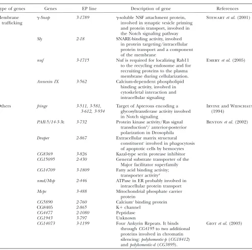
Membrane trafficking
g-Snap 3-1789 g-soluble NSF attachment protein, involved in synaptic vesicle priming and protein transport, involved in the Notch signaling pathway
Stewartet al.(2001)
Sly 2-18 SNARE-binding activity, involved in protein targeting/intracellular protein transport and a component of the membrane
nuf 3-1715 Nuf is required for localizing Rab11 to the recycling endosome and for recruiting proteins to the plasma membrane during cellularization.
Emeryet al.(2005)
Annexin IX 3-562 Calcium-dependent phospholipid binding activity, involved in cytoskeletal interaction and intracellular signaling
Others fringe 3-511,3-581, 3-612,3-934
Target of Apterous encoding a glycosyltransferase activity involved in Notch signaling
Irvineand Wieschaus
(1994)
PAR-5/14-3-3e 3-732 Protein kinase activity/Ras signal transductionb/ anterior-posterior
polarization in Drosophila
Bentonet al.(2002)
Draper 2-867 Extracellular matrix structural
constituentcinvolved in phagocytosis
of apoptotic cells by hemocytes CG8369 3-826 Kazal-type serin protease inhibitor CG15095 2-430 General substrate transporter of the
Major facilitator superfamily CG14709 3-1809 Fatty acid binding activity;
transporter activityd
nmd/Msp 2-446 ATPase in ER probably involved in intracellular protein transport
Mcpc 3-488 Mitochondrial phosphate carrier
protein
CG5890 2-760 Calciumebinding protein
CG8405 2-865 K1channel
CG4477 2-1080 Peptidase
CG1943 3-797 Unknown
CG14073 3-1199 Four Ankyrin Repeats. It binds throughCG4195to two additional proteins involved in chromatin silencing:polyhomeotic-p(CG18412) andpolyhomeotic-d(CG3895).
Giotet al.(2003)
ahttp://flybase.bio.indiana.edu/cgi-bin/goreport?GO:0016944 bhttp://flybase.org/.bin/goreport?GO:0007265
c
http://flserver.gen.cam.ac.uk:7081/.bin/goreport?GO:0005201
d
http://fbserver.gen.cam.ac.uk:7081/.bin/goreport?GO:0005215
e
suppressed theBx1wing phenotype (Figure 8, A, B, and
D). Expression ofmiR-14increased Notch activity levels
at the boundary of Bx1 wing discs, as shown by the
expression of Wg (Figure 8, F and G). Overexpression of
miR-14 has been reported to suppress cell death
in-duced by multiple stimuli (Xuet al.2003). In this
con-text, it is interesting to note that loss of Notch activity
causes cell death in the wing disc (Yeand Fortini1999)
and it has been postulated that this cell death causes
defects in the adult wing margin (Adachi-Yamadaet al.
1999). We then analyzed the capacity of suppressed cell death to bypass the requirement for Notch in cell survival and, consequently, to rescue the wing-margin
defects ofBx1wings. For this purpose, we used the P35
baculovirus protein, which strongly inhibits caspase
enzymatic activity in Drosophila tissues (Hay et al.
1995) and the Drosophila inhibitor of caspasesDIAP1
(Wanget al.1999). Expression ofp35orDIAP1in the D
compartment ofBx1
flies did not rescue the wing-margin defects (Figure 8, C and E). Wg expression levels in boundary cells also were not increased (data not shown). Taken together, these results indicate that the capacity of
miR-14to rescue theBx1
wing-margin phenotype is not a consequence of suppressed cell death, and the loss of wing-margin structures in this mutant background is not a direct result of cell death. It appears then as if the wing-margin defects in the absence of Notch signaling might be a consequence of impaired patterning of the wing margin as well as a failure of growth and not a direct consequence of cell death.
In the last few years, several groups have carried out
computational identifications ofmicro-RNAtarget genes
by looking for target sites located in the 39-UTRs of the
mRNAs. Interestingly, Bx/dLMO was identified as a
potential miR-14 target gene (Stark et al. 2003). We
then monitored the capacity of miR-14 to reduce the
levels of dLMO protein in the wing disc as well as to
phenocopy a dLMO (held-up wing) loss-of-function
phenotype. The phentoype of apgal4
;UAS-miR-14 flies
resembled the held-up wing phenotype of dLMO
mu-tant flies (Figure 8, H and I), and the expression levels of dLMO protein were reduced by overexpression of
miR-14in the wing disc (Figure 8, J–L). We then
moni-tored the capacity ofmiR-14to regulate the expression
level of adLMO39-UTR sensor transgene consisting of
the dLMO 39-UTR cloned into the
tubulin-promoter-EGFP reporter plasmid. ThedLMO39-UTR sensor
trans-gene was expressed uniformly in the wing imaginal disc
(Figure 8N). Gal4-dependent expression ofmiR-14
re-duced expression of thedLMO39-UTR sensor transgene
(Figure 8M). AlthoughmiR-14is expressed during larval
stages (Grunet al.2005), loss ofmiR-14did not show any Figure6.—lilliputianas a suppressor of the theBeadex1
phe-notype. (A and B) Cuticle preparations ofBx1/Y; apgal4/EP-27
(A) andBx1/1; lilli632/1(B) adult wings. (C–F) Clones of cells
mutant forlilliputian(lilli4u5) and labeled by the absence of
the GFP marker (green). Wingless (Wg) protein expression is shown in red (top) or white (bottom). Note reduced levels of Wg protein expression in clones abutting the DV boundary (white arrows) when compared to the endogenous level (red arrowhead) of Wg expression.
Figure 5.—master of thickveins
as a suppressor of the theBeadex1
phenotype. (A–C) Cuticle prepa-rations of Bx1/1; mtv6/1 (A),
Bx1/Y; apgal4/EP-473 (B), and
Bx1/Y; apgal4/uas-mtv (C) adult
wings. (D) Bx1/Y; apgal4/uas-mtv
wing imaginal discs labeled to vi-sualize Gal4 (green) and Wingless (Wg, red in the top and white in the bottom) protein expression. (E) Clones of cells mutant for master of thickveins (mtv6) and
la-beled by the absence of the GFP marker (green). Cut protein ex-pression is shown in red (top) or white (bottom). Note loss of Cut expression in clones abutting the DV boundary. (F and G) Clones of cells mutant formaster of thickveins(mtv6), labeled by theforked(f36a) cuticle marker and
overt wing phenotype nor did it enhance the wing-margin defects of Bx1
/1 wings. Thus, the direct regulation of
dLMO protein levels by miR-14 might be required in
other developmental contexts in which dLMO activity is involved (Tsaiet al.2004).
Membrane fusion and vesicle trafficking: In many
signal transduction pathways, vesicle trafficking of ligands or receptors is a key regulatory event (reviewed
in Gonzalez-Gaitan 2003). SNARE proteins play a
central role in intracellular membrane fusion and
ves-icle trafficking (reviewed in Jahnand Scheller2006).
The interaction of SNAREs present on two opposing membranes is generally believed to provide the driving force to initiate membrane fusion. We identified two genes involved in SNARE-dependent membrane fusion
in our screen:g-solubleN-ethylmaleimide-sensitive
fac-tor (NSF) attachment protein (SNAP) (3-1789) and Slh
(2-18, Figure 4A), the Drosophila ortholog of the Sec1p/ Munc18 protein. SNAPs are highly conserved proteins that participate in intracellular membrane fusion and vesicular trafficking. They recruit NSF to the membrane after being bound to specific membrane receptors termed SNAREs (SNAP receptor). The complex, which is then disrupted upon ATP hydrolysis by NSF, is a pre-requisite of membrane fusion. Sec1/Munc18 proteins are required for the controlled assembly of SNARE com-plexes and are essential for membrane fusion at the plasma membrane. The nature of the suppression of the
Bx1 phenotype by g-SNAP or Slh overexpression may
rely on elevated levels of transmembrane or secreted proteins involved in Notch signaling and/or DV bound-ary formation in the plasmatic membrane.
Rab11 is involved in controlling vesicular protein transport through recycling endosomes to the plasma
membrane (Pfeffer and Aivazian 2004).
Dominant-negative forms of Rab11 inhibit the recycling of endocy-tosed transmembrane proteins to the plasma membrane, thereby suggesting that Rab11 regulates trafficking of vesicular cargo through the recycling endosomal compar-tment. Nuf (nuclear fallout) is a homolog of arfophilin-2, an ADP ribosylation factor effector that binds Rab11 and influences recycling endosome (RE) organization
(Hickson et al. 2003). Nuf and Rab11 are mutually
required for their localization to the RE. Delta has been shown to pass through the recycling endosome which is marked by Rab 11 and Nuf, an essential step for its activity as a Notch ligand (Emeryet al.2005). We identifiednufas
a gain-of-function suppressor of theBx1phenotype (line
3-1715) and loss-of-function alleles ofnufenhanced the
wing-margin defects ofBx1/1heterozygous flies (Table 1).
Figure7.—CG11399, the Drosophila phosphorylated
C-ter-minal domain interacting factor, suppresses theBeadex1
phe-notype. (A) Genomic map of theCG11399region. Exons are shown as boxes, and the ORF is marked in black.CG11399was identified as a suppressor of theBeadex1phenotype by the
EP-3-28 insertion (black arrowhead). Two other EP lines (GS11380 and EY11352, black arrowheads) are shown. (B– D)In situhybridization toapgal4/1; EP-28/1(B) andwild-type
(C and D) wing imaginal discs with an anti-sense (B and C) and sense (D)CG11399RNA probe. (E–L) Cuticle prepara-tions of Bx1/Y; apgal4/EP-28 (E), Bx1/1; EY11352/1 (F),
EY11352 (G), EY11352/Df(3L)ri-79c(H), NND1/Y (I),NND1/Y;
EY11352/1 ( J), dxENU/Y(K), and dxENU/Y; EY11352/1 (L)
adult wings. Note ectopic vein tissue marked by a black arrow in G and H. (M–O) Cuticle preparations ofwild-type(M),ap
-gal4/1; EP-28/1(N), andapgal4/1; GS11380/1(O) adult nota.
Clones of cells mutant for nuf did not affect Notch activity levels at the DV boundary, as shown by the ex-pression levels of Wg (supplemental Figure S1 at http:// www.genetics.org/supplemental/). Thus, enhanced re-cycling of endosomal Delta, and probably other proteins involved in DV boundary formation, might increase, directly or indirectly, Notch signal and rescue the
wing-margin defects ofBx1
adult wings. This recycling might be required to modulate Notch activity levels at the DV boundary but does not appear to be a strict requirement for this process.
Finally, we identified Annexin IX (3-562, Figure 4M)
as a gain-of-function suppressor ofBeadex. Annexin IX is
a member of the annexin family of intracellular Ca21
-dependent lipid-binding proteins. Interestingly, some members of this family are found in apical transport vesicles in Madin–Darby canine kidney cells and may be
involved in apical delivery of trans-Golgi
network-de-rived vesicles (reviewed in Harder and Simons1997).
Taken together, the finding of genes as gain-of-function
suppressors of the Bx1 wing-margin phenotype and
encoding for proteins involved in membrane fusion, endosome recycling, and vesicle trafficking indicates that the relative levels at the membrane of transmem-brane proteins or secreted molecules are crucial and probably modulated during wing development.
Conclusions:Here we show that a gain-of-function
EP-based screen in aBx1-sensitized background to search for suppressors of the wing-margin phenotype is efficient in identifying known and new genes involved in DV boundary formation as well as in the regulation of
Beadex/dLMO gene activity. Dominant genetic interac-tions ofBx1
with loss-of-function alleles of the suppressor genes identified have demonstrated that the vast majority are involved in wing development. This is in contrast with classic EP screens based on the gain-of-function pheno-type of candidate genes, in which the number of genes not participating in the developmental context of in-terest is relatively higher. We have shown that many of the
Bx1suppressors involved in DV boundary formation are
not essential during wing development (i.e.,taranis,nmd,
nuf,draper, andcabut; supplemental Figure S1 at http:// www.genetics.org/supplemental/). This observation sug-gests that these suppressors share redundant activities with other gene products. The EP gain-of-function ap-proach has also been shown to be extremely efficient in unraveling new roles for the recently identified
micro-RNAs (miRs, e.g., Brennecke et al. 2003; Nairz et al.
2006). Loss-of-function-based forward genetic screenings have not been as productive in this respect, probably because of the reduced size of these miRs or their redundant activities. Taken together, a suppressor
EP-Figure8.—miR-14as a suppressor of theBeadex1phenotype: genomic map of themiR-14region (blue box).miR-14was identified
as a suppressor of theBeadex1phenotype by four EP insertions (blue arrowheads). (B–E) Cuticle preparations ofBx1/Y; apgal4/EP-235
(B),Bx1/Y; apgal4/uas-p35(C),Bx1/Y; apgal4/uas-miR-14(D), andBx1/Y; apgal4/EP-DIAP1(E) adult wings. (F and G)Bx1/Y; apgal4/1(F)
andBx1/Y; apgal4/ EP-235(G) wing discs labeled to visualize Gal4 (red) and Wingless (Wg, blue) protein expression. (H) Cuticle
preparation of aapgal4/uas-miR-14adult wing. (I) apgal4/uas-miR-14 adult fly. Note theheld-up wing phenotype. ( J–L)ptcgal4
/uas-miR-14( J and K) andwild-type(L) wing discs labeled to visualize GFP (red) and dLMO (green, top, or white, bottom) protein ex-pression. Note reduced levels of dLMO protein in the GFP domain (white arrow). (M and N) Expression of thedLMO39-UTR sensor transgene (green or white) inptcgal4/uas-miR-14(M) or wild-type wing discs (N). The sensor transgene was downregulated bymiR-14
based gain-of-function screen in a sensitized background provides a suitable combination to identify new genes, including miRs and redundant genes, involved in a given process.
Redundancy and regulatory feedback loops contrib-ute to the robustness of gene regulatory networks
(Stelling et al.2004). Classical loss-of-function-based
forward genetic screenings have been highly productive in identifying genes that behave as hubs in these
networks (Friedman and Perrimon 2007). Essential
genes in yeast are among those most highly connected
(Barabasi and Oltvai 2004). However, forward
ge-netic screenings are not as effective in identifying redundant genes or regulators of these feedback loops, whose loss of function might not show any overt
phenotype. More quantitative in vivo genetic
screen-ings, such as the one done recently in Drosophila for bristle number (Norgaet al.2003), or, alternatively, cell
culture-based RNAi quantitative screenings have been
more efficient in this regard (Friedmanand Perrimon
2007). Our results indicate that an EP-based
gain-of-function in vivo genetic screen in a sensitized
back-ground is a strong alternative for the identification of redundant genes or regulators of feedback loops in-volved in developmental gene regulatory networks.
Here we have identified, characterized, and discussed four classes of genes in the context of DV boundary formation or dLMO activity: chromatin organization genes, transcription factors, miRs, and proteins involved in vesicle trafficking and membrane fusion. Several conclusions can be drawn. Among the genes involved in
chromatin organization,Osabinds Chip and modulates
the expression of Ap target genes (Mila´ net al. 2004).
Several transcription factors involved in other signaling pathways during wing development have also been shown to act asBx1suppressors, suggesting that Notch and these pathways share common elements or that these pathways collaborate with Notch in boundary formation. The finding of genes encoding for proteins that participate in distinct aspects of vesicle trafficking and membrane fusion indicates that the sorting of sufficient levels of certain molecules, including Notch and its ligand Delta, toward the plasma membrane is especially critical to reach appropriate levels of Notch activity at the DV boundary. Consistent with this, it is interesting to note that overexpression of these genes in an otherwise wild-type background does not show any overt wing pheno-type, suggesting that the activity of the Notch pathway is finely regulated and buffered during boundary forma-tion (Rulifsonet al.1996; Herranzet al.2006).
The screen was designed and performed to find new genes involved in Ap and/or dLMO activity, as well as new Ap target genes involved in DV boundary forma-tion. Although genes known to participate in DV
boundary formation, like fringe or osa, were scored
several times, we did not identify new transmembrane proteins or cell adhesion molecules involved in the
generation of an affinity difference between D and V
cells. P elements are known for their preferential
insertion in certain regions of the genome called hot spots. The gene or genes involved in this process might be located in the so-called cold spots, thus suggesting that a distinct transposable element, like the
lepidop-teranpiggyBac, with a different profile of hot spots and
cold spots (Thibaultet al.2004), is a good candidate to
search, on a similar suppressor gain-of-function basis, for thesekind of genes.
We thank S. Cohen, in whose lab this project was started, and G. Broenner, S. Cohen, J.C. Eissenberg, E. Hafen, B. Hay, N. Paricio, the Bloomington Stock Center, and the Developmental Hybridoma Bank for reagents. We thank A. Olza for technical help, and J. Brennecke for providing us with unpublished reagents. We thank two anonymous reviewers for their comments, which helped to improve the final version of the manuscript. Work in M.M.’s laboratory is funded by a grant from the Direccio´n General de Investigacio´n Cientı´fica y Te´cnica (BFU2004-00167/BMC), a European Union research contract LSHM-CP-2003-503330 (APOPIS), a grant from the Generalitat de Catalunya (2005 SGR 00118), and Institute for Research in Biomedicine intra-mural funds.
LITERATURE CITED
Adachi-Yamada, T., K. Fujimura-Kamada, Y. Nishida and K. Matsumoto, 1999 Distortion of proximodistal information causes JNK-dependent apoptosis in Drosophila wing. Nature
400:166–169.
Aurora, K., H. Dai, S. G. Kazuko, J. Jamal, M. B. O’Connoret al., 1995 The Drosophilaschnurrigene acts in the Dpp/TGFb sig-nalling pathway and encodes a transcription factor homologous to the human MBP family. Cell81:781–790.
Bang, A. G., A. M. Baileyand J. W. Posakony, 1995 Hairless pro-motes stable commitment to the sensory organ precursor cell fate by negatively regulating the activity of theNotchsignaling pathway. Dev. Biol.172:479–494.
Barabasi, A. L., and Z. N. Oltvai, 2004 Network biology: understand-ing the cell’s functional organization. Nat. Rev. Genet.5:101–113. Bejarano, F., L. Perez, Y. Apidianakis, C. Delidakisand M. Mila´ n, 2007 Hedgehog restricts its expression domain in the Drosoph-ila wing. EMBO Rep.8:778–783.
Benton, R., I. M. Palaciosand D. StJohnston, 2002 Drosophila 14–3-3/PAR-5 is an essential mediator of PAR-1 function in axis formation. Dev. Cell3:659–671.
Bray, S. J., 1997 Expression and function of Enhancer of split bHLH proteins during Drosophila neurogenesis. Perspect. Dev. Neurobiol.4:313–323.
Brennecke, J., D. R. Hipfner, A. Stark, R. B. Russelland S. M. Cohen, 2003 bantam encodes a developmentally regulated mi-croRNA that controls cell proliferation and regulates the proa-poptotic gene hid in Drosophila. Cell113:25–36.
Bruckner, K., L. Perez, H. Clausen and S. Cohen, 2000 Gly-cosyltransferase activity of Fringe modulates Notch-Delta interac-tions. Nature406:411–415.
Calgaro, S., M. Boube, D. L. Cribbsand H. M. Bourbon, 2002 The Drosophila gene taranis encodes a novel trithorax group mem-ber potentially linked to the cell cycle regulatory apparatus. Genetics160:547–560.
Collins, R. T., T. Furukawa, N. Tanese and J. E. Treisman, 1999 Osa associates with the Brahma chromatin remodeling complex and promotes the activation of some target genes. EMBO J18:7029–7040.
deCelis, J. F., and A. Garcia-Bellido, 1994 Modifications of Notch function byAbruptexmutations inDrosophila melanogaster.Genetics
136:183–194.
mediate only a subset of Notch activities during imaginal devel-opment. Development122:2719–2728.
deCelis, J. F., A. Garcia-Bellidoand S. J. Bray, 1996b Activation and function ofNotchat the dorsal-ventral boundary of the wing imaginal disc. Development122:359–369.
deCelis, J. F., S. Brayand A. Garcia-Bellido, 1997 Notch signalling regulates veinlet expression and establishes boundaries between veins and interveins in the Drosophila wing. Development124:
1919–1928.
Diaz-Benjumea, F. J., and S. M. Cohen, 1993 Interaction between dorsal and ventral cells in the imaginal disc directs wing develop-ment in Drosophila. Cell75:741–752.
Diaz-Benjumea, F. J., and S. M. Cohen, 1995 Serrate signals through Notch to establish a Wingless-dependent organizer at the dorsal/ventral compartment boundary of theDrosophilawing. Development121:4215–4225.
Doherty, D., G. Fenger, S. Younger-Shepherd, L.-Y. Janand Y.-N. Jan, 1996 Dorsal and ventral cells respond differently to the Notch ligandsDeltaandSerrateduring Drosophilawing develop-ment. Genes Dev.10:421–434.
Eissenberg, J. C., J. Ma, M. A. Gerber, A. Christensen, J. A. Kennisonet al., 2002 dELL is an essential RNA polymerase II elongation factor with a general role in development. Proc. Natl. Acad. Sci. USA99:9894–9899.
Emery, G., A. Hutterer, D. Berdnik, B. Mayer, F. Wirtz-Peitzet al., 2005 Asymmetric Rab 11 endosomes regulate delta recycling and specify cell fate in the Drosophila nervous system. Cell
122:763–773.
Fan, H., K. Sakuraba, A. Komuro, S. Kato, F. Harada et al., 2003 PCIF1, a novel human WW domain-containing protein, interacts with the phosphorylated RNA polymerase II. Biochem. Biophys. Res. Commun.301:378–385.
Fernandez-Funez, P., C. H. Lu, D. E. Rincon-Limas, A. Ga´ rcia -Bellidoand J. Botas, 1998 The relative expression amounts of apterous and its co-factor dLdb/Chip are critical for dorso-ventral compartmentalization in the Drosophila wing. EMBO J.
17:6846–6853.
Friedman, A., and N. Perrimon, 2007 Genetic screening for signal transduction in the era of network biology. Cell128:225–231. Funakoshi, Y., M. Minamiand T. Tabata, 2001 mtv shapes the
ac-tivity gradient of the Dpp morphogen through regulation of thickveins. Development128:67–74.
Garcı´a-Bellido, A., and P. Santamaria, 1972 Developmental anal-ysis of the wing disc in the mutant engrailed of Drosophila mel-anogaster. Genetics72:87–104.
Garcı´a-Bellido, A., P. Ripoll and G. Morata, 1973 Develop-mental compartDevelop-mentalisation of the wing disk ofDrosophila.Nat. New Biol.245:251–253.
Giot, L., J. S. Bader, C. Brouwer, A. Chaudhuri, B. Kuanget al., 2003 A protein interaction map of Drosophila melanogaster. Science302:1727–1736.
Giraldez, A. J., and S. M. Cohen, 2003 Wingless and Notch signal-ing provide cell survival cues and control cell proliferation dur-ing wdur-ing development. Development130:6533–6543.
Gonzalez-Gaitan, M., 2003 Signal dispersal and transduction through the endocytic pathway. Nat. Rev. Mol. Cell Biol.4:213–224. Greider, N. C., D. Nellen, R. Burke, K. Baslerand M. Affolter, 1995 schnurriis required for Drosophila Dpp signalling and encodes a zinc finger protein similar to the mammalian tran-scription factor PRDII-BF1. Cell81:791–800.
Grienenberger, A., B. Miotto, T. Sagnier, G. Cavalli, V. Schramkeet al., 2002 The MYST domain acetyltransferase Cha-meau functions in epigenetic mechanisms of transcriptional re-pression. Curr. Biol.12:762–766.
Grun, D., Y. L. Wang, D. Langenberger, K. C. Gunsalusand N. Rajewsky, 2005 microRNA target predictions across seven Drosophila species and comparison to mammalian targets. PLoS Comput. Biol.1:e13.
Harder, T., and K. Simons, 1997 Caveolae, DIGs, and the dynamics of sphingolipid-cholesterol microdomains. Curr. Opin. Cell Biol.
9:534–542.
Hay, B. A., D. A. Wassarmanand G. M. Rubin, 1995 Drosophila homologs of baculovirus inhibitor of apoptosis proteins function to block cell death. Cell83:1253–1262.
Heitzler, P., and P. Simpson, 1991 The choice of cell fate in the epidermis of Drosophila. Cell64:1083–1092.
Heitzler, P., L. Vanolst, I. Biryukovaand P. Ramain, 2003 Enhancer-promoter communication mediated by Chip during Pannier-driven proneural patterning is regulated by Osa. Genes Dev.17:591–596. Herranz, H., E. Stamataki, F. Feiguinand M. Mila´ n, 2006 Self-refinement of Notch activity through the transmembrane pro-tein Crumbs: modulation of gamma-secretase activity. EMBO Rep.7:297–302.
Hickson, G. R., J. Matheson, B. Riggs, V. H. Maier, A. B. Fielding et al., 2003 Arfophilins are dual Arf/Rab 11 binding proteins that regulate recycling endosome distribution and are related to Drosophila nuclear fallout. Mol. Biol. Cell14:2908–2920. Hsu, T., and R. A. Schulz, 2000 Sequence and functional
proper-ties of Ets genes in the model organism Drosophila. Oncogene
19:6409–6416.
Irvine, K., and E. Wieschaus, 1994 fringe, a boundary specific sig-nalling molecule, mediates interactions between dorsal and ven-tral cells during Drosophila wing development. Cell79:595–606. Irvine, K. D., and C. Rauskolb, 2001 Boundaries in development: formation and function. Annu. Rev. Cell Dev. Biol.17:189–214. Jahn, R., and R. H. Scheller, 2006 SNAREs engines for membrane
fusion. Nat. Rev. Mol. Cell Biol.7:631–643.
Janody, F., Z. Martirosyan, A. Benlali and J. E. Treisman, 2003 Two subunits of the Drosophila mediator complex act to-gether to control cell affinity. Development130:3691–3701. Kennison, J. A., and J. W. Tamkun, 1988 Dosage-dependent
modi-fiers of polycomb and antennapedia mutations in Drosophila. Proc. Natl. Acad. Sci. USA85:8136–8140.
Lawrence, P. A., and G. Morata, 1976 Compartments in the wing of Drosophila: a study of the engrailed gene. Dev. Biol.50:321–337. Matsuno, K., R. J. Diederich, M. J. Go, C. M. Blaumuellerand S.
Artavanis-Tsakonas, 1995 Deltex acts as a positive regulator of Notch signaling through interactions with the Notch ankyrin repeats. Development121:2633–2644.
Mila´ n, M., and S. M. Cohen, 1999 Regulation of LIM homeodomain activity in vivo: a tetramer of dLDB and apterous confers activity and capacity for regulation by dLMO. Mol. Cell4:267–273. Mila´ n, M., and S. M. Cohen, 2000 Temporal regulation of apterous
activity during development of the Drosophila wing. Develop-ment127:3069–3078.
Mila´ n, M., and S. M. Cohen, 2003 A re-evaluation of the contribu-tions of Apterous and Notch to the dorsoventral lineage restric-tion boundary in the Drosophila wing. Development130:553– 562.
Mila´ n, M., S. Campuzano and A. Garcia-Bellı´do, 1996 Cell-cycling and patterned cell proliferation in the wing primordium ofDrosophila.Proc. Natl. Acad. Sci. USA93:640–645.
Mila´ n, M., F. J. Diaz-Benjumeaand S. M. Cohen, 1998 Beadex enc-odes an LMO protein that regulates Apterous LIM-homeodo-main activity in Drosophila wing development: a model for LMO oncogene function. Genes Dev.12:2912–2920.
Mila´ n, M., U. Weihe, L. Perezand S. M. Cohen, 2001a The LRR proteins capricious and Tartan mediate cell interactions during DV boundary formation in the Drosophila wing. Cell106:785–794. Mila´ n, M., U. Weihe, S. Tiong, W. Bender and S. M. Cohen, 2001b msh specifies dorsal cell fate in the Drosophila wing. Development128:3263–3268.
Mila´ n, M., T. T. Phamand S. M. Cohen, 2004 Osa modulates the expression of Apterous target genes in the Drosophila wing. Mech. Dev.121:491–497.
Moloney, D. J., 2000 Fringe is a glycosyltransferase that modifies Notch. Nature406:357–358.
Mun˜ oz-Descalzo, S., J. Teroland N. Paricio, 2005 Cabut, a C2H2 zinc finger transcription factor, is required during Drosophila dorsal closure downstream of JNK signaling. Dev. Biol. 287:
168–179.
Munro, S., and M. Freeman, 2000 The notch signalling regulator fringe acts in the Golgi apparatus and requires the glycosyltrans-ferase signature motif DXD. Curr. Biol.10:813–820.
Nairz, K., C. Rottig, F. Rintelen, E. Zdobnov, M. Moseret al., 2006 Overgrowth caused by misexpression of a microRNA with dispensable wild-type function. Dev. Biol.291:314–324. Norga, K. K., M. C. Gurganus, C. L. Dilda, A. Yamamoto, R. F.