DOI: 10.1534/genetics.107.076091
Misregulation of the Kinesin-like Protein Subito Induces Meiotic Spindle
Formation in the Absence of Chromosomes and Centrosomes
Janet K. Jang, Taslima Rahman, Vanessa S. Kober, Jeffry Cesario and Kim S. McKim
1Waksman Institute and Department of Genetics, Rutgers, the State University of New Jersey, Piscataway, New Jersey 08854-8020 Manuscript received May 17, 2007
Accepted for publication July 15, 2007
ABSTRACT
Bipolar spindles assemble in the absence of centrosomes in the oocytes of many species. InDrosophila melanogasteroocytes, the chromosomes have been proposed to initiate spindle assembly by nucleating or capturing microtubules, although the mechanism is not understood. An important contributor to this process is Subito, which is a kinesin-6 protein that is required for bundling interpolar microtubules located within the central spindle at metaphase I. We have characterized the domains of Subito that regulate its activity and its specificity for antiparallel microtubules. This analysis has revealed that the C-terminal domain may interact independently with microtubules while the motor domain is required for maintaining the interaction with the antiparallel microtubules. Surprisingly, deletion of the N-terminal domain resulted in a Subito protein capable of promoting the assembly of bipolar spindles that do not include centrosomes or chromosomes. Bipolar acentrosomal spindle formation during meiosis in oocytes may be driven by the bundling of antiparallel microtubules. Furthermore, these experiments have revealed evidence of a nuclear- or chromosome-based signal that acts at a distance to activate Subito. Instead of the chromosomes directly capturing microtubules, signals released upon nuclear envelope breakdown may activate proteins like Subito, which in turn bundles together microtubules.
T
WO events are required to ensure that the pairsof homologous chromosomes properly segregate among two daughter cells at the first meiotic division. First, microtubules must assemble into a bipolar spin-dle, a process that includes the critical events of estab-lishing bipolarity and the attachment of microtubules to the kinetochores. Second, the homologs must be linked by chiasmata, the result of a crossover formed earlier during prophase. By linking together two ho-mologs, the chiasmata coordinate attachment of homol-ogous kinetochores to microtubules emanating from opposite poles. Since the chiasmata are holding the two homologs together, and the kinetochore microtubules are working to pull them apart, the arrangement re-mains stable until cohesion is released on the arms of each chromatid, allowing the movement of homolog pairs to the poles.
One mechanism to establish bipolar spindles, and the one commonly observed in mitotically dividing cells, is through centrosome-containing microtubule organiz-ing centers (MTOC). Microtubules groworganiz-ing from the MTOCs at the poles can either attach to a kinetochore (kinetochore microtubules) or interdigitate with micro-tubules from the opposite pole (interpolar micromicro-tubules). Another mechanism, responsible for forming the bipolar
spindle in the absence of centrosomes, is observed in the
oocytes of many species (Compton 2000; Karsenti
and Vernos2001; Wadsworthand Khodjakov2004).
In these situations, the chromosomes play an impor-tant role in the assembly of microtubules into the meiotic spindle. In Drosophila oocytes, for example, nuclear envelope breakdown (NEB) is followed by the accumulation of microtubules around the chromo-somes (Theurkaufand Hawley1992; Matthieset al.
1996). Subsequent bundling and tapering of these mi-crotubules by motor proteins results in a bipolar spindle. The kinesins are a large family of motor proteins that promote unidirectional movement of a cargo along microtubules and several Drosophila kinesin proteins have been shown to play important roles in spindle assembly (Goshimaand Vale2003). For example, the
kinesin-4 or chromokinesin proteins are able to interact with microtubules while attached to chromosomes as their cargo (Mazumdar and Misteli 2005). Another
three groups within the kinesin family can bundle and slide parallel or antiparallel microtubules. The first is the kinesin-14 family that includes minus end-directed motors such as NCD in Drosophila. NCD and the minus end-directed motor Dynein have been proposed to bundle and taper microtubules to establish mitotic (Walczaket al.1998; Goshimaet al.2005) and meiotic
(Matthieset al.1996; Endowand Komma1997; Skold et al.2005) spindle poles in the absence of centrosomes. The second is the kinesin-5 family, including Klp61F in
1Corresponding author: Waksman Institute, Rutgers University, 190
Frelinghuysen Road, Piscataway, NJ 08854. E-mail: mckim@rci.rutgers.edu
Drosophila, which are plus end-directed motors that function to maintain bipolar spindle assembly and elon-gation at anaphase. The activity of these proteins may antagonize the forces of the kinesin-14 family during spindle assembly (Kwonand Scholey2004; Taoet al.
2006). The third is the kinesin-6 family that includes Subito and Pavarotti in Drosophila. As shown for human MKLP1, kinesin-6 proteins are thought to be plus end-directed motors that slide antiparallel microtubules (Nislowet al.1992). Examination of these proteins in
human cells (Neefet al. 2003), Caenorhabditiselegans
(Raichet al.1998), and Drosophila (Adamset al.1998;
Cesarioet al.2006) has shown they are usually
associ-ated with interpolar microtubules in the middle region of the spindle and are important for cytokinesis. During anaphase, the interpolar microtubules overlap in anti-parallel arrays in the spindle midzone, an area that typi-cally accumulates proteins important for cytokinesis (D’avino et al. 2005). Unlike the kinesin-5 and -14
proteins, most studies of kinesin-6 proteins have not implicated them in prometaphase spindle assembly.
In Drosophila, however, the kinesin-6 protein Subito has been shown to have a role in spindle assembly.subito
encodes the Drosophila homolog of MKLP2 and has an important role in organizing the meiotic acentrosomal ( Janget al.2005) and mitotic spindles (Cesarioet al.
2006). The Drosophila meiotic spindle develops a minent bundle of interpolar microtubules during pro-metaphase, referred to as the metaphase I central spindle, which is a critical part of the acentrosomal spin-dle assembly pathway ( Janget al.2005). Insubito null
mutant oocytes, the central spindle is absent ( Janget al.
2005) and there are an abnormal number of spindle poles and high levels of meiotic nondisjunction (Giunta et al.2002). Thus, Subito, and by inference the central spindle, is required to organize the acentrosomal spindle during Drosophila female meiosis. Interestingly, the cen-tral spindle forms before the microtubules are organized into a bipolar spindle and may function to direct the kinetochore microtubules toward one of the two poles. During mitotic metaphase, Subito may also organize interpolar microtubules but the effect of its absence is much more dramatic in meiosis, possibly because Subito activity is more critical in the absence of centrosomes.
Subito first appears on prometaphase meiotic spin-dles, suggesting it functions as the microtubules are recruited to the spindle. Just how the microtubules are recruited to surround the chromosomes, however, is poorly understood. The chromosomes could directly interact with microtubules via chromokinesin mole-cules (Mazumdar and Misteli 2005). Alternatively,
the chromosomes could be the source of a signal, such as RanGTP (Clarke2005), which could activate
micro-tubule assembly factors such as motor proteins. In either case, regulating kinesin proteins like Subito could be particularly important when centrosomes are absent and motor proteins may provide most of the organizing
activity. We have characterized the role of the N-, motor, and C-terminal coiled coils domains of Subito and found that regulating Subito activity is a critical compo-nent of organizing the acentrosomal spindle. Deregu-lation of Subito leads to the assembly of microtubules into multiple spindles in the absence of chromosomes or centrosomes. Furthermore, Subito appears to be activated by NEB, suggesting there is a diffusible signal that promotes the bundling of microtubules in oocytes.
MATERIALS AND METHODS
Genetics ofsuballeles:The isolation and genetic analysis of mostsuballeles has been described previously (Giuntaet al.
2002). These alleles ofsubinclude female sterile mutantssub1, subHM26 (Schupbach and Wieschaus 1989), and sub131
(Giunta et al. 2002); one dominant allele, subDub (Moore
et al.1994); and a fertile hypomorph,sub1794(Giuntaet al.
2002). Two of these mutants,sub1andsub131, are protein null alleles ( Janget al.2005).
Generation and initial analysis of transgenic lines: Full-length and deletion derivatives of subito were amplified by PCR. The clones were verified by sequencing and then the fragments were fused to GFP from pEGFP (Clonetech) at the N terminus using aXhoI orSalI site engineered into the beginning of thesubitocoding region. The full construct was then subcloned into the pUASP vector (Rorth1998). In some
cases, the Gateway system was used to generate the pUASP clones (T. Murphy, personal communication).
To measure fertility and chromosome segregation during meiosis, females were crossed to y w/BsY males. The non-disjunction frequency was calculated as 2(BS$1B1#) /½B1$1 BS#12(BS$1B1#). Ovary protein levels were assayed by Western blot. Whole ovaries were dissected from yeasted females in PBS and then ground and boiled in SDS gel load-ing buffer. Protein from2 to 3 ovaries was loaded per lane. Primary antibodies were rat anti-SUB, rat-anti HA ‘‘high af-finity’’ (Roche, clone 3F10), and mouse anti-GFP (Chemicon, clone JL-8), all used at 1:5000; the secondary HRP-conjugated antibodies (Jackson Labs) were used at 1:5000. The secondary was detected using ECL reagents (Amersham, Piscataway, NJ).
Antibodies and immunofluorescent microscopy:Two meth-ods to isolate oocytes: ‘‘mass isolation’’ and ‘‘dissection’’ were used. In the mass isolation protocol, oocytes were collected by physical disruption. Stage 14 oocytes were collected from 50 to 200 3- to 7-day-old yeast fed females by physical disruption in a common household blender. The details have been described previously (Theurkaufand Hawley1992), but in short, the
oocytes were fixed in modified Robb’s media and cacodylate/ formaldehyde fixative for 8–10 min and then their outer membranes were removed by rolling the oocytes between the frosted part of a slide and a coverslip. In the dissection pro-tocol, oocytes were hand dissected from 3- to 7-day-old yeast fed females and then fixed using the buffer A protocol (Belmont
et al.1989). In this method, the stage 14 oocytes retain their outer membranes and chorion, blocking the entry of anti-bodies. The advantage of the dissection protocol, however, is that we were able to isolate a range of oocyte stages. In contrast, the mass isolation procedure resulted in only mature stage 14 oocytes. Thus, for the analysis ofP{GAL4TVP16-nos.UTR} MVD1/P{UASP:GFP-subDNT}mutant oocytes, the dissection pro-cedure was more effective than the mass isolation propro-cedure at isolating oocytes shortly after NEB.
solution in Robb’s buffer for 1 hr prior to fixation. Control oocytes were incubated for 1 hr in buffer (Robb’s) without colchicine.
When examining sub mutant oocytes, heterozygotes for protein null allelessub1/sub131were used. Oocytes were stained for DNA with Hoescht and for microtubules with anti-tubulin monoclonal antibody DM1A (at 1:50), in some cases directly conjugated to FITC (Sigma, St. Louis). Heterozygotes were used to eliminate potential genetic background effects. The rat anti-SUB antibody was used at 1:75 combined with either a Cy3 or Cy5 anti-rat secondary antibody absorbed against a range of mammalian serum proteins including mouse and rabbit ( Jackson Labs). Additional primary antibodies were the rat anti-HA (Roche, clone 3F10) (1:25), rabbit anti-TACC (1:75) (Gergelyet al.2000), rabbit anti-AurB (1:250), rabbit
anti-INCENP (1:250) (Adams et al. 2001), and mouse
anti-RCC1 (1:20) (Frasch1991) with Cy3 conjugated secondary
antibodies ( Jackson Labs). Images were collected on a Leica TCS SP confocal microscope with a 633, NA 1.3 lens. Images are shown as maximum projections of complete image stacks followed by merging of individual channels and cropping in Adobe Photoshop.
Western blotting: Total ovary protein was isolated by dissecting whole ovaries from 10 yeasted females in PBS and then grinding and boiling them in SDS gel loading buffer. Protein from approximately one ovary was loaded per lane. The rat anti-HA (Roche, clone 3F10) or mouse anti-GFP (Clonetech, clone JL-8) primary antibodies were used at 1:5000 and the secondary anti-rat-HRP antibody ( Jackson
Labs) was used at 1:5000. The secondary was detected using ECL reagents (Amersham).
RESULTS
We have proposed that acentrosomal spindle assem-bly depends on the bundling of interpolar micro-tubules. Experiments to test this hypothesis were undertaken with a functional analysis of the Subito protein. By analyzing mutations of Subito, the goal was to identify what determines the specificity for interpolar microtubules and what controls when and how Subito interacts with the meiotic spindle.
Subito localization in oocytes is dependent on microtubules: As oocytes enter stage 14, the nuclear envelope breaks down and microtubules immediately begin to assemble around the chromosomes (Matthies et al.1996). There is no congression of bivalents since the chromosomes begin prometaphase in a single condensed mass or karyosome. The meiotic spindle assembles around this structure. Subito colocalizes with meiotic spindle microtubules from the earliest stages of spindle assembly, although always immediately adjacent to the karyosome ( Janget al.2005) (Figure 1).
Figure 1.—Subito localization
Since Subito colocalizes with microtubules but only adjacent to the chromosomes, we tested their relative importance by treating stage 14 oocytes with colchicine to depolymerize the microtubules. All control oocytes (7/7) incubated in buffer without colchicine had nor-mal spindle structure, in which the brightest tubulin staining was in the central spindle region (Figure 1A). The bright tubulin staining was most likely due to the overlap of interpolar microtubules, which also coloc-alize with Subito staining. Colchicine treatment of stage 14 oocytes most frequently resulted in partial loss of the microtubules (Figure 1, B and C). While colchicine treatment will completely depolymerize spindle tubules in larval brains, the partial loss of the micro-tubules in colchicine-treated live oocytes could have been due to limited permeability since they still pos-sessed their vitelline membrane and chorion. Despite partial depolymerization, the central spindle went from the brightest part of the spindle to the dimmest in all the examined oocytes (12/12), suggesting that interpolar microtubules were more sensitive to colchicine than the kinetochore microtubules. In addition, and regardless of whether there was a bipolar spindle, none of the colchicine-treated oocytes had Subito staining visible in the usual pattern. In some colchicine-treated oocytes, faint Subito staining was localized along the length of the spindle but it was never concentrated in the center as in untreated oocytes. Thus, Subito localization de-pends on the microtubules and may localize to kineto-chore microtubules when interpolar microtubules are absent. What restricts Subito to interpolar microtubules and adjacent to the chromosomes is the subject of the remaining experiments.
Analysis of Subito function using epitope-tagged transgenes: To investigate what controls Subito locali-zation, we examined the function of mutated derivatives ofsub(Figure 2). Subito contains a central motor do-main flanked by two poorly conserved dodo-mains. Trans-genes were made by cloning wild-typesubsequence or
variants containing a mutation in one of the domains into the pUASP vector, which contains multiple copies of the UAS sequence in the promoter. This permitted expression in Drosophila that was controlled with a second transgene containing GAL4 fused to a Drosoph-ila promoter (Rorth1998). In all the experiments
dis-cussed below, theUASP:subtransgenes were expressed using the P{GAL4TVP16-nos.UTR}MVD1 driver, which hasGAL4fused to thenanospromoter and induces the expression ofUAScontaining transgenes in the female germline (Rorth1998). For each transgene, at least two
and usually more insertion lines were examined in case expression levels from different insertion sites were dif-ferent. In most cases, with the exceptions noted below, differences in expression levels as assayed by Western blot were minimal and not the explanation for mutant phenotypes (supplemental Figure S1 at http://www. genetics.org/supplemental/).
The phenotype of wild-type and mutant sub trans-genes was characterized using genetic and cytological assays. Genetics assays were performed to examine two functions of Subito in meiotic chromosome segregation and early embryonic development. By expressing the transgenes in a genetic background with a null allele (sub1or
sub131, herein referred to as
subnull) and scoring
the fertility of the females, we were able to determine if a mutation affected the Subito embryonic function. Females homozygous forsubnull alleles are sterile due to a maternal requirement for Subito early in embryo-genesis (Schupbachand Wieschaus1989; Giuntaet al.
2002). By expressing the transgenes in a genetic back-ground with a sub hypomorphic allele (sub1794
/subnull
) and measuring the frequency of X chromosome
non-disjunction (see materials and methods), we were
able to determine if a mutation affected the Subito meiotic chromosome segregation function.Sub1794
is a hypomorph that is fertile but exhibits a high frequency of meiotic nondisjunction when homozygous or het-erozygous with a null allele (Table 1). Similarly, by
Figure 2.—Structure of
expressing a transgene in wild-type females and mea-suring fertility and the frequency of X chromosome nondisjunction, dominant effects on chromosome seg-regation could be detected. Transgenes expressing the full-length Subito protein (P{UASP:sub1
}) fused to GFP or an HA tag almost completely rescued the meiotic
(Table 1) and sterile (Tables 2 and 3) sub mutant
phenotypes.
Cytological assays were performed on the same wild-type and mutantsubtransgenes to examine their effects on spindle assembly and Subito localization. Using tu-bulin staining, we could determine if a mutant affected spindle organization. For example,submutant oocytes typically exhibit monopolar, tripolar, and frayed spin-dles (Giunta et al. 2002; Jang et al. 2005). Protein
expressed from the full-length Subito transgene (P{UASP:GFP-sub1
}) fused to GFP or an HA tag localized to the same region of the meiotic metaphase I spindle in stage 14 oocytes as the endogenous Subito protein (e.g.
Figure 3A) and rescued the spindle organization defects ofsubmutants (see below).
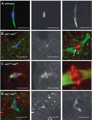
The C-terminal domain colocalizes with spindle mi-crotubules: The function of the C-terminal domain of Subito was investigated with construct P{UASP:GFP-subCT
}, which contained only the C-terminal domain of
sub fused to GFP (Figure 2). Immunofluorescence of
P{UASP:GFP-subCT}stage 14 oocytes revealed that the
C-terminal fragment of Subito colocalized with the micro-tubules (Figure 3). Therefore, the C-terminal domain appears to have microtubule-binding activity indepen-dent of the motor domain. However, the intensity of GFP-SubitoCT staining varied depending on the
pres-ence or abspres-ence of wild-type protein. In the prespres-ence of wild-type Subito, the staining that colocalized with tu-bulin was weak (Figure 3B). In addition, there was also a small dot of stronger staining that colocalized with the chromosomes. The nature of this structure is not known but it did appear to associate with microtubule fibers. In the absence of wild-type Subito (e.g.,subnullfemales),
GFP-SubitoCTstaining with the microtubules was more
intense (Figure 3C), suggesting the substrate that GFP-SubitoCT binds to may be more abundant in subnull
oocytes. SubitoCTand the wild-type protein may compete
for binding on the spindle or a particular type of bind-ing site may be more abundant insubmutants, such as free microtubule plus ends ( Janget al.2005).
Interest-ingly, GFP-SubitoCTwas often enriched on the
micro-tubules closest to the chromosomes, indicating that the C-terminal domain could specify a preference for micro-tubule plus ends. The C-terminal domain may also be important for protein stability since protein expression from a construct lacking this domain,P{UASP:GFP-subDCT
},
TABLE 1
Rescue of meiotic nondisjunction phenotype bysubtransgenes
Transgenea
sub genotype
Regular progeny
Nondisjunction progeny
Female parents
Progeny/female parent
Nondisjunction (%)
None sub1794/subnull 1888 391 146 15.6 29.3
P{UASP:GFP-sub1
}35 sub1794/subnull 2161 26 30 72.9 2.3
1/1 1074 16 20 54.5 3.0
P{UASP:GFP-subCT}43 1/1 243 13 38 6.7 9.7
subnull/1 209 13 14 15.8 11.1
sub1794/subnull 0 0 30 0 —
P{UASP:GFP-subDNT}31 1/1 0 0 40 0 —
subnull/1 60 1 40 1.5 3.2
sub1794/subnull 180 3 56 3.3 3.2
For each transgene, data for one example insertion are shown. At least one additional insertion for each transgene gave similar results.
a
Each transgene was expressed by crossing toP{GAL4TVP16-nos.UTR}MVD1.
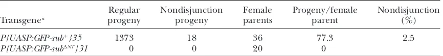
TABLE 2
Rescue of sterility incn sub1bw/sub131bwmutants bysubtransgenes
Transgenea
Regular progeny
Nondisjunction progeny
Female parents
Progeny/female parent
Nondisjunction (%)
P{UASP:GFP-sub1
}35 1373 18 36 77.3 2.5
P{UASP:GFP-subDNT}31 0 0 20 0
Control females of the genotype cn sub1bw/sub131bw; P{GAL4
TVP16-nos.UTR}MVD1/1or cn sub1bw/ sub131bw;1/P{UASP:GFP-sub1
}35 were sterile. For each transgene, data for one example insertion are shown. At least one additional insertion for each transgene gave similar results.
aEach transgene was expressed by crossing to
was not detected by either Western blot or immunofluo-rescence (supplemental Figure S1 at http://www.genetics. org/supplemental/).
Given the absence of the motor domain, it was not surprising thatUASP:GFP-subCT
did not rescue sub
mu-tant phenotypes (Table 1). More interestingly, expres-sion of the GFP-SubitoCTfragment had dominant effects
on chromosome segregation and fertility. In otherwise wild-type females, expression of GFP-SubitoCT caused
high levels of X chromosome nondisjunction (Table 1).
TABLE 3
Dominant effects ofsubmotor domain mutants
Transgenea
sub genotype
Regular progeny
Nondisjunction progeny
Nondisjunction (%)
P{UASP:sub1HA}31 1/1 1254 1 0.2
sub1/sub131 2518 6 0.5
sub1/1 1932 1 0.1
P{UASP:subATP}4 1/1 2483 173 12.2
sub1/1 608 166 32.8
P{UASP:subL6}10 1/1 4550 0
sub1/1 2189 100 8.4
P{UASP:GFP-subNT}28 sub1/1 1900 8 0.8
Control females of the genotype cn sub1bw/sub131bw; P{GAL4
TVP16-nos.UTR}MVD1/1or cn sub1bw/ sub131bw;1/P{UASP:sub1HA}31 were sterile.
a
Each transgene was expressed by crossing toP{GAL4TVP16-nos.UTR}MVD1.
Figure 3.—Localization of full-length
GFP-tagged Subito and C-terminal domain proteins in stage 14 oocytes. In all the experiments dis-cussed below, the transgenes were expressed us-ing the P{GAL4TVP16-nos.UTR}MVD1 driver. DNA is in blue, Subito or GFP-Subito in red, and tubulin in green. The second column shows the Subito signal from GFP (A–C) or antibody staining (D). Tubulin, along with Subito in some cases, is shown in the last column. (A) Full-length Subito fused to GFP (P{UASP:GFP-sub1
GFP-SubitoCTalso affected fertility, causing reduced
pro-geny numbers insub1794
/1females and sterility insub1 / sub174females. These results suggest that the C-terminal
fragment can interfere with the activity of the wild-type protein.
The dominant effects of SubitoCTon chromosome
seg-regation were mirrored by the effects of GFP-SubitoCTon
spindle morphology. When GFP-SubitoCTwas expressed
in a wild-type background, 8/10 oocytes had abnormal microtubule organization such as tripolar, monopolar, or frayed spindles or spindles lacking interpolar micro-tubules, effects similar to, although milder than, those found in the null mutants. To determine if the GFP-SubitoCT dominant phenotypes resulted from effects
on the wild-type protein, the oocytes expressing GFP-SubitoCTwere stained for the wild-type protein.
Locali-zation of wild-type Subito to the central spindle was reduced or not detected in 5 of the 10 spindles (Figure 3D). In contrast, all 10 control oocytes (expressing wild-type GFP-Subito) localization of the endogenous Subito protein was not affected. Thus, GFP-SubitoCT may
in-terfere with the localization and function of wild-type Subito, resulting in loss-of-function phenotypes.
In summary, the C-terminal domain of Subito local-izes with spindle microtubules. It may also be able to compete with the wild-type protein for binding sites, resulting in the formation of abnormal spindles and elevated rates of chromosome segregation errors.
The Subito motor domain is required for localiza-tion to the central spindle: To investigate the
impor-tance of the motor activity for Subito function, we characterized four point mutations and one internal deletion in the motor domain ofsubito (sub1794,
subhm26, SubDub,
subATP, and
subL6, Figure 2). In
sub1794and subhm26
mutants, the Subito localization pattern was similar to wild type, although Subito protein levels were reduced insubhm26mutants. This was confirmed on a Western blot
(data not shown), suggesting the Subitohm26 protein
could localize but was unstable. In subDub
, an invariant amino acid of the motor do-main is changed (Figure 2) (Giuntaet al.2002),
caus-ing a dominant meiotic nondisjunction phenotype in both males and females (Moore et al. 1994) and
de-fective spindle assembly (Giunta et al. 2002). SubDub
homozygotes are lethal but subDub
/subnull
females are viable, allowing for the analysis of meiosis in females where the only source of Subito protein was from the
Dub allele. Using the polyclonal antibody, the majority of subDub/subnull stage 14 oocytes had abnormal Subito
staining. In 10/18 oocytes, the SubitoDub protein was
either weakly distributed along the spindle or concen-trated toward the poles. In the remaining 8/18 oocytes, no Subito staining was detected (Figure 4B) even though the protein was readily detected on a Western blot (data not shown). Most (13/18) of these oocytes had abnor-mal spindle morphology. These results indicate that the
subDub
mutation affected the localization of the protein to interpolar microtubules on the spindle.
Since the biochemical defect of subDub
has not been characterized, we generated a mutant in the ATP-binding
Figure4.—Localization of Subito in stage 14
oocytes of motor domain mutants. In all the ex-periments discussed below, the transgenes were expressed using the P{GAL4TVP16-nos.UTR} MVD1 driver. DNA is in blue, Subito in red, and tubulin in green. The Subito channel is shown below each color image. (A) Subito de-tected using an anti-Subito antibody in a wild-type oocyte. (B) Subito detected using an anti-Subito antibody in a subDub/sub1oocyte. (C) Subito de-tected using an antibody to the HA tag in an oo-cyte expressing wild-type sub fused to an HA epitope tag (P{UASP:sub1HA}) or (D) a similar HA-fusion protein with a mutation in the ATP binding domain (P{UASP:subATP}) in a wild-type background or (E)subATPin asubnullmutant back-ground. (F) Subito detected using an anti-HA an-tibody in an oocyte expressing a HA-fusion protein lacking 60 amino acids present in the mo-tor domain of all kinesin-6 family members (P{UASP:subL6}) in a wild-type background. (G) Subito detected using an anti-Subito antibody in sub1
; P{UASP: sub1HA}or (H)sub1
domain (Figure 2) with the objective of eliminating motor activity but not microtubule binding (e.g., Zhu
and Jiang2005). This mutant,subATP, did not rescue the
sterility ofsubnull mutants and had similar dominant phenotypes assubDubmutants. For example, expression
of the mutant protein in a wild-type background re-sulted in chromosome segregation errors, suggesting that the mutant had a dominant negative effect (Table 3). Consistent with these genetic results, analysis of the meiotic spindles in subATP
females revealed abnormal albeit mild defects in microtubule organization. A total of 16/38 spindles were judged to be abnormal because they were frayed, lacked interpolar microtubules, were unusually curved, or contained asymmetries such as large knob structures. In contrast, 7/7 control oocytes (expressing the wild-type P{UASP-sub1HA
} transgene) had normal bipolar spindles. Similar tosubDub,
microtu-bule-associated staining of the mutant SubitoATPprotein
was reduced; only faint staining coincided with the microtubules of the meiotic spindle (Figure 4, D and E). A Western blot indicated that the SubitoATPprotein was
expressed at normal levels (supplemental Figure S1 at http://www.genetics.org/supplemental/), suggesting that ATP hydrolysis is essential for protein localization. The effect on interpolar spindles in these oocytes suggests that the SubitoATPprotein may have disrupted
the function of the wild-type protein. To test this, we stainedsubATP
; sub1
oocytes with our polyclonal antibody for the endogenous Subito protein and found it was reduced by expression of SubitoATP(Figure 4H). Among
the 38 oocytes characterized above for spindle mor-phology, 22 lacked endogenous Subito staining and in many others Subito staining was weak. These results indicated that SubitoATP negatively impacted on the
localization of wild-type protein, which was probably a contributing factor in the dominant effects on chromo-some segregation.
Motor function was also investigated with the analysis of a mutation (subL6
), which removed 60 amino acids present in the motor domain of all kinesin-6 family members but no other kinesin-like proteins (Figure 2). Even though none of the motor domain sequences conserved in other kinesins were deleted in thesubL6
mutation, there was little evidence of microtubule binding. Spindle staining by immunofluorescence was absent (Figure 4F) even though the protein was easily detected by Western blotting (supplemental Figure S1 at http://www.genetics.org/supplemental/). Therefore, this portion of the motor domain appears to be essential for localization to the spindle. The mutant transgenes did not rescue the sterility ofsubnull alleles and when expressed in a wild-type background did not cause meiotic nondisjunction, suggesting the mutant protein was nonfunctional. However, expressingsubL6
in a back-ground where wild-typesubitodosage was reduced (e.g.,
subnull
/1) resulted in increased meiotic nondisjunction (Table 3), suggesting that Subito was present in limiting
amounts in the oocyte. Similarly, thesubATPmutant
phe-notype was more severe whensubdosage was reduced. In summary, three mutants affecting different parts of the motor domain had qualitatively similar effects. They caused dominant negative phenotypes, including chro-mosome segregation errors, spindle assembly defects, and reduced localization of the wild-type protein. The mutant proteins also failed to localize correctly on the metaphase spindle, but the dominant negative pheno-types suggested that the mutant motor domain mutant proteins could interact with the spindle in ways that interrupt localization of the wild-type protein.
The N-terminal domain regulates Subito localization:
Two constructs were made to investigate the function of the N-terminal domain. The first, P{UASP:GFP-subNT
}, contained the N-terminal domain without the motor. When crossed toP{GAL4TVP16-nos.UTR}MVD1, protein was not detected in stage 14 oocytes by immunofluores-cence. Thus, the N-terminal domain lacks sequences that direct localization. In addition, expression of this frag-ment did not have effects on meiotic chromosome segregation or fertility (Table 3). Since the protein levels were reduced in this mutant (supplemental Figure S1 at http://www.genetics.org/supplemental/), however, it is possible that GFP-subNT
was not expressed at a high enough level to act as a dominant negative.
The second construct,P{UASP:GFP-subDNT
}, was a de-letion of the N-terminal domain. Immunofluorescence analysis in oocytes demonstrated that this fragment of Subito had a dramatic effect on spindle formation. Stage 14 oocytes expressing P{UASP:GFP-subDNT
} con-tained a large number of bundled microtubules that appeared like bipolar spindles (Figure 5A). On the basis of Western blot analysis, the presence of ectopic spin-dles cannot be attributed to higher expression levels. This phenotype was identical in the presence or absence of wild-type Subito protein and was observed in all oocytes examined but never in control oocytes. Further-more, most of these ‘‘ectopic’’ spindles were not asso-ciated with chromosomes (Figure 5B). Indeed, the karyosome had usually prematurely split into at least two masses of chromosomes that were scattered in the oocyte cytoplasm. Thus, microtubules assembled into bi-polar spindles without chromosomes and centrosomes. These results suggest that the N-terminal domain reg-ulates Subito activity and as a consequence spindle for-mation. The N-terminal domain is required to ensure that microtubules are only assembled around the chro-mosomes. The SubitoDNTprotein may promote the
un-regulated bundling of microtubules. In support of this conclusion, we observed the GFP-SubitoDNTprotein in
Aurora B were also found at the center of the ectopic spindles, suggesting the complex of proteins that forms in the meiotic central spindle of wild-type oocytes was also forming on these ectopic spindles (Figure 5C).
Another feature of meiotic spindles is the localization of TACC and MSPS to the poles even though centro-somes are absent (Cullenand Ohkura2001). To
ex-amine the structure of the poles in the ectopic spindles assembled as a result of GFP-SubitoDNTactivity, we stained P{GAL4TVP16-nos.UTR}MVD1; P{UASP:GFP-subDNT} oo-cytes with an antibody to TACC, but no staining was detectable at the poles (Figure 5, D and E). The absence of TACC (and presumably other proteins like MSPS) at the poles may be the reason the spindles assembled as a result of GFP-SubitoDNTactivity were often splayed at the
poles. From a sample of 32 spindles (i.e., 64 poles), 70% were splayed while 30% were finely tapered similar to wild type. The two poles of a spindle were often differ-ent, with 34% having one splayed and one tapered pole.
To examine the time course of ectopic spindle for-mation in P{UASP:GFP-subDNT
}oocytes, we isolated oo-cytes from all stages of development (see materials and methods). A key event is NEB, which occurs in
mature oocytes at the beginning of stage 14. In stage 13 or earlier stage oocytes, which is prior to NEB, ectopic spindles were not observed (Figure 5F). Although the mutant GFP-SubitoDNTprotein and microtubules were
abundant in the oocyte cytoplasm prior to stage 14, they apparently did not interact. After NEB, spindle assembly in the earliest stage 14 oocytes was restricted to the vicinity around the chromosomes (Figure 5, G and H). In addition, these early stage 14 oocytes often had mul-tiple chromosome masses, suggesting that spindle as-sembly in the presence of GFP-SubitoDNT caused the
karyosome to split prematurely, which is not usually observed until anaphase I. This would explain the ob-servation that the chromosomes appeared scattered in the oocyte cytoplasm in older stage 14 oocytes. The
Figure 5.—Subito lacking the N-terminal
do-main promotes formation of ectopic spindles. In all the experiments discussed below, the transgenes were expressed using the P{GAL4TVP16-nos. UTR}MVD1driver. DNA is blue; Subito-GFP, TACC (D and E), or Aurora B (C) is red, and tubulin is green. (A) Stage 14 oocyte expressing Subito with a deletion of the N-terminal domain fused to GFP (P{UASP:GFP-subDNT}). The locations of chro-mosome masses are shown by the arrow and arrow-heads. While these images are taken from oocytes also expressing wild-type Subito protein, similar ef-fects were observed in oocytes lacking wild-type Su-bito protein. (B) The same oocyte as shown in A with the region around the arrow magnified. (C) GFP-subDNTstage 14 oocyte stained for Aurora B (red). (D) In wild-type metaphase TACC (red) is visible at the poles. (E) InGFP-subDNTectopic spin-dles TACC (red) is absent. (F) InGFP-subDNTstage 10 oocytes, there are no ectopic spindles. The oo-cyte karyosome is shown by an arrow. The cortical green staining is from microtubule growth that is nucleated from many organizing centers along the cortex of the Drosophila oocyte prior to NEB as part of the axis specification during oogenesis (Theurkaufet al.1992; Chaet al.2002; Steinhauer
and Kalderon2006). (G) EarlyGFP-subDNTstage 14
oocyte with only one spindle, as indicated by a sin-gle patch of Subito staining (arrow). In the method used to prepare this oocyte, tubulin staining was not possible (seematerials and methods). (H)
simplest interpretation of these data is that the activity of the GFP-SubitoDNT protein is dependent on NEB.
Shortly after NEB, GFP-SubitoDNT protein and
micro-tubules begin to assemble into spindles in the vicinity of the chromosomes. The ectopic spindles continue to form such that in older stage 14 oocytes, they become numerous and less restricted in position. They also were observed in embryos, which in wild type is after the time when anaphase I and meiosis II occur (supplemental Figure S2 at http://www.genetics.org/supplemental/).
As described in the discussion, the
chromosome-associated protein RCC1 could be one of the factors that promotes spindle assembly following NEB. In the pre-sence of RCC1, Ran is converted into its active form (GTP bound), which can then promote microtubule assembly. Drosophila RCC1 has been reported to be present in the oocyte nucleus (Frasch 1991) and we
have confirmed it is chromosome associated after NEB (Figure 5I).
GFP-subDNT did not rescue the sterile phenotype of subnullfemales (Table 2). Indeed,GFP-subDNThad a
dom-inant effect on fertility, the severity of which was de-pendent on the dosage of wild-typesubgene product. Females expressing GFP-SubitoDNT in a subnull
/1 or a 1/1 background had lower fertility than in asub1794
/ subnull
background. In contrast, genetic assays measured a surprisingly low frequency of meiotic nondisjunction when GFP-SubitoDNT was expressed in a sub1794
/subnull
mutant (Table 1). It is possible, however, that this experi-ment scored a selected set of oocytes since expression of
GFP-subDNThad a dominant effect on female fertility.
While the motor and C-terminal domains are involved in the specificity of Subito localization, the N-terminal domain of Subito appears to regulate its activity in bundling microtubules. Spindle assembly in the pres-ence of this mutant form of Subito is not limited to a single structure adjacent to the karyosome. The activity of this protein also appears to cause the karyosome to prematurely separate.
DISCUSSION
Subito is a kinesin motor protein that contributes to acentrosomal meiotic spindle formation (Giuntaet al.
2002), possibly by stabilizing the overlap of antiparallel microtubules located in the central spindle during meiotic metaphase ( Janget al.2005). The object of this
study was to investigate the characteristics of Subito that facilitate spindle formation (Figure 6). The results have stimulated a model for acentrosomal spindle assembly that emphasizes the bundling of antiparallel micro-tubules without direct contacts with the chromosomes.
Localization of Subito depends on the C-terminal do-main:When a fragment containing only the C-terminal domain of Subito was expressed, the protein localized to the spindle microtubules. It is also likely that that there is competition between the C-terminal domain and full-length Subito for binding sites. This is based on the observation that when the C-terminal domain was ex-pressed in a wild-type background, there was less wild-type protein localized to the central spindle at metaphase I and an increased incidence of chromosome segregation
Figure 6.—Model for
function of Subito in spin-dle assembly. (A) Schematic of Subito and summary of the functions attributed to each domain. (B) Prior to NEB, Subito does not inter-act with the microtubules. After NEB, Subito associates with microtubules early in prometaphase. Initially, the majority of microtubules may only interact laterally with the chromosomes (Skoldet al. 2005). There
may also be kinetochore mi-crotubules forming concur-rently ( Janget al.2005) but
the relative timing is not known. In a model for acen-trosomal spindle assembly that involves the capture and bundling of microtu-bules, stabilized plus and mi-nus ends would be expected throughout the spindle, not just near the chromosomes ( Janget al.2005). Indeed, there is experimental evidence for both microtubule plus and minus ends
localized throughout meiotic spindles (Elliottet al.2005; Burbanket al.2006). (C) A model for the structure of the spindles when
errors. Similarly, the C-terminal domain of MKLP2 has been shown to have microtubule binding activityin vitro
(Echardet al.1998) andin vivo(Gruneberget al.2004).
These results indicate that Subito, and other MKLP2 paralogs, have two microtubule-binding domains, one each in the C-terminal and motor domains. This feature of the MKLP2 proteins may enable them to form cross-bridges between antiparallel microtubules (Nislow et al.1992). Specificity may also reside in the protein– protein interactions involving Subito. Similar to MKLP2, the C-terminal domain of Subito may interact with the Passenger proteins and CDC14 (Gruneberget al.2004).
Indeed, the passenger proteins and Subito or MKLP2 may be obligate partners during meiosis ( Jang et al.
2005) and mitosis (Cesarioet al.2006; Neefet al.2006). Subito motor domain is required to maintain the interaction with microtubules: We characterized two mutations that affect conserved amino acids in the mo-tor domain and a third that affects a momo-tor domain sequence specific to kinesin-6 proteins. All three mu-tants exhibited dominant nondisjunction and weak spindle staining, suggesting they had similar defects in motor activity. ThesubDub
mutation changes an E to K at position 385. Although this mutation is outside of the microtubule-binding region (Valeand Fletterick
1997), it is the last residue in a group of seven amino acids that are invariant in all kinesin-like proteins. This mutant has a dominant nondisjunction phenotype (Mooreet al.1994) and the protein fails to accumulate
in the central spindle. The failure to localize to micro-tubules is surprising because similar mutants (E/A) in minus end-directed motors such as Kar3 or NCD bind strongly to microtubules but lack a microtubule-stimulated ATPase activity (Yunet al.2001). Thus, the
SubitoDubprotein is predicted to bind microtubules but
have an inactive motor. A similar array of phenotypes was observed when we generated a mutation that changes the three invariant amino acids GKT in the ATP-binding domain to AAA (subATP
). This change has been made in other kinesins (e.g., Zhuand Jiang2005) and
simi-lar changes of the GKT sequence have been made in Pavarotti (EKT) (Minestriniet al.2002) and the kinesin-5
homolog Eg5 (GKN, GKI) (Blangy et al.1998) or
ki-nesin heavy chain (GKN) (Nakata and Hirokawa
1995). Most of these mutants exhibited ‘‘rigor’’ binding phenotypes associated with excessive binding of micro-tubulesin vivo. In contrast, the SubitoATPprotein failed
to localize strongly to meiotic spindles.
Despite the weak localization of the motor domain mutants, several observations indicate these motor domain mutant proteins interact with the spindle mi-crotubules. First, these mutants have dominant effects on meiotic chromosome segregation and spindle orga-nization. Second, these mutants cause reductions in the localization of wild-type protein to the spindle. Indeed, interfering with the localization of wild-type Subito pro-tein could be the cause of the dominant nondisjunction
phenotype. Third, at least one of the motor defective proteins (SubitoATP) localizes strongly to metaphase
mi-crotubules in mitotic cells, although not to the central spindle like wild-type protein ( J. Cesarioand K. McKim,
unpublished data). These observations suggest that in the presence of motor domain defective proteins, wild-type Subito engages in interactions that lead to its removal from the spindle.
Motor defective Subito protein may be able to initially associate with the microtubules but then be rapidly dis-placed toward the poles. This would explain the observa-tion that motor domain mutant proteins fail to localize on the spindle despite containing an intact C-terminal domain that can independently interact with the spin-dle (compare Figure 3C and Figure 4D). Such a ‘‘polar wind’’ has been implicated in previous studies of Droso-phila oocytes (Carpenter1991; Cullenand Ohkura
2001). It is also possible that the motor-inactive Subito proteins could be dislodged from the spindle by another mechanism. Whatever the mechanism by which the mo-tor domain mutant proteins fail to remain on the spindle, these results suggest that the motor domain is required to retain Subito on the spindle in oocytes.
The N-terminal domain is one of at least two factors that regulate Subito activity:We have identified two fac-tors that regulate Subito, by characterizing stage 14 oo-cytes expressing a Subito mutant lacking the N-terminal domain (SubitoDNT). The first regulator of Subito is
shown by the observation that thesubDNTmutant formed
a large number of ectopic spindles, indicating there is a mechanism to limit where Subito interacts with micro-tubules. The second regulator of Subito is shown by the observation that the unregulated microtubule bundling activity insubDNTmutants was dependent on NEB. This is
a different result from overexpressing the kinesin-6 member Pavarotti, which was observed to have effects on oogenesis prior to NEB (Minestriniet al.2002).
Pos-sibly, NEB releases a diffusible factor into the cytoplasm that activates Subito microtubule binding and bundling. Aside from being numerous, the most striking aspect of the ectopic spindles ofsubDNT
mutants was that they were not built around chromosomes (Figure 6C). We suggest that, through a still-unknown mechanism, the N-terminal domain regulates Subito activity to ensure that microtubules are bundled only in the direct vicinity of the chromosomes. The N-terminal domain could regulate Subito activity in a spatial manner. For exam-ple, this domain could promote interactions with a membranous sheath that has been proposed to
sur-round the developing spindle (Kramer and Hawley
2003). Interestingly, the initial studies of the Subito homolog MKLP2 demonstrated an interaction with Rab6, a Golgi-associated Rab protein, although through the C-terminal domain of MKLP2 (Echardet al.1998).
Rather than regulate when or where the motor is ac-tive, the N-terminal domain could affect the biochem-ical activity of the motor. For example, unregulated plus end-directed motor activity could lead to lengthening of the spindle through the sliding of antiparallel micro-tubules, causing the karyosome to be pulled apart and leaving the chromosomes scattered in the oocyte cyto-plasm. The scattered chromosomes could go through repeated cycles of stimulating microtubule assembly followed by detachment from the spindle to generate the ectopic spindles observed insubDNT
oocytes. More studies, including understanding the details of karyo-some structure and the biochemical properties of Subito and the SubitoDNT mutant, are needed to
dis-tinguish these possibilities.
Summary—the role of Subito in microtubule recruit-ment and assembly: We have suggested that the anti-parallel overlaps of microtubules in the central spindle play an important role early in spindle assembly ( Jang et al.2005) (Figure 6). Our results are also consistent with previous studies suggesting that interpolar microtubules are more sensitive to destabilizing agents like temperature and colchicine than kinetochore microtubules (Brinkley
and Cartwright1975; Salmonand Begg1980). Subito
is critical for the central spindle since it is required for the interpolar microtubules. Like other members of the kinesin-6 family (Nislow et al. 1992), Subito probably
cross-links antiparallel microtubules. Subito has two microtubule binding domains, which may cooperate to facilitate interactions with antiparallel microtubules. In addition, motor activity may play a role in the localization of Subito. However, previous studies have also suggested that spindle assembly in Drosophila oocytes involves the recruitment of microtubules by the chromosomes (Theurkaufand Hawley1992) and subsequent
bun-dling of parallel microtubules by motor proteins such as NCD (Matthieset al.1996; Skoldet al.2005).
Spindles appear in subDNT
mutant oocytes, however, that do not have direct contacts to the chromosomes. Therefore, conditions exist in the Drosophila oocyte cytoplasm in which recruitment and assembly of micro-tubules into a spindle may occur without direct contacts with the chromosomes. The concept of a cytoplasmic state permissive to spindle assembly has been proposed in Xenopus oocytes to explain how the injection of DNA can stimulate spindle assembly but only in cytoplasm from M phase eggs. Normally, however, chromosomes are needed to stimulate the process, leading to the idea that there is an ‘‘organizational field’’ around the chromosomes (Karsentiand Vernos2001). This
two-component model of acentrosomal spindle assembly is consistent with our observations. Alternatively, we can-not rule out that one signal in high concentration near the chromosomes is responsible for generating both the permissive cytoplasmic state for spindle assembly and the organizational field around the chromosomes. How-ever, the results from thesubDNT
mutant, which interacts
with microtubules but is not restricted to the chromo-somes, suggest activation is separable from restriction around the chromosomes.
Spindle assembly in Drosophila oocytes begins im-mediately following NEB (Matthieset al. 1996),
sug-gesting NEB somehow triggers the process. We have evidence that Subito activity is also regulated by NEB. Subito, even the unregulated form, does not bundle microtubules until after NEB. Nonetheless, spindle sembly is constrained such that microtubules only as-semble around the chromosomes. On the basis of the phenotype of thesubDNT
mutant, Subito may also be reg-ulated by proximity to the chromosomes. Since spindle assembly may be initiated by overlapping microtubules rather than direct contacts with the chromosomes (see below and Karsentiand Vernos2001), tightly
regulat-ing a protein like Subito that can bundle microtubules could be particularly important. In contrast, however, Subito is not essential for spindle assembly. There are probably several proteins or redundant mechanisms for recruiting microtubules to the spindle.
A key part of this model is that the chromosomes may not be essential for the polymerization of microtubules but may regulate the number and size of the spindles. A similar situation may occur during acentrosomal spin-dle formation in mammalian meiosis. Mouse oocyte microtubules can polymerize and be organized into bipolar spindles without the presence of chromosomes (Brunetet al.1998). Furthermore, several bipolar
spin-dles of varying sizes tended to form, indicating that chromosomes may be needed to control spindle forma-tion and growth (Brunet et al. 1999). In mouse or
Drosophila oocytes, therefore, it may be necessary to regulate the interaction of microtubules with motor proteins to occur in the vicinity of chromosomes. There are also other effects of the chromosomes. It is possible that the presence of chromosomes may promote local-ization of spindle pole proteins like TACC.
Interestingly, microtubules do not attach to kineto-chores in mouse oocytes throughout most of prometa-phase I. Instead, kinetochores are not competent to anchor and stabilize microtubule ends until the end of prometaphase I,8 hr after NEB (Brunetet al.1999).
Thus, in both Drosophila and mammal oocytes, spindle assembly may be initiated by the interaction of non-kinetochore microtubules with motor proteins. Inter-polar microtubules, which depend on Subito, play a critical role in organizing these bundles into the bipolar spindle. Kinetochore microtubules have a secondary role in spindle formation, being assimilated into the bipolar structure by interacting with the interpolar microtubules.
chromosomes and has been proposed to promote microtubule assembly around chromosomes (reviewed in Kahana and Cleveland 1999; Trieselmann and
Wilde2002; Shiand Skeath2004; Clarke2005). Of
relevance to our studies is the observation that the ad-dition of RCC1 or an activated form of Ran (RanG19V)
stimulated microtubule assembly in the absence of chromatin (Kalab et al. 1999). Drosophila RCC1, a
Ran cofactor, is found in the oocyte nucleus before and after NEB (Frasch1991; this work) and Ran has also
been suggested to have a role in meiotic spindle as-sembly in mouse oocytes (Cao et al. 2005) although
there is evidence for RanGTP-independent pathways as well (Dumontet al.2007). We are currently
investigat-ing if Ran signalinvestigat-ing is involved in meiotic spindle assembly of Drosophila oocytes and if it is responsible for cytoplasmic state permissive to spindle assembly or the ‘‘organizational field’’ around the chromosomes or both.
We are grateful to Li Nguyen and Erica Kolibas for technical assis-tance and Mar Carmena, Jordan Raff, and Manfred Frasch for pro-viding antibodies. Some stocks used in this study were obtained from the Bloomington Stock Center. This work was supported by a grant from the National Institutes of Health (GM 067142) to K.S.M.
LITERATURE CITED
Adams, R. R., A. A. Tavares, A. Salzberg, H. J. Bellenand D. M.
Glover, 1998 pavarottiencodes a kinesin-like protein required
to organize the central spindle and contractile ring for cytokine-sis. Genes Dev.12:1483–1494.
Adams, R. R., H. Maiato, W. C. Earnshaw and M. Carmena,
2001 Essential roles of Drosophila inner centromere protein (INCENP) and aurora B in histone H3 phosphorylation, meta-phase chromosome alignment, kinetochore disjunction, and chromosome segregation. J. Cell. Biol.153:865–880.
Belmont, A. S., M. B. Braunfeld, J. W. Sedatand D. A. Agard,
1989 Large-scale chromatin structural domains within mitotic and interphase chromosomes in vivo and in vitro. Chromosoma
98:129–143.
Blangy, A., P. Chaussepiedand E. A. Nigg, 1998 Rigor-type mutation
in the kinesin-related protein HsEg5 changes its subcellular locali-zation and induces microtubule bundling. Cell Motil. Cytoskeleton
40:174–182.
Brinkley, B. R., and J. Cartwright, Jr., 1975 Cold-labile and
cold-stable microtubules in the mitotic spindle of mammalian cells. Ann. NY Acad. Sci.253:428–439.
Brunet, S., Z. Polanski, M. H. Verlhac, J. Z. Kubiakand B. Maro,
1998 Bipolar meiotic spindle formation without chromatin. Curr. Biol.8:1231–1234.
Brunet, S., A. S. Maria, P. Guillaud, D. Dujardin, J. Z. Kubiaket al.,
1999 Kinetochore fibers are not involved in the formation of the first meiotic spindle in mouse oocytes, but control the exit from the first meiotic M phase. J. Cell. Biol.146:1–12. Burbank, K. S., A. C. Groen, Z. E. Perlman, D. S. Fisherand T. J.
Mitchison, 2006 A new method reveals microtubule minus
ends throughout the meiotic spindle. J. Cell. Biol.175:369–375. Cao, Y. K., Z. S. Zhong, D. Y. Chen, G. X. Zhang, H. Schattenet al.,
2005 Cell cycle-dependent localization and possible roles of the small GTPase Ran in mouse oocyte maturation, fertilization and early cleavage. Reproduction130:431–440.
Carpenter, A. T. C., 1991 Distributive segregation: Motors in the
polar wind? Cell64:885–890.
Cesario, J. M., J. K. Jang, B. Redding, N. Shah, T. Rahmanet al.,
2006 Kinesin 6 family member Subito participates in mitotic
spindle assembly and interacts with mitotic regulators. J. Cell Sci.119:4770–4780.
Cha, B. J., L. R. Serbus, B. S. Koppetschand W. E. Theurkauf,
2002 Kinesin I-dependent cortical exclusion restricts pole plasm to the oocyte posterior. Nat. Cell Biol.4:592–598. Clarke, P. R., 2005 Cell biology. A gradient signal orchestrates the
mitotic spindle. Science309:1334–1335.
Compton, D. A., 2000 Spindle assembly in animal cells. Annu. Rev.
Biochem.69:95–114.
Cullen, C. F., and H. Ohkura, 2001 Msps protein is localized to
acentrosomal poles to ensure bipolarity of Drosophila meiotic spindles. Nat. Cell Biol.3:637–642.
D’Avino, P. P., M. S. Savoianand D. M. Glover, 2005 Cleavage
fur-row formation and ingression during animal cytokinesis: a micro-tubule legacy. J. Cell Sci.118:1549–1558.
Dumont, J., S. Petri, F. Pellegrin, M. E. Terret, M. T. Bohnsack
et al., 2007 A centriole- and RanGTP-independent spindle as-sembly pathway in meiosis I of vertebrate oocytes. J. Cell. Biol.
176:295–305.
Echard, A., F. Jollivet, O. Martinez, J. J. Lacapere, A. Rousselet
et al., 1998 Interaction of a Golgi-associated kinesin-like protein with Rab6. Science279:580–585.
Elliott, S. L., C. F. Cullen, N. Wrobel, M. J. Kernanand H. Ohkura,
2005 EB1 is essential during Drosophila development and plays a crucial role in the integrity of chordotonal mechanosensory or-gans. Mol. Biol. Cell16:891–901.
Endow, S. A., and D. J. Komma, 1997 Spindle dynamics during
mei-osis in Drosophila oocytes. J. Cell. Biol.137:1321–1336. Frasch, M., 1991 The maternally expressed Drosophila gene
encod-ing the chromatin-bindencod-ing protein BJ1 is a homolog of the ver-tebrate gene Regulator of Chromatin Condensation, RCC1. EMBO J.10:1225–1236.
Gergely, F., D. Kidd, K. Jeffers, J. G. Wakefieldand J. W. Raff,
2000 D-TACC: a novel centrosomal protein required for normal spindle function in earlyDrosophilaembryo. EMBO J.19:241–252. Giunta, K. L., J. K. Jang, E. M. Manheim, G. Subramanianand K. S.
McKim, 2002 subitoencodes a kinesin-like protein required for
meiotic spindle pole formation inDrosophila melanogaster.Genetics
160:1489–1501.
Goshima, G., and R. D. Vale, 2003 The roles of microtubule-based
motor proteins in mitosis: comprehensive RNAi analysis in the Drosophila S2 cell line. J. Cell. Biol.162:1003–1016.
Goshima, G., F. Nedelecand R. D. Vale, 2005 Mechanisms for
fo-cusing mitotic spindle poles by minus end-directed motor pro-teins. J. Cell. Biol.171:229–240.
Gruneberg, U., R. Neef, R. Honda, E. A. Niggand F. A. Barr,
2004 Relocation of Aurora B from centromeres to the central spindle at the metaphase to anaphase transition requires MKlp2. J. Cell. Biol.166:167–172.
Jang, J. K., T. Rahmanand K. S. McKim, 2005 The kinesin-like
pro-tein Subito contributes to central spindle assembly and organiza-tion of the meiotic spindle in Drosophila oocytes. Mol. Biol. Cell
16:4684–4694.
Kahana, J. A., and D. W. Cleveland, 1999 Beyond nuclear
trans-port. Ran-GTP as a determinant of spindle assembly. J. Cell. Biol.
146:1205–1210.
Kalab, P., R. T. Puand M. Dasso, 1999 The ran GTPase regulates
mitotic spindle assembly. Curr. Biol.9:481–484.
Karsenti, E., and I. Vernos, 2001 The mitotic spindle: a self-made
machine. Science294:543–547.
Kramer, J., and R. S. Hawley, 2003 The spindle-associated
trans-membrane protein Axs identifies a membranous structure en-sheathing the meiotic spindle. Nat. Cell Biol.5:261–263. Kwon, M., and J. M. Scholey, 2004 Spindle mechanics and
dynam-ics during mitosis in Drosophila. Trends Cell Biol.14:194–205. Matthies, H. J., H. B. McDonald, L. S. Goldsteinand W. E. Theurkauf,
1996 Anastral meiotic spindle morphogenesis: role of thenon-claret disjunctionalkinesin-like protein. J. Cell. Biol.134:455–464. Mazumdar, M., and T. Misteli, 2005 Chromokinesins:
multital-ented players in mitosis. Trends Cell Biol.15:349–355. Minestrini, G., E. Matheand D. M. Glover, 2002 Domains of the
Moore, D. P., W. Y. Miyazaki, J. Tomkieland T. L. Orr-Weaver,
1994 Double or nothing: a Drosophila mutation affecting meiotic chromosome segregation in both females and males. Genetics
136:953–964.
Nakata, T., and N. Hirokawa, 1995 Point mutation of adenosine
triphosphate-binding motif generated rigor kinesin that selec-tively blocks anterograde lysosome membrane transport. J. Cell. Biol.131:1039–1053.
Neef, R., C. Preisinger, J. Sutcliffe, R. Kopajtich, E. A. Nigget al.,
2003 Phosphorylation of mitotic kinesin-like protein 2 by polo-like kinase 1 is required for cytokinesis. J. Cell. Biol.162:863–875. Neef, R., U. R. Klein, R. Kopajtichand F. A. Barr, 2006
Coop-eration between mitotic kinesins controls the late stages of cyto-kinesis. Curr. Biol.16:301–307.
Nislow, C., V. A. Lombillo, R. Kuriyama and J. R. McIntosh,
1992 A plus-end-directed motor enzyme that moves antiparallel microtubules in vitro localizes to the interzone of mitotic spin-dles. Nature359:543–547.
Raich, W. B., A. N. Moran, J. H. Rothmanand J. Hardin, 1998
Cy-tokinesis and midzone microtubule organization in Caenorhab-ditis elegans require the kinesin-like protein ZEN-4. Mol. Biol. Cell9:2037–2049.
Rorth, P., 1998 Gal4 in the Drosophila female germline. Mech.
Dev.78:113–118.
Salmon, E. D., and D. A. Begg, 1980 Functional implications of
cold-stable microtubules in kinetochore fibers of insect sperma-tocytes during anaphase. J. Cell. Biol.85:853–865.
Schupbach, T., and E. Wieschaus, 1989 Female sterile mutations
on the second chromosome ofDrosophila melanogaster.I. Maternal effect mutations. Genetics121:101–117.
Shi, W. Y., and J. B. Skeath, 2004 The Drosophila RCC1 homolog,
Bj1, regulates nucleocytoplasmic transport and neural differenti-ation during Drosophila development. Dev. Biol.270:106–121. Skold, H. N., D. J. Kommaand S. A. Endow, 2005 Assembly pathway
of the anastral Drosophila oocyte meiosis I spindle. J. Cell. Sci
118:1745–1755.
Steinhauer, J., and D. Kalderon, 2006 Microtubule polarity and
axis formation in the Drosophila oocyte. Dev. Dyn.235:1455– 1468.
Tao, L., A. Mogilner, G. Civelekoglu-Scholey, R. Wollman, J.
Evanset al., 2006 A homotetrameric kinesin-5, KLP61F,
bun-dles microtubules and antagonizes Ncd in motility assays. Curr. Biol.16:2293–2302.
Theurkauf, W. E., and R. S. Hawley, 1992 Meiotic spindle
assem-bly in Drosophila females: behavior of nonexchange chromo-somes and the effects of mutations in the nod kinesin-like protein. J. Cell. Biol.116:1167–1180.
Theurkauf, W. E., S. Smiley, M. L. Wongand B. M. Alberts,
1992 Reorganization of the cytoskeleton during Drosophila oo-genesis: implications for axis specification and intercellular trans-port. Development115:923–936.
Trieselmann, N., and A. Wilde, 2002 Ran localizes around the
mi-crotubule spindle in vivo during mitosis in Drosophila embryos. Curr. Biol.12:1124–1129.
Vale, R. D., and R. J. Fletterick, 1997 The design plan of kinesin
motors. Annu. Rev. Cell Dev. Biol.13:745–777.
Wadsworth, P., and A. Khodjakov, 2004 E pluribus unum:
to-wards a universal mechanism for spindle assembly. Trends Cell Biol.14:413–419.
Walczak, C. E., I. Vernos, T. J. Mitchison, E. Karsenti and R.
Heald, 1998 A model for the proposed roles of different
mi-crotubule-based motor proteins in establishing spindle bipolar-ity. Curr. Biol.8:903–913.
Yun, M., X. Zhang, C. G. Park, H. W. Parkand S. A. Endow, 2001 A
structural pathway for activation of the kinesin motor ATPase. EMBO J.20:2611–2618.
Zhu, C., and W. Jiang, 2005 Cell cycle-dependent
transloca-tion of PRC1 on the spindle by Kif4 is essential for midzone formation and cytokinesis. Proc. Natl. Acad. Sci. USA 102:
343–348.