ENNIS, GILDA E. Physiological Stress Responses Associated with Cognitive Challenge: Individual Differences and Relationship to Memory. (Under the direction of Shevaun D. Neupert, Ph.D.)
Roozendaal, McReynolds, & McGaugh, 2004).
Utilizing data from the Boston oversample of the second phase of the Midlife Development in the United States, a national survey of health and well-being funded by the National Institute on Aging, cortisol reactivity associated with an in-home cognitive challenge was examined to determine whether reactivity varied according to age and education, and the personality traits of extraversion, openness, and neuroticism. Whether the effects of personality were moderated by age, education or gender was also investigated. Analyses were also
conducted to determine whether cortisol increases associated with completed cognitive tests hindered subsequent performance on declarative and working memory assessed at the end of the testing battery. Whether these effects were dependent upon age and time of day of testing were also explored. In the case of working memory, sympathetic arousal, as measured by increased heart rate and sweat production, was also considered as an additional moderator.
Findings suggested that highly educated older and middle-aged adults did not express an increased cortisol response over time. Of the personality variables tested, extraversion was associated with an increased cortisol response. This was further qualified by an Age X
who did not experience a cortisol increase in the afternoon and 2) same aged-participants who did experience a cortisol increase in the morning. Older adults who experienced a cortisol response in the afternoon tended to be highly educated.
Differences and Relationship to Memory
by
Gilda Edwards Ennis
A thesis submitted to the Graduate Faculty of North Carolina State University
in partial fulfillment of the requirements for the Degree of
Master of Science
Psychology
Raleigh, North Carolina
2009
APPROVED BY:
____________________________ ____________________________ Jason Allaire, Ph.D Thomas M. Hess, Ph.D
____________________________ Chair of Advisory Committee
BIOGRAPHY
Gilda Edwards Ennis received a B.S. in Medical Technology from the University of North Carolina at Chapel Hill. After working as a staff technologist and supervisor, Gilda taught for fourteen years within the department of Medical Laboratory Technology at Wake Technical Community College in Raleigh, North Carolina. Seeking to
ACKNOWLEDGEMENTS
I would first like to acknowledge and thank my advisor and Master’s Thesis committee chair, Dr. Shevaun Neupert. I am very grateful for her mentorship and continued guidance and support, all of which have helped me successfully complete my Master’s degree.
I would also like to thank my committee members, Dr. Jason Allaire and Dr. Thomas Hess, for their valued suggestions and feedback.
TABLE OF CONTENTS
LIST OF TABLES……….. vii
LIST OF FIGURES………... ix
INTRODUCTION……….. 1
REVIEW of LITERATURE Definition of Stress……… 4
The Physiological Stress Response………... 6
Age and Education Differences in the Acute Cortisol Stress Response to a Cognitive Testing Battery ………... 8
Personality Differences in the Acute Cortisol Stress Response ………...… 12
Effects of Acute Cortisol Elevations and Sympathetic Arousal upon Declarative Memory……….… 17
Effects of Acute Cortisol Elevations Upon Immediate or Briefly Delayed Recall……….…. 19
Effects of Acute Cortisol Elevations and Sympathetic Arousal upon Working Memory……….……… 22
Effects of Naturalistic Stressors upon Working Memory………..………... 27
Effects of Stress upon Memory in Aging Adults……….…………. 29
RESEARCH QUESTIONS and HYPOTHESES………..…….. 31
METHOD Participants………..……….. 33
Procedure………..………. 34
Measures Salivary Cortisol……….….……… 35
Psychophysiological Measures……….…….. 36
Declarative Memory……….…….. 37
Working Memory………..……….. 38
Covariates………..……….. 39
ANALYSES Research Questions and Hypotheses 1-4………..………. 41
RESULTS
Determining Covariates………. 44
Characteristics of Excluded Participants………... 47
Research Questions and Hypotheses 1-4……….... 47
Research Question and Hypothesis 5 and Research Question 6……….……… 49
Analyses Addressing Research Questions and Hypotheses 1-4……… 51
Exclusion of Outliers………... 51
Demographic Characteristics……….. 51
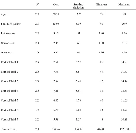
Descriptive Statistics………... 52
Covariates……….... 52
Multilevel Models………... 53
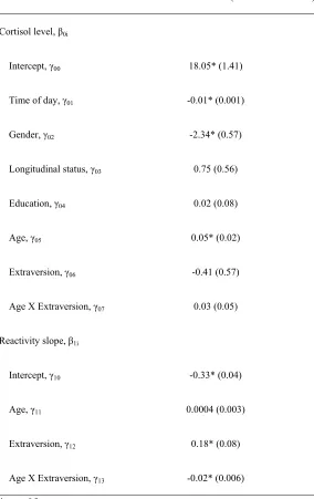
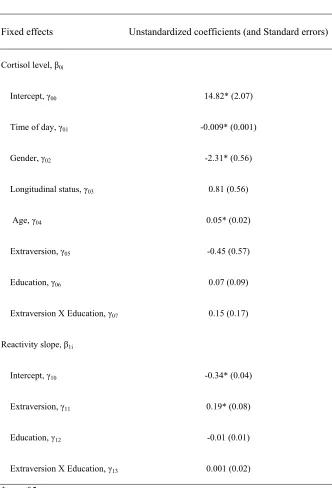
Model for Age X Extraversion……….... 55
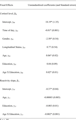
Model of Age X Education………. 57
Remaining Models……….. 58
Analyses Addressing Research Question and Hypothesis 5……….. 59
Demographic Characteristics……….. 59
Descriptive Statistics………... 59
Covariates……….... 60
Moderated Regression………... 61
Analyses Addressing Research Question 6………... 66
Exclusion of Outliers………... 66
Demographic Characteristics……….. 66
Descriptive Statistics………... 66
Covariates……… 66
Measures of Sympathetic Arousal………... 66
Moderated Regression………... 67
DISCUSSION………..…… 70
Research Questions and Hypotheses 1-4………. 72
Research Question and Hypothesis 5………..…… 78
Research Question 6………..…….. 87
LIMITATIONS……….... 90
CONCLUSIONS……….……… 92
APPENDICES………. 107
Appendix A: List of In-Home Procedures………..… 108
Appendix B: Declarative Memory Testing………. 109
LIST OF TABLES
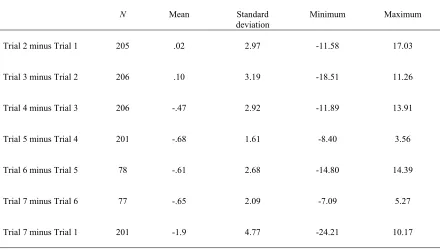
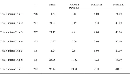
Table 1. Descriptive statistics for research questions and hypotheses 1-4…………... 111 Table 2. Descriptive statistics for trial to trial change in cortisol (nmol/L)……...… 112 Table 3. Descriptive statistics for time in minutes between each cortisol collection…. 113 Table 4. Unstandardized coefficients (and standard errors) of multilevel model of age
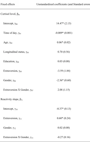
and extraversion differences in cortisol reactivity………... 114 Table 5. Unstandardized coefficients (and standard errors) of multilevel model of age
and education differences in cortisol reactivity……… 115 Table 6. Unstandardized coefficients (and standard errors) of multilevel model of age
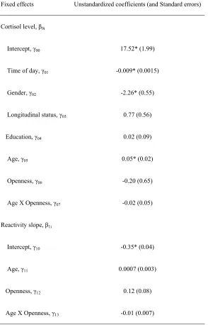
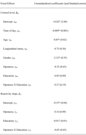
and openness differences in cortisol reactivity……… 116 Table 7. Unstandardized coefficients (and standard errors) of multilevel model of age
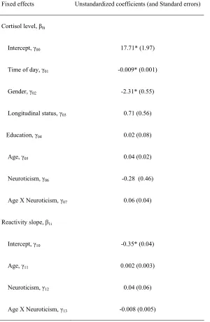
and neuroticism differences in cortisol reactivity……… 117 Table 8. Unstandardized coefficients (and standard errors) of multilevel model of
extraversion and education differences in cortisol reactivity………...118 Table 9. Unstandardized coefficients (and standard errors) of multilevel model of
extraversion and gender differences in cortisol reactivity………119 Table 10. Unstandardized coefficients (and standard errors) of multilevel model of
openness and education differences in cortisol reactivity……… 120 Table 11. Unstandardized coefficients (and standard errors) of multilevel model of
openness and gender differences in cortisol reactivity………...………. 121 Table 12. Unstandardized coefficients (and standard errors) of multilevel model of
neuroticism and education differences in cortisol reactivity……… 122 Table 13. Unstandardized coefficients (and standard errors) of multilevel model of
Table 14. Descriptive statistics for research question and hypothesis 5
where letter-number sequencing was the dependent variable………...124 Table 15. Descriptive statistics for research question and hypothesis 5
where a test of declarative memory was the dependent variable………..125 Table 16. Results from moderated regression analysis where cortisol level was a
predictor and logical memory immediate and delayed were dependent
variables………126 Table 17. Results from moderated regression analysis where cortisol change was a
predictor and logical memory immediate and delayed were dependent
variables………127 Table 18. Moderated regression analyses where letter-number sequencing was the
dependent variable……… 128
Table 19. Moderated regression analyses where cortisol change (from baseline to
trial 5) was the dependent variable………... 129 Table 20. Descriptive statistics for research question 6………... 130 Table 21. Moderated regression analyses where heart rate and cortisol level were
predictors of letter-number sequencing……… 131 Table 22. Moderated regression analyses where heart rate and cortisol change were
predictors of letter-number sequencing……… 132 Table 23. Moderated regression analyses where skin conductance and cortisol level
were predictors of letter-number sequencing………...… 133 Table 24. Moderated regression analyses where skin conductance and cortisol change
LIST OF FIGURES
Figure 1. Working memory performance at baseline, and during the stress and recovery periods of the TSST for cortisol responders and non-responders
from Elzinga and Roelofs (2005; p. 101)... 135 Figure 2. Predicted age and extraversion differences in cortisol reactivity, controlling
for testing time, gender, longitudinal groups status and education…………. 136 Figure 3. Predicted age and education differences in cortisol reactivity across trials,
controlling for testing time, gender, and longitudinal group status…………. 137 Figure 4. Conceptualization 1 of Age X Cortisol Change X Time of Day interaction... 138 Figure 5. Conceptualization 2 of Age X Cortisol Change X Time of Day interaction... 139 Figure 6. Conceptualization 3 of Age X Cortisol Change X Time of Day interaction... 140 Figure 7. Predicted change in cortisol from baseline to trial 5 as a function of
education, time of day, and age, controlling for gender and longitudinal
status……… 141
Figure 8. Predicted performance on letter-number sequencing as a function of cortisol response and mean skin conductance level, controlling for age, education, time of day of testing, smoking status, and hypertensive status……….. 142 Figure 9. Predicted performance on letter-number sequencing as a function of cortisol
response and standard deviation of skin conductance level, controlling for age, education, time of day of testing, smoking status, and hypertensive
Introduction
For some individuals, the task of taking cognitive tests can cause a physiological
stress response, where the hypothalamic-pituitary-adrenocortical (HPA) axis is activated,
resulting in the increased release of cortisol from the adrenal cortex. This physiological
reaction to cognitive testing seems more likely to occur in older adults than younger
adults (Steptoe, Kunz-Ebrecht, Wright, & Feldman, 2005), and may be moderated by
education, where older adults with advanced education have a more significantly elevated
cortisol response during and after testing than older adults with less education (Neupert,
Miller, & Lachman, 2006).
Activations of the HPA axis tend to occur when individuals interpret situations as
threatening (McEwen, 2000). The process of completing cognitive tests may be
threatening to older adults if they perceive that such testing may reveal declining
intellectual capacity. This threat could be exaggerated in older adults with advanced
education, since cognitive capabilities may be especially valued and loss of such abilities
may be seen as a loss of social esteem and social status (Dickerson & Kemeny, 2004).
In addition to age and education, personality differences may also contribute to
the intensity of a cortisol response to cognitive testing. Although researchers have not
examined whether personality differences contribute to varying cortisol levels in response
to completing a battery of cognitive tests, some have investigated whether exposure to a
laboratory based psychosocial stressor (e.g. public speaking) would elicit varying cortisol
openness and extraversion demonstrated greater cortisol elevations than those ranking
low in these two personality characteristics (Oswald, Mathena, & Wand, 2004). In
another analysis, extraversion in men (but not women) was significantly related to
cortisol increase following exposure to a psychosocial stressor (Oswald et al., 2006). In
this same study, women scoring high in the trait of neuroticism experienced only a slight
increase in cortisol to the laboratory based psychosocial stressor, while the relationship
between cortisol response and neuroticism in men was not significant (Oswald et al.,
2006). Thus, personality and gender may interact to influence the cortisol response to
psychosocial stress.
Analyzing cortisol elevations in response to a battery of cognitive tests is
important, because cortisol increases may have an influence upon assessments of
declarative and working memory at the end of the testing battery. Experimental studies
(e.g. Kirschbaum, Wolf, May, Wippich, & Hellhammer, 1996; Lupien et al., 1997;
Lupien, Gillin, & Hauger, 1999) have suggested that cortisol elevations - not related to
the to-be-remembered material - may generally have impairing effects upon declarative
and working memory, cognitive processes dependent upon the glucocorticoid receptor
rich regions of the hippocampus and prefrontal cortex (Eichenbaum, 2001; Lupien,
Maheu, Tu, Fiocco, & Schramek, 2007). The effects of cortisol upon memory, however,
are complex and may be dependent upon whether elevations occur in the morning or
Cortisol levels follow a diurnal pattern. For individuals who awake in the morning
and return to sleep during the night, cortisol levels peak in the morning and slowly
decline in the afternoon until reaching a trough in the evening and during sleep (Hennig,
Kieferdorf, Moritz, Huwe, & Netter, 1998; Lupien et al. 2007). Due to the greater
elevation of cortisol in the morning compared to the afternoon and evening, at least for
those who follow regular waking and sleeping patterns and have a normal diurnal cortisol
rhythm, stress-induced elevations in cortisol may be more likely to impair memory
function during the AM period (Het, Ramlow, & Wolf, 2005; Lupien, et al., 2002).
Studies investigating time of day of effects (e.g. Het et al., 2005, Lupien et al., 2002)
have primarily focused upon declarative memory. Whether these same effects influence
working memory performance deserves further investigation (Het et al., 2005).
In regards to working memory, cortisol induced impairments may also be
dependent upon activation of the sympathetic nervous system (Elzinga & Roelofs, 2005;
Roozendaal, McReynolds, & McGaugh, 2004), which can also occur in response to
threatening situations. Sympathetic arousal results in the classic fight or flight response,
due to the release of noradrenaline and adrenaline, resulting in elevated heart rate,
increased sweat production, and higher blood pressure (Gunnar & Quevedo, 2007).
Many studies examining the effects of acute cortisol elevations upon declarative
and working memory performance (e.g. Elzinga & Roelofs, 2005; Kirschbaum et al.
1996) and the combined effects of cortisol and sympathetic arousal upon working
addressed whether the acute physiological response to stress has similar effects upon
cognitive performance in middle-aged and older adults.
This study examined whether an increased cortisol response to a battery of cognitive
tests differed according to age, education, and the personality traits of openness, extraversion,
and neuroticism. Whether elevated cortisol was associated with interactions of age and
education, personality and gender, personality and age, and personality and education was also
explored. Since experimental studies (e.g. Kirschbaum et al. 1996; Lupien et al. 1997; Lupien
et al., 1999) have indicated that cortisol elevations not related to the to-be-remembered
material may generally have impairing effects upon declarative and working memory, analyses
were conducted to determine whether cortisol increases associated with completed cognitive
tests hindered subsequent performance on declarative and working memory assessed at the end
of a testing battery. Whether these effects were dependent upon age and time of day of testing
were also explored. In the case of working memory, sympathetic arousal, as measured by
increased heart rate and sweat production, was also considered as an additional moderator.
Review of Literature
Definition of stress
McEwen (2000) defined stress as “an event or events that are interpreted as
threatening to an individual and which elicit physiological and behavioral responses” (p.
173). Through a process of cognitive appraisal, individuals may first determine the
degree of risk or potential threat that a situation presents (Lazarus & Folkman, 1984).
Folkman (1984), may be followed by a “secondary appraisal” that determines whether
resources, skills, or abilities are adequate to cope with the potentially threatening
situation. When resources for coping are seen as sufficient to offset the pressing demands
of a particular event or situation, then that event or situation may be appraised as
challenging; however, when resources are perceived as inadequate, the event or situation
tends to be seen as a threat.
Completing a battery of cognitive tests may be perceived as threatening to some
older adults, if they believe that they do not have adequate skills or abilities to perform
well on such tests. This perception of threat may be accentuated if the participants believe
that poor performance will be judged negatively by others, resulting in a loss of social
esteem or status. According to Dickerson and Kemeny (2004), individuals possess a
social self-preservation system that is sensitive to conditions of social evaluative threat or
occasions where “an important aspect of the self-identity is or could be negatively judged
by others” (p. 358). Actual or anticipated negative social evaluation was suggested by
Dickerson and Kemeny as capable of activating a physiological response causing the
release of the stress hormone cortisol.
This study defined stress as an event or events that provoke a physiological
response through activation of the HPA axis, causing the adrenocortical release of
cortisol (McEwen, 2000). The cognitive testing situation was interpreted as a stressful
event when the cortisol concentration became 2.5 nmol/L higher than the level prior to
also be a physiological response to perceived threats or stressors (Gunnar & Quevedo,
2007), was not considered as reflecting a psychological stress response, but did receive
consideration as a potential moderator of cortisol’s effects upon working memory.
The physiological stress response
Physiological responses to stress are partly mediated by the HPA axis, a system
that reacts more slowly to a perceived threat or challenge than the instantaneous
reactivity of the sympathetic nervous system. Stimulation of the HPA axis begins with
the hypothalamus, which releases corticotropin-releasing hormone (CRH), causing
pituitary gland secretion of adrenocorticotropin (ACTH). This latter hormone travels
through the blood stream to trigger the adrenal cortex to release glucocorticoids, which
include cortisol as their major constituent (Lupien et al. 2007). Following the perception
of a stressor, cortisol levels may peak in about 15 to 30 minutes and then decline slowly
to pre-stressor levels 60 to 90 minutes later due to cortisol’s negative feedback on the
HPA axis (de Kloet, Joels, & Holsboer, 2005).
Cortisol increases the availability of glucose for energy production, while also
reducing the inflammatory effects of adrenaline and noradrenaline released from
sympathetic activation (de Kloet et al., 2005). Being liposoluble, cortisol easily crosses
the blood brain-barrier where it subsequently binds to glucocorticoid receptors that are
located predominantly in the hippocampus and prefrontal cortex, brain regions involved
in learning and memory (Lupien, Buss, Schramek, Maheu & Pruessner, 2005; Lupien et
Perceptions of threatening situations can also result in the activation of the
sympathetic nervous system, causing the synaptic release of noradrenaline and the
indirect release of both noradrenaline and adrenaline from the adrenal medulla into the
blood stream. These catecholamine neurotransmitters prepare the body for the metabolic
demands of the classic fight or flight response by mobilizing immune and inflammatory
responses, increasing sweat production, elevating heart and respiratory rates, and
increasing the flow of blood to the brain and muscles (Gunnar & Quevedo, 2007).
Because of the effects of catecholamines upon the heart and sweat glands, sympathetic
arousal was operationalized as increased level and variability in heart rate and sweat
production, with the latter being determined through skin conductance measures.
Although neither type of catecholamine can cross the blood brain barrier,
peripheral adrenaline stimulates β-adrenoreceptors in sensory vagal afferents, which
terminate in a structure within the medulla known as the nucleus of the solitary tract
(NTS) (van Stegeren, Wolf, Everaerd, Scheltens, Barkhof, & Rombouts, 2007). Neural
projections from the NTS continue into the amygdala and then into other forebrain
regions, leading to the activation of the noradrenergic system through postsynaptic
binding of noradrenaline to adrenoreceptors in these areas (LaBar & Cabeza, 2006;
McGaugh, 2000; Roozendaal, Okuda, De Quervain, & McGaugh, 2006; Roozendaal,
Okuda, Van der Zee, & McGaugh, 2006; van Stegeren et al., 2007). According to animal
studies, noradrenergic activation of the amygdala appears to regulate glucocorticoids’
as glucocorticoids’ effects upon declarative long-term memory consolidation and
retrieval. Glucocorticoids appear to interact with noradrenergic activation to enhance
declarative long-term memory consolidation and inhibit declarative long-term memory
retrieval (Roozendaal, Hahn, Nathan, de Quervain, & McGaugh, 2004; Roozendaal,
Okuda, Van der Zee et al., 2006).
Such interactive effects, however, do not appear to play a role in declarative
memory testing where recall is assessed immediately or within 30 minutes of material
presentation. In this case, impairments seem to be associated primarily with
glucocorticoid elevations, particularly in the morning when naturally-occurring levels are
high (Wolf, 2008). The relationship of stress-induced cortisol elevations upon working
memory and declarative memory tested via immediate and briefly delayed recall was the
focus of the present study. The potential interactive effects of cortisol and sympathetic
arousal upon working memory were also explored.
Age and education differences in the acute cortisol stress response to a cognitive test battery
As previously discussed, a situation may achieve salience as a stressor when it is
perceived as threatening. According to the social self-preservation system hypothesis
(Dikerson & Kemeny, 2004), physiological stress responses can be provoked when
individuals believe that actual or anticipated social evaluation may reveal the absence of
social evaluative threat and encompasses a situation where “an important aspect of the
self-identity is or could be negatively judged by others” (p. 358).
In light of the social self-preservation system, cognitive testing may be more
threatening to individuals who value having above average cognitive abilities and believe that
such testing could demonstrate some loss of this cherished capacity. Older adults who possess
a bachelor’s or more advanced degree may be particularly susceptible to the social evaluative
threat that cognitive testing may present, especially if they are concerned that such testing
might reveal a decline in the valued ability of intellectual functioning.
Neupert et al. (2006) investigated whether a series of cognitive tests evaluating the
domains of vocabulary, short-term working memory, speed, and reasoning would elicit a
physiological stress response in 74 adults, ages 25 to 74, from the Boston oversample of the
first phase of the Midlife in the United States (MIDUS) Survey. Findings revealed that
middle-aged and older adults having higher levels of education (i.e. a four year college degree or more)
produced a significantly more positive cortisol slope throughout the testing occasions than the
less educated participants and younger educated adults.
A somewhat similar investigation also examined whether the intensity of the stress
response to cognitive challenge would vary depending upon education in an older adult sample,
aged 65 to 80 years, selected from the London area (Steptoe et al. 2005). The higher educated
group possessed educational qualifications extending from high school certificates to university
degrees. The less educated group had completed only elementary school and had no additional
also compared to a younger adult group, aged 27 to 42 years, who held at a minimum a high
school certificate.
Unlike the findings from Neupert et al. (2006), there were no differences in cortisol
reactivity to cognitive challenge between the older adult group with higher education and the
group with lower education. It should be noted, however, that the educational qualifications
within the higher educated groups from both studies may not be comparable. The Steptoe et al.
(2005) study’s older higher educated group included participants who had only completed high
school with participants who had university degrees; the Neupert et al. (2006) higher educated
older adult sample did not include those with a high school or two-year post-secondary degree.
Rather, it was composed of those older adults who had a bachelor’s degree or more.
Differences in sample composition related to the differing aims of both research teams.
Neupert et al. (2006) hypothesized that the cortisol response to cognitive testing would be
higher in the educated older adult sample because of social evaluative threat; thus a greater
focus upon those with a bachelor’s and more advanced degree was necessitated. Steptoe et al.
(2005), however, hypothesized that cortisol increase to cognitive testing would be greater in
the lower educated sample, since individuals with lower socioeconomic status (SES) were
thought to have greater activation of physiological stress processes to perceived threats (e.g.
McEwen & Seeman, 1999; Steptoe & Marmot, 2002). Thus, Steptoe et al. sought to separate
participants from the lowest SES (i.e. those who had no education beyond elementary school)
from the rest of the group, leaving the higher educated group comprised of participants who
In the Steptoe et al. (2005) study, both older adult groups, regardless of educational
attainment, demonstrated greater cortisol increases compared to the younger educated sample.
Thus, it is possible that the cognitive testing seemed more threatening to the older adult group,
since it evoked a greater cortisol response.
These findings suggest that older adults may experience greater cortisol elevations to
cognitive testing than younger adults. According to Neupert et al. (2006), education may
moderate this relationship for middle-aged and older adults, whereby increased cortisol
depends upon middle-aged and older adults having at least a bachelor’s degree. This study
examined whether middle-aged and older adults experienced a greater cortisol response to
cognitive testing than younger adults, and whether education moderated this relationship.
Additional studies (Oswald et al., 2004, 2006) discussed in the next section have
indicated that personality may also play a role in the acute cortisol response to a psychosocial
stressor. The stressor utilized in these studies, however, was not a cognitive challenge as in the
Neupert et al. (2006) or Steptoe et al. (2005) research. A laboratory-based stressor known as
the Trier Social Stress Test (TSST; involves the public performance of five minutes of free
speech and five minutes of mental arithmetic, see Kirschbaum, Pirke, & Hellhammer, 1993)
was employed. Application of the TSST procedure provoked different cortisol responses
depending upon personality trait (Oswald et al., 2004, 2006). There is limited research
investigating the relationship between personality and potential cortisol responses to cognitive
response to cognitive testing and whether this relationship was dependent upon gender, age or
education.
Personality differences in the acute cortisol stress response
Oswald et al. (2006) found that in a sample of adults (aged 18-30 years; males: M = 21.7, SD = 2.8; females: M = 21.4, SD = 2.8), openness was positively associated with an increased cortisol response to the TSST, with those scoring high on this personality dimension
having higher cortisol responses than those scoring low. This positive association was
primarily explained by high scores on the openness subscales of actions and ideas.
Previous research has also shown a positive relationship between openness and an
increased cortisol response to the TSST (Oswald et al., 2004). Although the potential
mechanisms explaining this relationship are not clear, examining the characteristics of those
scoring high in openness may provide some potential clues. These individuals tend to “have a
rich and complex emotional life,” are imaginative, and enjoy novelty (Costa & McCrae, 1992,
p.6). Perhaps high emotional engagement, an active imagination, and novelty seeking
predispose those having high openness to increased cortisol responses when exposed to
psychosocial stressors. Although further research is needed to understand what mediates the
relationship between high openness and an increased cortisol response to psychosocial
stressors, this was not the focus of the present study.
Neither agreeableness nor conscientiousness was significantly associated with cortisol
responses to the stressor in the Oswald et al. (2006) study. However, significant interactions
to the TSST. These interactions were identified when the group’s cortisol-time curve was
deconstructed into the component parts of baseline cortisol, peak cortisol, fold stimulation
(peak/baseline), and delta (peak minus baseline).
Fold stimulation and delta were found to correlate with extraversion and neuroticism in
a gender-dependent manner. Specifically, a significant positive relationship was reported
between fold stimulation and extraversion in men (p = .04), while a trend towards a negative relationship between fold stimulation and neuroticism was found in women (p = .06).
Correlation of fold stimulation with the subscales for extraversion (i.e. warmth, gregariousness,
assertiveness, activity, excitement, and positive emotion) explained that the positive
association between fold stimulation and extraversion in men was accounted for primarily by
warmth, activity, and positive emotions. Thus, those men scoring highest on the warmth,
activity, and positive emotions subscales had the greatest increase between baseline and peak
cortisol levels following the psychosocial stressor.
It is not clear why men scoring high in extraversion had a greater cortisol response to
the stressor than those scoring low in this personality dimension. Previous research by Oswald
et al. (2004) reported that high extraversion and high scores on the extraversion subscales of
gregariousness, activity, and excitement-seeking were positively correlated with increased
cortisol fold stimulation in the study group as a whole. Individuals who are defined as
extraverted tend to be sociable and enjoy activity and excitement (Costa & McCrae, 1992), and
these traits may predispose them to a more labile HPA response than those who are low in
needed to explore why extraversion may be related to increased cortisol responses to
psychosocial stressors.
Extraverted females did not experience an increased cortisol response to the stressor in
the Oswald et al. (2006) research, while no gender differences were found in the earlier Oswald
et al. (2004) study. The demographic characteristics in the Oswald et al. (2004) research (age:
M = 21.6; SD = 3.6) were similar to that in the Oswald et al. (2006) study and the assessment of personality was the same (i.e. the Revised NEO Personality Inventory; see Costa and
McCrae, 1992). Sample sizes, however, were smaller in the Oswald et al. (2004) study (males:
N = 9; females: N = 5) compared to the Oswald et al. (2006) analysis (males: N = 43; females:
N = 25). Thus, the detection of moderation effects may not have been possible in Oswald et al. (2004) due to the smaller sample size.
Gender differences in the Oswald et al. (2006) research may have been due to testing
women who were in the follicular phase of their menstrual cycle. Women in the follicular
phase of their menstrual cycle tend to produce smaller cortisol responses to a laboratory based
psychosocial stressor than men (Kajantie & Philips, 2006). Exposure to a stressor during the
luteal phase of the menstrual cycle results in cortisol responses similar to those found in men
(Kajantie & Philips, 2006); thus, it is possible that if testing had been carried out while women
were in the luteal phase of their menstrual cycle, there may have been no differences in the
cortisol response of extraverted men and women.
As mentioned previously, a trend towards a negative relationship between fold
difference would have remained if women were tested in the luteal phase of their menstrual
cycle is also not known. This study investigated whether to control for women who were in the
follicular phase of their menstrual cycle in order to equate as far as possible the potential
stress-induced cortisol responses of men and women.
Correlation of fold stimulation with the neuroticism subscales (i.e. anxiety, anger,
depression, self-consciousness, impulsiveness, and vulnerability) indicated that the initial
association between fold stimulation and neuroticism in women was accounted for by a
significant and negative relationship between cortisol fold stimulation and depression and
self-consciousness. No significant associations were found between cortisol and the other
neuroticism subscales. Thus, those women scoring highest on the depression and
self-consciousness subscales had the smallest increase between their baseline and peak cortisol
levels following the psychosocial stressor. It is possible that women scoring high in
neuroticism had high baseline cortisol levels that did not increase much following the
psychosocial stressor. Initial analyses revealed that participants “with higher pre-stress cortisol
also had higher peak levels during the TSST than subjects with lower pre-stress cortisol”
(Oswald et al., 2006, p. 1586). Additionally, “baseline cortisol levels were negatively
associated with fold stimulation; subjects who had higher pre-stress cortisol levels had less
relative change in levels than subjects whose initial levels were lower” ( p. 1586). Data
supporting that pre and post stressor levels of cortisol remained high in women scoring high in
Since neuroticism is characterized by increased perceptions of stress and increased
negative affect to stressors (Mroczek & Almeida, 2004), it may seem paradoxical that the
female participants scoring high in trait neuroticism within Oswald et al.’s (2006) study
experienced only a slight increase in cortisol following stressor exposure. Increased emotional
reactivity to minor daily problems has also been found in male neurotics (Suls, Green, & Hillis,
1998), and yet there was no significant association between high neuroticism in males and
cortisol response following the psychosocial stressor in either of the Oswald et al. (2004, 2006)
studies. Further, there was no significant association between neuroticism and cortisol response
to the stressor in either men or women in the Oswald et al. (2004) study.
The absence of an increased HPA response to the psychosocial stressor within
neurotic participants could have been due to pre-existing HPA axis dysregulation. Adults
scoring high in neuroticism have been found to have significantly higher cortisol levels
30 to 60 minutes after awakening than those scoring low in this personality dimension.
This difference was not dependent upon gender or age, when testing young to middle
aged adults only (range = 21-57 years) (Portella, Harmer, Flint, Cowen, & Goodwin,
2005). Following 60 minutes after awakening, however, there were no significant
differences in cortisol levels between the low and high neuroticism groups.
Dysregulation of the HPA axis has been found in adult men who test high for trait
neuroticism and lack a history of psychiatric illness (Zobel et al., 2004). After supplying
a small dose of a synthetic glucocorticoid (i.e. dexamethasone) to adults with an average
neuroticism produced a cortisol response, indicating that the HPA axis was unable to
respond to the glucocorticoid by dampening cortisol release as is typically expected in
adults with normal HPA function. This same effect was not found in women scoring high
in trait neuroticism, however, and whether dysregulation was associated with a blunted
cortisol response to a psychosocial stressor was not explored.
The current study sought to examine whether increases in cortisol over time, while
completing a battery of cognitive tests, differed according to the personality traits of openness,
extraversion, and neuroticism. To gain an understanding of changes in within-person
trajectories of cortisol over time, multilevel modeling was utilized instead of the calculation of
fold stimulation and delta, which cannot provide intraindividual fluctuation/change
information. Multilevel modeling was employed to determine whether intraindividual changes
in cortisol trajectories were dependent upon the person-level factor of personality. Whether
within-person increases in cortisol over time were also dependent upon between-person
differences in gender, age or education was also investigated. Analyzing the degree of cortisol
increase to cognitive testing is important, because acute cortisol elevations to completed tests
may have a negative influence upon subsequent tests examining declarative and working
memory (e.g. Kirschbaum et al. 1996; Lupien et al. 1997; Lupien et al., 1999; Tops, van der
Pompe, Baas, Mulder, Boer, Meijman, & Korf, 2003).
Effects of acute cortisol elevations and sympathetic arousal upon declarative memory
Acute increases in cortisol have not been associated with impairments in
various effects upon declarative or explicit memory, (Kirschbaum et al., 1996; Lupien et
al. 1997; Lupien et al., 2007; Wolf, 2008; LaBar & Cabeza, 2006), defined here as the
conscious recollection of previously learned facts or observed events (Eichenbaum,
2001). Elevated cortisol levels have differing effects depending upon the phase of
memory tested (i.e. encoding/consolidation or retrieval) and the length of time between
presented material and recall (i.e. immediate or a brief delay of 10 to 30 minutes vs. a
long-term delay of hours to days). Sympathetic arousal appears to play a role when
testing long-term consolidation or long-term retrieval. (Abercrombie, Speck, &
Monticelli, 2006; Cahill, Gorski, & Le, 2003; Roozendaal, Hahn, Nathan, de Quervain, &
McGaugh, 2004; Roozendaal, Okuda, Van der Zee et al., 2006; Wolf, 2008). However,
the effects of sympathetic activation are less clear when declarative memory is tested
through immediate or briefly delayed (10-30 minutes) recall, when phase of memory
effects cannot be easily determined (Wolf, 2008).
Cortisol elevations appear to interact with sympathetic activation to enhance
declarative long-term memory consolidation (Abercrombie et al., 2006; Cahill, Gorski, &
Le, 2003; Roozendaal Okuda, Van der Zee et al. 2006) and impair declarative long-term
memory retrieval (Roozendaal, Hahn et al., 2004; Wolf, 2008). These effects seem to be
strongest when testing memory for emotionally arousing information (Wolf, 2008). When
analyzing the effects of cortisol increase on the immediate or slightly delayed recollection
of material, recall of neutral items appears to be more impaired than the recollection of
This study examined whether cortisol elevations, associated with the completion
of previous cognitive tests, hindered immediate and briefly delayed recall tested towards
the end of a cognitive testing battery. Since consolidation and retrieval processes could
not be analyzed specifically, sympathetic arousal was not investigated as a potential
moderator of cortisol-associated declarative memory impairment. Rather, potential
moderators included time of day of testing and age, and these will be addressed in the
next sections.
Effects of acute cortisol elevations upon immediate or briefly delayed recall. To examine the effects of acute endogenous, or naturally-occurring, cortisol elevations upon
memory in humans, cortisol can be manipulated by exposing participants to a laboratory
stressor (e.g. the TSST). After exposure to the stressor, some participants (i.e. the
‘responders’) produce a significant increase in cortisol compared to baseline levels, while
others (i.e. the ‘non-responders’) do not.
When testing for the immediate and briefly delayed recall of generic or neutral
word lists, cortisol responders remembered fewer words than cortisol non-responders
(Kirschbaum et al., 1996; Lupien et al., 1997). This impairing effect was reported in
young adult cortisol responders (undergraduate students), who were exposed to the
laboratory stressor immediately prior to learning (Kirschbaum et al., 1996), and elderly
adult responders (62-83 years of age), who were exposed to the stressor after learning and
The impairing effects of cortisol upon immediate recall have been demonstrated
in a pharmacological study involving male undergraduate students (18-27 years of age).
In this study, cortisol was orally administered two hours prior to the immediate free
recollection of neutral, pleasant, and unpleasant words (Tops et al., 2003). The average
cortisol level in the treatment group at the time of cognitive testing was approximately
22.0 nmol/L, a value only slightly higher than the mean value observed in the cortisol
responders in the Kirschbaum et al. (1996) and Lupien et al. (1997) studies (17.7 nmol/L
and 19.5 nmol/L, respectively). Participants in the treatment group recalled fewer
pleasant and neutral words than those in the placebo group. There were no significant
differences between groups in the recollection of unpleasant words.
Additional evidence suggests that the effects of cortisol upon delayed recall may
also be dependent upon time of day of testing (Het et al., 2005, Lupien et al., 2002).
Cortisol levels peak in the morning and slowly decline in the afternoon until reaching a
trough in the evening and during sleep, at least for healthy individuals who awake in the
morning and return to sleep during the night (this pattern may reverse in some who stay
awake through the night and sleep during the day; see Hennig, et al. 1998). Due to the
greater elevation of cortisol in the AM phase than in the PM, stress-induced cortisol
elevations may be more likely to impair memory function in the morning than in the
afternoon (Het et al., 2005, Lupien et al., 2002).
According to a meta-analysis examining the cognitive effects of the
or objects, morning administration of cortisol significantly impaired delayed recall (d = -.40), while afternoon dosing produced a small, but significant memory enhancement (d = .22) (Het et al., 2005). The cognitive effect of cortisol dosing in each of the studies was
compared to a placebo group, and each study reviewed was conducted either in the
morning or afternoon. Effect sizes represented averages, and there was substantial
variability of effects for those studies conducted in the morning (-.77 ≤ d ≤ -.03, N = 4) and in the afternoon (.02 ≤ d ≤ .41, N = 8). This may have been due to the use of different memory tasks, varying time delays in the testing of memory lasting from 3 minutes to 1
week, and different pharmacological doses of cortisol. Time of day effects were not
analyzed for studies where cortisol was given prior to the retrieval of already learned
material, since impairing effects were found for all studies (N = 4) examined. The average effect size for these latter studies was moderate (d = -.49) and significantly different from zero.
The present study investigated whether endogenous cortisol elevations to
previously completed cognitive tests had an impairing effect upon declarative memory
tested via immediate and delayed (approximately 30 minutes) recall of subsequently
learned material. Whether effects were dependent upon age and time of day of testing
was also analyzed. Although it was recognized that acute cortisol increase appears to
hinder memory for neutral items rather than emotionally arousing ones (Tops et al.,
differentiate whether recall of neutral items was more impaired than the recollection of
emotionally arousing ones.
Effects of acute cortisol elevations and sympathetic arousal upon working memory
Working memory is a form of memory where information is “actively maintained
and/or manipulated in conscious awareness over a short period of time” (LaBar &
Cabeza, 2006, p. 56). It is a memory function that is dependent upon the prefrontal cortex
(Owen, Downes, Sahakian, Polkey, & Robbins, 1990), a cortical region rich in
glucocorticoid receptors (Lupien et al., 2007). Studies examining cortisol’s effects upon
working memory have produced mixed findings. In selected research, where participants
were undergraduate students randomly assigned to a stress or control condition, the
working memory of cortisol responders in the stress group was either: 1) not affected
(Kuhlmann, Piel, & Wolf, 2005), 2) impaired at high working memory loads, but not at
low loads (Oei, Everaerd, Elzinga, Van Well, & Bermond, 2006), or 3) impaired only
during a period of acute sympathetic activation during exposure to the laboratory stressor
(Elzinga & Roelofs, 2005). Factors contributing to differences in these findings might
include variations in stress-induced peak cortisol concentrations, type of working
memory test employed, and whether cortisol elevations occurred concurrently with
sympathetic arousal.
Prior to working memory assessment, peak cortisol levels in the stress group
reached an average of 34.4 nmol/L in the Oei et al. (2006) study. In contrast, peak
significant effects were found, were lower. Oei et al. utilized a test of working memory
(i.e. the Sternberg item recognition task; Sternberg, 1966) that assessed low and high
working memory processing load, and found that working memory was significantly
impaired in the stress group at high working memory loads, but not at low loads,
compared to the control group. Kuhlmann et al. used a working memory task (i.e. the
forward and backward digit span test; Weschler, 1987) that may not have produced high
working memory loads for the participants. This combined with the lower cortisol levels
in their research might explain why a significant association between stress-induced
cortisol increase and working memory performance was not found.
Elzinga and Roelofs (2005), unlike the two previously discussed studies
(Kuhlmann et al. 2005; Oei et al. 2006), examined whether the influence of cortisol upon
working memory was dependent upon sympathetic arousal, as measured by increased
heart rate and blood pressure. To assess this interaction, participants assigned to the stress
condition were tested with three parallel versions of the forward and backward digit-span
test during three separate conditions: one at baseline prior to the TSST, a second
following the TSST and in front of the TSST audience, and a third after the stress task
when no audience was present.
Although cortisol responders in the stress condition demonstrated peak cortisol
levels (i.e. 16 – 18 nmol/L) comparable to those in research where no significant effects
between cortisol increase and working memory were reported (Kuhlmann et al., 2005),
(2005) study declined significantly compared to baseline measures. This dysfunction in
working memory, however, happened only during sympathetic activation (as measured
by increased heart rate and blood pressure) occurring during working memory testing in
front of the TSST audience. When measures of sympathetic arousal returned to baseline
during testing without the audience being present, there were no significant differences
between the cortisol responder and non-responder groups in working memory
performance, even though cortisol levels remained high in the cortisol responder group
(see Figure 1).
Sympathetic arousal leads to noradrenergic activation through the postsynaptic
binding of noradrenaline to adrenoreceptors in the amygdala and other forebrain regions
(McGaugh, 2000; van Stegeren et al., 2007). Elzinga and Roelofs’ (2005) findings that
sympathetic arousal appears to interact with cortisol elevations to impair working
memory seem congruent with previous animal research, suggesting that glucocorticoids’
impairing effects upon working memory is dependent upon noradrenergic activation, and
perhaps noradrenergic activation within the amygdala specifically (Roozendaal,
McReynolds et al. 2004).
When corticosterone (i.e. the major glucocorticoid in rats) was infused into rats
with drug-induced lesions of the amygdala (specifically the basolateral amygdala) and
into rats with functional amygdala, working memory performance was impaired in the
rats with intact amygdala, but performance was not impaired in rats with amygdala
impairment associated with corticosterone and that an intact basolateral amygdala was
needed for this impairment (Roozendaal, McReynolds et al., 2004). Although not tested
specifically, corticosterone infusions in the rats with functional amygdala may have
resulted in noradrenergic activation within the amygdala, which helped to modulate the
corticosterone memory impairment. Glucocorticoid infusions can increase levels of
noradrenaline in a rat’s brain and facilitate noradrenergic activation (Roozendaal, Okuda,
De Quervain et al., 2006). The importance of noradrenergic activation in working
memory impairment was addressed in the Roozendaal, McReynolds et al. (2004) research
by infusing a drug that dampens sympathetic arousal by inhibiting activation of
adrenoreceptors (i.e. the β- adrenoreceptor antagonist propranolol). When this drug and
corticosterone were injected into rats prior to testing, the impairing effects of
corticosterone upon working memory were prevented, thus implying that corticosterone
induced impairment of working memory also involves noradrenergic activation, and
perhaps noradrenergic activation in a functional basolateral amygdala (Roozendaal,
McReynolds et al., 2004.) Thus, the Roozendaal, McReynolds et al. (2004) research
provides a possible explanation for why the Elzinga and Roelofs’ (2005) undergraduate
participants, who had stress-induced elevations in cortisol and increased sympathetic
arousal, may have experienced working memory impairment. Elevated cortisol and
noradrenergic activation in the amygdala, due to increased arousal, may have worked
Whether the effects of cortisol elevations upon working memory are also
dependent upon the time of day of cortisol increase has not been sufficiently investigated
(Het et al., 2005). Lupien, Buss et al. (2005) have suggested that high pharmacological
doses of glucocorticoids given in the AM phase may have more impairing effects upon
attentional/working memory abilities than similar doses administered during the PM.
This suggestion, however, was based upon two studies, where attention (and not working
memory specifically) was tested in the PM in one study (Hsu, Garside, Massey, &
McAllister-Williams, 2003) and working memory was analyzed in the AM in another
(Lupien, Gillin, & Hauger, 1999). Although attentional processes appear to play a role in
working memory function (Cowan, 2000), it is uncertain from these studies whether
working memory is differentially affected by cortisol increases in the morning and
afternoon, since only one study tested working memory specifically.
In the research reviewed here, working memory testing occurred around 10 AM
in two studies (Elzinga & Roelofs, 2005; Oei et al., 2006) where impairing effects were
found, and at around 12 PM in another (Kuhlmann et al., 2005) where no significant
effects were reported. As discussed previously, differences in these findings may have
been due to variations in peak cortisol levels, type of working memory test employed,
and sympathetic arousal levels. Further studies are needed to determine whether
differences in working memory performance are also dependent upon time of day of
The current study investigated whether endogenous cortisol elevations associated
with previously completed cognitive tests had an impairing effect upon working memory.
Whether effects were dependent upon age, time of day of testing and sympathetic arousal
occurring during testing was also analyzed. Because β-adrenoreceptor antagonists may
potentially influence working memory performance, and since this type of medication is
commonly used to treat hypertension (Fisher & Williams, 2005), hypertensive status was
used as a proxy for participants taking β-adrenergic receptor antagonists and was
controlled in analyses where sympathetic arousal was examined as a moderator of
working memory performance.
Effects of naturalistic stressors upon working memory
All of the studies reviewed in this proposal regarding cortisol’s effects upon
working and declarative memory have come from the experimental literature, where
participants were exposed to a laboratory-based psychosocial stressor. Few studies have
examined memory effects due to cortisol elevations in response to naturalistic stressors.
One exception comes from Lewis, Nikolova, Chang, and Weekes (2008), who examined
the effects of high examination stress upon working memory in undergraduate students.
A within-subject comparison of working memory performance during a period of high
examination stress (i.e. 3 or more tests or assignments due that week) and low
examination stress (i.e. when no classes were in session) was conducted. Examination
Participants experienced no significant change in forward digit span measures
between the low and high stress times; however, they did experience an improvement in
the backward digit span during high examination stress. The undergraduate participants
perceived significantly more stress in the high stress period and also demonstrated a
significant increase in cortisol (M = 0.11 ug/dL or 3.0 nmol/L) compared to levels during the low stress period (M = .09 ug/L or 2.5 nmol/L). Thus, the undergraduates during the high examination stress period, when cortisol levels were elevated, exhibited enhanced
working memory capacity, at least for backward digit span. This stands in contrast to
some of the previously discussed experimental studies (e.g. Elzinga & Roelofs, 2005; Oei
et al., 2006), where stress was associated with working memory impairment.
The varying findings may be explained by considering the differences in the
concentration of cortisol during stress. The mean cortisol level in the Lewis et al. (2008)
study was far lower than peak levels measured in the Oei et al. (2006) or Elzinga and
Roelofs (2005) study (i.e. 34.4 nmol/L and 16 – 18 nmol/L, respectively). Although
speculative, time of day effects may also have played a role. Lewis et al. participants
were not tested in the morning as in the Oei et al. and Elzinga and Roelofs research.
Sessions began at around 3:30 PM or 5:30 PM and lasted for approximately two hours.
Slight increases in cortisol during the afternoon and evening when circulating cortisol
levels are lower than those in the morning may have contributed to working memory
assessing participants in the morning and the afternoon would have been an important
contribution to the Lewis et al. research.
There continues to be a need to investigate the effects of cortisol upon memory in
a more naturalistic setting by taking into consideration the time of day of testing, and
sympathetic arousal in the case of working memory, which was also not addressed in the
Lewis et al. (2008) study. Although the present study did not examine naturalistic
stressors per se, it did explore whether participants who completed a battery of cognitive
tests within a naturalistic setting (i.e. their home) experienced a stress response, and
whether working and declarative memory performance was dependent upon this
response, as well as time of day of testing and age.
Effects of stress upon memory in aging adults
Much of the research regarding the effects of acute increases in endogenous
cortisol upon memory has utilized participants who are young adults. Few studies have
examined effects upon middle-aged and older individuals. The Lupien et al. (1997)
research discussed in the section on declarative memory was one exception, and findings
appeared to indicate that the impairing effects of endogenous cortisol upon recall found
in young adults (Kirschbaum et al., 1996) can also be replicated in older adults. Although
not exploring the effects of cortisol and sympathetic arousal specifically, other research
has shown that naturally-occurring daily stress has similar effects upon working memory
Results indicated that the within-person reaction times of young and older adults were
slower on a 2-back working memory task on days when at least one stressor was
reported, compared to days when no stressors were reported. Cortisol and sympathetic
arousal were not measured in this research, so it is not known whether potential
stress-induced elevations of cortisol might have interacted with sympathetic arousal to influence
cognitive performance deficits in the young and older adult participants.
Since aging in general may play a role in the performance of declarative and
working memory tasks, it would be important to determine whether the effects of acute
increases in cortisol upon declarative memory are dependent upon age, and whether the
effects of the interaction of cortisol and sympathetic arousal upon working memory are
also dependent upon age. Testing of age differences in working memory capacity has
revealed that older adults (age, M > 60 years) perform more poorly than younger adults (age, M < 30 years) (Bopp & Verhaeghen, 2005). Further, cross-sectional studies have reported a linear decline in performance beginning in the decade of the 20s and 30s for a
variety of working memory tasks (Borella, Caretti, & De Beni, 2008) and for the
Weschler (1997) letter-number sequencing task (Myerson, Emery, White & Hale, 2003),
which is a working memory test that was employed in the present analysis. Age
differences have also been reported for declarative memory performance; however,
declining function was not manifested in a longitudinal study until after 60 years of age
(Rönnlund, Nyberg, Nilsson, & Bäckman, 2005). Similar results were found in a
This study analyzed young, middle-aged, and older adults to determine whether
the effects of acute increases in cortisol upon working and declarative memory differed
by age and time of day of testing. Whether differences in working memory performance
were dependent upon sympathetic arousal was also addressed.
Research Questions and Hypotheses
Unless hypotheses specifically suggested investigating the time of day of testing as
an independent variable, all analyses controlled for time of day of testing to take into
consideration the diurnal variation of cortisol.
1) Is cortisol reactivity (defined as a change in cortisol slope over time) that is
associated with completing a cognitive testing battery dependent upon an
interaction between age and education?
a. Hypothesis: Middle-aged and older adults with higher education (i.e.
bachelor’s degree or more) will experience a greater cortisol response, as
demonstrated by a more positive cortisol slope, to the cognitive testing battery
than younger adults and less educated middle-aged and older adult participants
(Neupert et al., 2006).
2) Is cortisol reactivity that is associated with completing a cognitive testing battery
dependent upon the personality traits of extraversion, openness, or neuroticism?
a. Hypothesis: Participants scoring high in the traits of extraversion and
positive cortisol slope, than participants scoring low in these personality
dimensions (Oswald et al., 2004, 2006).
b. Because of mixed results in the literature regarding the cortisol responses
of neurotics to psychosocial stressors (Oswald et al., 2004, 2006), this study
explored whether the cortisol response of those scoring high in neuroticism was
different than those scoring low in neuroticism. No predictions were made
concerning whether those scoring high in neuroticism would have a blunted
cortisol response or an increased cortisol response compared to those scoring low
in this personality dimension.
3) Is cortisol reactivity that is associated with completing a cognitive testing battery
dependent upon an interaction between personality trait (i.e. extraversion,
openness, or neuroticism) and gender (Oswald et al., 2006)?
4) Is cortisol reactivity that is associated with completing a cognitive testing battery
dependent upon an interaction between: a) age and the personality traits of
extraversion, openness, or neuroticism, or b) education and the personality traits
of extraversion, openness, or neuroticism (Neupert et al., 2006; Oswald et al.,
2006)?
5) Are working and declarative memory performance decrements dependent upon
cortisol elevations, age, and time of day of testing?
a. Hypothesis: When controlling for the effects of education, participants
the working and declarative memory tasks will perform less well on the working
and declarative memory tests than those showing little or no cortisol response to
the previous testing (e.g. Kirschbaum et al. 1996; Lupien et al. 1997; Lupien et
al., 1999; Tops et al., 2003).
b. No specific predictions were made regarding whether working and
declarative memory performance decrements are dependent upon an interaction
between age, cortisol elevations, and time of day of testing.
6) Are working memory performance decrements dependent upon cortisol
elevations, age, and sympathetic arousal, when controlling for education and
hypertensive status?
Method
Participants
Participants were from the Boston oversample of the second phase of the Midlife
Development in the United States (MIDUS II), a national survey of health and well-being
funded by the National Institute on Aging. This sample included longitudinal participants
(N = 104) from the original MIDUS I Boston subsample, a probability sample of the Greater Boston area (see Lachman & Firth, 2004), as well as an additional probability
sample of non-longitudinal participants from the same Boston area (N = 309). Although longitudinal participants were assessed previously in the MIDUS I, data from the first
wave were not utilized. Out of this total sample of 413 participants, the longitudinal
non-longitudinal participants completed cortisol assessments 1-5 and 7 (see Appendix A).
Age and education data were not available for participants who chose not to have their
cortisol collected.
All data for the current study were collected between 2004 and 2007 and each
participant was tested only once. Participants ranged in age from 34 to 88 (M = 59.93; SD
= 12.80; N = 257) Those with a history of stroke, diabetes, neurological disorders, or who did not report English as their language spoken at home when growing up were excluded
from the analyses.
Procedure
Participants were administered 15 cognitive tests while in their home (see
Appendix A). Ten tests evaluating working memory, vocabulary, reasoning, and speed
were completed prior to the assessment of immediate recall (i.e. logical memory
immediate; Wechsler, 1997). Following this test, two more tests were completed prior to
another assessment of working memory (i.e. letter-number sequencing; Wechsler, 1997).
The test of briefly delayed recall (i.e. logical memory delayed; Wechsler, 1997) was
administered immediately after this working memory task.
Salivary cortisol samples were collected over an average of 95.42 minutes (SD = 20.71 minutes) during cognitive testing. Samples were taken seven times from the
longitudinal participants and six times from the non-longitudinal participants (sample #6
Psychophysiological indicators of sympathetic arousal (e.g. heart rate and skin
conductance level due to amount of perspiration on the finger) were measured using a
MEDAC System/3TM on the longitudinal participants only during the logical memory
immediate, letter-number sequencing, and logical memory delayed tests. The instrument
was manufactured by NeuroDyne Medical Corporation (Cambridge, MA) and is referred
to as “Neurodyne” in Appendix A.
Following the completion of all cognitive tests, a diary, demographic
questionnaire, photograph, and peak expiratory flow testing were taken or given to the
participants.
The personality traits of extraversion, openness, and neuroticism were measured using
the Revised Midlife Development Inventory (MIDI) Personality Scale (Lachman & Weaver,
1997; Addendum for MIDI Personality Scales, 2005) via a mail survey prior to the in-home cognitive testing procedure.
Measures
Salivary cortisol. Participants were instructed to avoid caffeinated products and to collect samples before they ate, drank, or brushed their teeth, since these activities may
elevate cortisol levels. Salivary cortisol was collected via Sarstedt Salivette, a cotton-like
swab that participants chewed on for about one minute. The saturated swab was then
placed into a plastic container, which was stored in a Ziploc bag and refrigerated until