Copyright1999 by the Genetics Society of America
A New Mouse Insertional Mutation That Causes Sensorineural Deafness
and Vestibular Defects
K. N. Alagramam,*
,1H. Y. Kwon,
†,1N. L. A. Cacheiro,
†L. Stubbs,
‡C. G. Wright,
§L. C. Erway** and R. P. Woychik*
*Department of Pediatrics, Rainbow Babies and Children’s Hospital, Case Western Reserve University, Cleveland, Ohio 44106,†Life Sciences
Division, Oak Ridge National Laboratory, Oak Ridge, Tennessee 37831-8080,‡Human Genome Center, Lawrence Livermore National
Laboratory, Livermore, California 94550;§Department of Otorhinolaryngology, University of Texas Southwestern Medical Center,
Dallas, Texas 75235-9035 and **Department of Biological Sciences, University of Cincinnati, Cincinnati, Ohio 45221
Manuscript received January 5, 1999 Accepted for publication April 22, 1999
ABSTRACT
This article describes a new recessive insertional mutation in the transgenic line TgN2742Rpw that causes deafness and circling behavior in mice. Histologic analysis revealed virtually complete loss of the cochlear neuroepithelium (the organ of Corti) in adult mutant mice. In association with the neuroepithelial changes, there is a dramatic reduction of the cochlear nerve supply. Adult mutants also show morphological defects of the vestibular apparatus, including degeneration of the saccular neuroepithelium and occasional malformation of utricular otoconia. Audiometric evaluations demonstrated that the mice displaying the circling phenotype are completely deaf. Molecular analysis of this mutant line revealed that the transgenic insertion occurred without creating a large deletion of the host DNA sequences. The mutant locus was mapped to a region on mouse chromosome 10, where other spontaneous, recessive mutations causing deafness in mice have been mapped.
I
NHERITED inner ear abnormalities in humans and balance disturbances that frequently result from inner ear dysfunction (Steel 1995).mice can be grouped into several categories,
includ-ing morphogenetic inner ear defects in which relatively The inner ear is a complex organ in terms of its development, morphology, and function. As many as a gross changes in overall form of the labyrinth occur,
cochleo-saccular defects involving disorders of inner ear hundred genes have been implicated in nonsyndromic hearing loss in humans (Morton1991). In mice, the fluid homeostasis, and neuroepithelial defects, in which
number of loci associated with hearing loss is thought the sensory epithelia are directly affected. Mutations
to be.60 (Nadeauet al. 1991). Genes associated with
with primary effects in the latter category are thought
the shaker-1 locus (Gibson et al. 1995) and the Snell’s
to be a major cause of inner ear defects in humans
waltzer locus (Avraham et al. 1995) were among the
(Fraser1976). Therefore, mouse mutants with
neuro-first two murine genes associated with deafness to be epithelial defects are potentially important models for
identified. While a few of the genes associated with such disorders in humans.
human deafness have been cloned, many are yet to be Neuroepithelial defects of the inner ear are often
identified (Van CampandSmith). Some of the human associated with partial or complete degeneration of the
genes associated with deafness have been identified re-sensory neuroepithelia of the cochlea and/or the
vestib-cently through the analysis of mouse mutants. For exam-ular apparatus. The organ of Corti, as well as the
macu-ple, neuroepithelial defects in the shaker-1 mouse are lae and cristae of the vestibular apparatus, may all be
caused by a mutation in the myosin VIIA gene (Myo7a; affected. The degenerative changes are due to primary
Gibson et al. 1995). A mutation in the human
homo-disorders of the neuroepithelia, rather than being
sec-logue, MYO7A, is involved in Usher syndrome 1B (con-ondary to alterations in tissues that maintain
homeosta-genital balance and hearing loss coupled with progres-sis of the inner ear fluids. As a consequence of the
sive retinitis pigmentosa; Weil et al. 1995), as well as
sensory cell loss that occurs in these disorders, neural
nonsyndromic deafness (Liu et al. 1997; Weil et al.
degeneration is usually also seen. In mice, this type of
1997). Similarly, the gene causing neuroepithelial de-inner ear pathology is associated with several recessive
fects in the mouse shaker-2 locus was recently identified mutations. These mutants often show the deafness and
as coding for an unconventional myosin (Myo15; Probst et al. 1998), and the human homologue
(MYO15) mapped to the nonsyndromic deafness locus
Corresponding author: Richard P. Woychik, Parke-Davis Laboratory DFNB3 (Wanget al. 1998).
for Molecular Genetics, 1501 Harbor Bay Pkwy., Alameda, CA 94502.
In humans, nonsyndromic deafness is more common E-mail: [email protected]
1These authors contributed equally to this work. than syndromic deafness (Schuknecht1993). Among
this method, each temporal bone provided a series of sequen-the prelingual forms of deafness, autosomal recessive
tial sections that included the full extent of the cochlea and nonsyndromic deafness accounts forz85% of the cases
vestibular apparatus. The mice used for histological analysis (KalatzisandPetit1998). Furthermore, the autoso- were between 10 and 12 mo old. Results from mutant mice mal recessive forms of hearing loss are often more severe were always compared to results obtained from wild-type
lit-termates. (compared to autosomal dominant or X-linked forms)
ABR and behavioral testing:Auditory function was evaluated and are almost exclusively sensorineural, due to defects
by performing auditory-evoked brain stem response (ABR) in the cochlea (Schuknecht1993;KalatzisandPetit
tests. For our experiments, ABR responses were obtained ac-1998). Therefore, characterization of a new mouse mu- cording to the methodology described previously (Erwayet tation causing nonsyndromic, recessive neuroepithelial al. 1993). In brief, acoustic stimuli were presented (20/sec) to the external auditory canals of anesthetized mice. Each deafness will be useful for studying this form of inner
stimulus elicits, from a normal ear, a volley of action potentials ear defect. Here we report the identification of such a
over the auditory nerve with subsequent activation of auditory mutation, the recessive insertional mouse mutation in
centers in the brain stem. The brain stem responses (ABRs) the transgenic line TgN2742Rpw that exhibits auditory from these stimuli were averaged from 128 to 1024 repetitions and vestibular defects associated with the inner ear neu- of the stimulus. Mouse ABR recordings exhibit four or five peaks, each with a well-defined latency (1–6 msec) and ampli-roepithelium. Additionally, we provide evidence that
tude (1–10mV). The threshold for the ABR was determined the organ of Corti develops normally in the mutant
for clicks and puretone pips of 8, 16, and 32 kHz. mice and subsequently undergoes degeneration, and we
Vestibular function was evaluated by performing a swim test. describe the molecular characterization and mapping The mice were placed in a clear plastic mouse cage half-filled of the mutant locus to mouse chromosome 10. These with warm (30–358) water. While in the water the mice were observed to determine if they showed “normal” swimming studies suggest a potential relationship between our new
behavior. Normal swimming behavior is defined as follows: mutant and other deafness mutations that have been
after being placed in water, wild-type mice quickly resurfaced mapped to the central region of chromosome 10. On
with a “whipping” motion of their tails, managed to keep their the basis of its phenotype, the TgN2742Rpw transgenic noses and tails above the surface of water, and swam toward mutant strain provides an excellent model for studies the side of the cage.
of nonsyndromic human deafness. Cloning the transgene insertion site: Sequences flanking the transgene insertion site were identified using a plasmid rescue procedure that was originally used for cloning the limb
deformity locus (Woychiket al. 1985). For the plasmid rescue
MATERIALS AND METHODS
procedure, we used a modified version of the cosmid vector c2RB (BatesandSwift1983), where the ampRgene on the
Mice:The transgenic line TgN(Imw)2742Rpw was generated
cosmid vector was inactivated, to generate a cosmid library of as part of an insertional mutagenesis program in the Life
partial Sau3A genomic fragments. The library was screened Sciences Division at the Oak Ridge National Laboratory.
by plating on medium containing ampicillin (Sambrooket al.
Transgenic mice were generated by microinjection of an
1989). Cosmid clones that grew on ampicillin plates were those 8-kb linearized plasmid construct containing a rat lectin cDNA
with an insert containing the ampR gene derived from the under the control of the humanb-actin promoter into
fertil-integrated transgene. The presence of the transgene within ized mouse embryos from the FVB/N strain. The transgenic
the ampRclones was confirmed by hybridization with a radiola-line was generated and maintained in the FVB/N strain; this
beled probe corresponding to the transgene sequence. The strain was used for all experiments described in this article.
host flanking sequences were identified by hybridization with
Identification of transgenic mice:Genomic DNA from tail
a radiolabeled probe composed of total mouse genomic DNA biopsies was isolated and quantified by fluorometry, and equal
(Woychiket al. 1985). Of these ampRclones, some contained amounts of DNA were digested with EcoRI. The digested DNA
only a portion of the transgene, while others contained a was size fractionated on 0.8% agarose gels and Southern
portion of the transgene along with sequences flanking the blotted to GeneScreen nylon membrane (Ausubelet al. 1991).
transgene integration site. One such clone, labeled cosmid-9, Mutant mice were identified by hybridization to sequences
was analyzed further. The genomic fragment in cosmid-9 was from within the transgene. Initially, quantitative Southern
screened for unique sequences (single-copy sequences devoid analysis was used to distinguish heterozygotes from
homozy-of any repetitive sequences/elements) by isolating several dif-gotes. Later, when unique copy-flanking sequences were
ferent restriction fragments and testing each of those frag-cloned (End-clone A and B, Figure 4A), these sequences were
ments by hybridization to a Southern blot containing wild-used to confirm the recessive nature of the TgN2742Rpw
muta-type mouse genomic DNA. One such unique sequence, F9, tion: mice that showed the phenotype were homozygous for
hybridized to a unique band in wild-type DNA. the transgene (Tg/Tg), and mice that were normal were either
To clone the wild-type region corresponding to the mutant heterozgous for the transgene (Tg/1) or wild type (1/1).
Histological analysis:Morphological studies involved histo- locus, probe F9 was used to screen a wild-type mouse genomic bacterial artificial chromosome (BAC) library according to logical examination of inner ear cross-sectional anatomy
utiliz-ing specimens embedded in glycol methacrylate. Five wild- the protocol supplied by Research Genetics (Birmingham, AL). End-cloning of the BAC insert was done as described type adult (FVB/N) mice and five adult mutant mice were
prepared for histological studies. Tissue specimens from all previously (Ausubelet al. 1991). One end-clone fragment was
called “end-clone A” (closer to the T7 site on the vector) and mice were processed as follows. The inner ear was fixed by
perilymphatic perfusion of 2.5% phosphate-buffered glutaral- the other was referred to as “end-clone B” (closer to the SP6 site on the vector). The size of each end-clone wasz0.6 kb. dehyde. After several days in fixative, the temporal bones were
decalcified in 0.35mEDTA and embedded in glycol methacry- Long-range restriction fragment analysis:Genomic DNA was prepared from spleen cells derived from wild-type, heterozy-late resin. They were then sectioned at 2–5mm and the sections
TABLE 1
Description of mapped gene markers and variant fragments
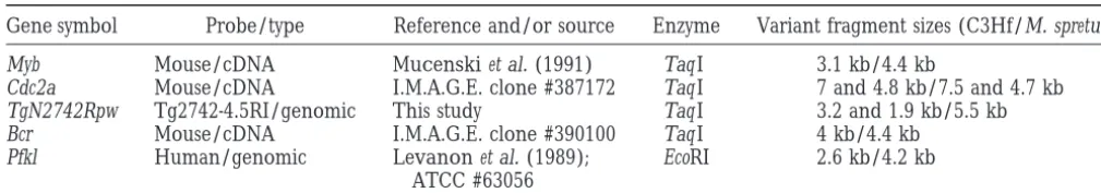
Gene symbol Probe/type Reference and/or source Enzyme Variant fragment sizes (C3Hf/M. spretus)
Myb Mouse/cDNA Mucenskiet al. (1991) Taq I 3.1 kb/4.4 kb
Cdc2a Mouse/cDNA I.M.A.G.E. clone #387172 Taq I 7 and 4.8 kb/7.5 and 4.7 kb
TgN2742Rpw Tg2742-4.5RI/genomic This study Taq I 3.2 and 1.9 kb/5.5 kb
Bcr Mouse/cDNA I.M.A.G.E. clone #390100 Taq I 4 kb/4.4 kb
Pfkl Human/genomic Levanonet al. (1989); EcoRI 2.6 kb/4.2 kb ATCC #63056
et al. 1990). The genomic DNA was restriction digested for the TgN2742Rpw line (data not shown). On the basis 12–15 hr with rare cutting enzymes under conditions sug- of the recessive genetics of the mutant trait, coupled gested by the manufacturer (New England Biolabs, Beverly,
with the fact that none of the numerous other lines MA). The protocols for pulsed-field gel electrophoresis
that have been prepared with the same transgene DNA (PFGE), Southern blotting, probing, and washings were
car-ried out as described previously (Stubbset al. 1994). construct showed a deaf-circling phenotype, we
con-Interspecific backcross mapping: Southern blots carrying clude that the phenotype in the TgN2742Rpw line arose
restriction-digested DNA from 160 interspecific backcross from a mutation caused by the integration of the progeny (IB;C3Hf/Rl-MgfSl2ENURg/1 3Mus spretus)3C3Hf/
transgene sequences into the host genomic DNA. Rl;Stubbset al. 1996) were prepared, and the blots hybridized
Histological examination: Histological examination with radiolabeled probes as previously described (Stubbset
al. 1990). Probes representing the Cdc2a and Bcr loci were of inner ear specimens from adult mutant mice showed cDNA clones identified by searching the dBEST database inner ear abnormalities affecting both the cochlear and (Bouguski et al. 1993) for mouse expressed sequence tags vestibular portions of the membranous labyrinth.
Analy-(ESTs) that matched published sequences of the two genes
sis of homozygotes at z1 yr of age revealed that the most closely linked to the mutant region (mouse Cdc2a,
Gen-organ of Corti is almost entirely missing throughout all bank accession no. M38724; Human BCR cDNA, Genbank
accession no. U0700). The cDNA clones were obtained from cochlear turns; that is, in most areas no inner or outer the I.M.A.G.E. consortium collection through Research Ge- hair cells or supporting cells are present (Figure 1, netics, Inc. (Huntsville, AL). The Pfkl cDNA was obtained
A–D). However, as illustrated in Figure 1D, scattered from the American Type Culture Collection [(ATCC),
Rock-remnants of the organ of Corti are occasionally noted ville, MD]. The mouse Myb cDNA was kindly provided by
and these are most often present in the apical cochlear Dr. Michael Mucenski. Data were stored and analyzed and
standard errors calculated using the Map Manager data analy- turn. In areas where some sensory epithelium remains, sis program (Manly1993). Probes representing other Mmu10 recognizable fluid spaces can be seen within the organ gene markers and information regarding variant fragments
of Corti. The spiral ganglion shows severely reduced that were used to follow their segregation are summarized in
numbers of neurons, which probably reflects ganglion Table 1.
cell degeneration secondary to loss of the inner hair cells. Although scattered strial atrophy affecting primar-RESULTS ily the apical cochlear turn has been noted, the stria vascularis in these mutants appears mostly normal. In
Generation of theTgN(Imw)2742Rpwmutant line:We
addition, Reissner’s membrane and the membranous identified a new recessive insertional mutation in mice
wall of the saccule are in normal positions. that confers a deafness and a circling phenotype. This
The TgN2742Rpw mutant mice also show vestibular mutation is designated as TgN(Imw)2742Rpw (Im,
inser-abnormalities, including degeneration of the saccular tional mutation; w, waltzer), in accordance with
stan-neuroepithelium and occasional malformation of utric-dard nomenclature rules (Gordon1992), and will be
ular otoconia. In adult mice, the neuroepithelium of abbreviated as TgN2742Rpw. The transgene in the
the saccular macula is completely absent, with no
recog-TgN2742Rpw line, which is composed of a rat lectin
nizable hair cells or supporting cells (Figure 2, A and cDNA under the control of the human beta-actin
pro-B). The neuroepithelia of the utricle and semicircular moter, segregates in a simple Mendelian fashion. Mice
ducts appear normal. However, sparsely distributed ab-heterozygous for the transgene in this line are
phenotyp-normally large otoconia have been noted overlying the ically normal. Of the offspring,z25% arising from a
utricular macula in some specimens (Figure 2, C and D). cross between heterozygous transgenic parents develop
ABR and behavioral analyses: We initially observed the phenotype, and these progeny are all homozygous
that the mutant mice did not respond to sharp metallic for the transgene (data not shown). Cytogenetic analysis
sounds, which suggested that they have impaired hear-of a homozygote and a heterozygote showed no
detect-ing ability. To quantify their auditory function, ABR was able chromosomal abnormality, indicating that no gross
Figure1.—Histological anal-ysis of the cochlea from normal and TgN2742Rpw mutant mice atz1 yr of age. (A) Low-power view of normal mouse cochlear duct in the upper basal turn showing major anatomical fea-tures: Reissner’s membrane (RM), stria vascularis (SV), or-gan of Corti (arrow), spiral ganglion (G). Bar, 100mm. (B) Low-power view of cochlear duct in the upper basal turn from an adult TgN2742Rpw mu-tant. Reissner’s membrane (RM) and the stria vascularis (SV) appear normal; however, the organ of Corti is entirely absent (arrow) and the spiral ganglion (asterisk) shows a greatly reduced neuronal pop-ulation. Bar, 100mm. (C) Nor-mal mouse organ of Corti in the upper basal turn illustrat-ing the two types of auditory sensory cells: inner hair cells (arrow) and three rows (1,2,3) of outer hair cells with underly-ing supportunderly-ing cells. T, tector-ial membrane. Bar, 50mm. (D) Degenerating organ of Corti in the apical cochlear turn of a TgN2742Rpw mutant. Fluid spaces are present (asterisk) within the organ of Corti, although no intact inner or outer hair cells can be identified. T, tectorial membrane. Bar, 50mm.
We tested the mice between 18 and 24 days postnatal, tivity. Results from one such evaluation are shown in Figure 3. ABR recordings indicated that the mice dis-since this represents a time period during which the
mouse inner ear matures to achieve adult hearing sensi- playing the circling phenotype were totally deaf at all
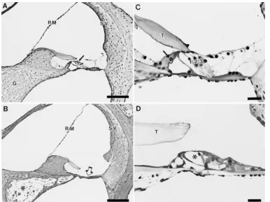
Figure 2.—Morphology of
the corresponding wild-type region. For this purpose, a genomic cosmid library was prepared from the
TgN2742Rpw mutant mice and screened using a plasmid
rescue procedure that was originally used for cloning the limb deformity locus (Woychiket al.1985; see materi-als and methodsfor details). The cosmid clones ob-tained were screened for the presence of mouse geno-mic DNA flanking the transgene insertion site. One such clone, cosmid-9, containedz25 kb of mouse geno-mic DNA in addition to approximately three copies of the transgene (≈20 kb) (Figure 4A). Within the 25 kb of mouse genomic DNA on cosmid-9, we identified a unique copy fragment, F9 (Figure 4A), and used this fragment as a probe to detect a restriction fragment length polymorphism (RFLP) associated with the mu-tant allele. To accomplish this, F9 was hybridized to Southern blots of restriction-enzyme-digested wild type, heterozygous, and homozygous DNA that had been solved by pulsed-field gel electrophoresis. We used re-striction enzymes that cut within the transgene (such as SalI) as well as enzymes that do not cut within the transgene (such as SfiI). In both cases, we were able to detect a unique RFLP in DNA containing the mutant
Figure 3.—Representative ABR waveforms from
allele (Figure 4, B and C). Therefore, the F9 probe
TgN2742Rpw/1 (Tg/1) and TgN2742Rpw/TgN2742Rpw
corresponds to the region flanking the transgene inser-(Tg/Tg) adult mice in response to a click stimulus.
Character-istic waveform (five peaks) was obtained for TgN2742Rpw/1 tion site. Furthermore, data from the SfiI restriction mice at the various intensities tested (40–70 dB), while no analysis suggest thatz30 copies of the transgene are response was elicited for TgN2742Rpw/TgN2742Rpw mice present at the site of insertion (Figure 4, B and C). even at 100 dB. The stimulus was initiated at time 0 (x axis)
To further characterize the mutant locus, a wild-type and the ABR waveforms were detected within 5–7 msec.
Ampli-BAC mouse genomic library was screened using the F9 tude (along the y axis) scale bars are indicated on the right
(in microvolts). sequence as a probe. Three clones were isolated with
an average insert size of 110 kb. One of these clones (BAC36) spans the transgene insertion site on the frequencies tested; the noncircling siblings, whether TgN2742Rpw mutant allele (Figure 4A). We made this
they were wild type or heterozygous for the mutation, determination by utilizing end-clones derived from the had thresholds in the normal range. Mutant mice older insert DNA contained in the BAC36 (labeled “A” and than 3.5 wk also showed no signs of auditory function “B” in Figure 4A) as probes on Southern blots con-(data not shown). taining genomic DNA digested with an enzyme that cuts To test for vestibular dysfunction in the TgN2742Rpw within the inserted transgene DNA (SalI). The genomic mutant mice, we conducted swimming tests. Mice from DNA samples that were used for this experiment were three litters derived from heterozygous parents were prepared from mice that were either wild type, heterozy-tested at 24–30 days of age. A total of 9 wild-type (1/1), gous, or homozygous for the TgN2742Rpw mutation. 15 heterozygous (TgN2742Rpw/1), and 8 homozygous Analysis of the data revealed that end-clones A and B (TgN2742Rpw/TgN2742Rpw) mice were tested. All the hybridize to the same-sized SalI fragment (680 kb) in
1/1and TgN2742Rpw/1mice displayed normal swim- the wild-type DNA, but each hybridized to a unique ming behavior resulting in proper orientation of the SalI fragment in the mutant allele; i.e., end-clone A
mice with respect to the water surface (seematerials hybridizes with a 415-kb fragment, and end-clone B hy-and methods). However, all of the TgN2742Rpw/ bridized with a 375-kb fragment (Figure 4D). The fact
TgN2742Rpw mice showed abnormal swimming behav- that both end-clones hybridize to the mutant DNA indi-ior associated with lack of orientation: when placed in cates that there is not a large deletion (.110 kb, the water, the mutants spiraled underwater (in a corkscrew size of the BAC) associated with the transgene insertion fashion) unable to maintain their noses and tails above site.
the water surface. Mutants needed to be rescued Mapping the TgN2742Rpwmutant locus:To localize promptly to prevent drowning. the TgN2742Rpw transgene insertion site on the genetic
Cloning the mutant locus:To characterize the muta- map, we used a 4.5-kb EcoRI fragment from the region tion in the TgN2742Rpw line at the molecular level, flanking the transgene insertion as a probe to analyze DNA from a previously described M. musculus 3 M.
Figure4.—Characterization of the TgN2742Rpw mutant locus. (A) Gross physical map of the mutant locus in the TgN2742Rpw line and its corresponding wild-type region based on pulsed-field gel electrophoresis. The inserted transgene DNA is designated as a hatched box. The unique sequence fragments [F9, end-clone A (represented by letter “A”), and end-clone B (represented by letter “B”)] used as probes are shown as black boxes. Numbers refer to DNA fragment sizes in kilobases. Figure not to scale to show structure of clones. Precise end points of the BAC clone have not been determined, and the SalI sites have not been mapped with respect to the SfiI sites. Restriction sites: S, SalI; Sf, SfiI. (B–D) Southern blots of spleen DNA from wild-type (1/
sites are tightly linked on mouse chromosome 10 (within 0–2.9 cM at 99% confidence limits).
DISCUSSION
A new mouse mutation with inner ear defects: We have identified a new mouse mutation that causes deaf-ness and balance disorders associated with morphologi-cal defects of the inner ear. Phenotypic characterization of the mutant mice revealed characteristic changes in the neuroepithelium of the inner ear. Additionally, in-vestigation of the hearing function using the ABR proce-dure showed that the TgN2742Rpw mutation causes total deafness. Since the mutants are deaf at all frequencies tested, it appears that the defect is not restricted to a particular region of the cochlea that is responsive to specific sound frequencies. Our histological examina-tion of inner ears from adult mutants revealed nearly complete absence of the organ of Corti together with loss of spiral ganglion cells in the cochlea. In addition, there is loss of the neuroepithelium of the saccule, to-gether with an occasional malformation of the otoconia
Figure 5.—Map location of TgN2742Rpw mutation. (A) overlying the utricular macula. These abnormalities are
Partial map of mouse chromosome 10 showing the position sufficiently severe to account for the auditory and vestib-of the TgN2742Rpw insertion site relative to other Mmu10
ular deficits observed during functional testing of the genes. The positions of human homologues of Myb, Cdc2a,
mice.
Bcr, and Pfkl are shown to the left of the map in the upper
portion of the figure. (B) Summary of the distribution of M. Although there is virtually a complete loss of the
co-spretus or M. musculus (C3Hf/Rl) alleles detected by Myb, chlear neuroepithelium in the mutant mice, the stria Cdc2a, Bcr, TgN2742Rpw, and Pfkl probes in 160 IB progeny. vascularis appears essentially normal and there is no
Each column of boxes represents a type of parental or
recom-evidence of reduced fluid volume in the cochlear duct binant chromosome; the number of backcross mice inheriting
or saccule. The inner ear pathology is, therefore, differ-a pdiffer-articuldiffer-ar type of chromosome differ-are listed differ-at the bottom of
each column. White boxes represent the inheritance of a ent from that found in the cochleo-saccular mutants, C3Hf/Rl allele for a gene, while black boxes denote the pres- which show strial degeneration and collapse of the mem-ence of M. spretus alleles. Recombination distances calculated branous walls of the labyrinth. In cochleo-saccular mu-from these data, given in centimorgans (cM) with standard
tants, such as the dominant spotting (W) or Steel (Sl) mice errors, are shown at right between rows representing adjacent
(Steel1995), degeneration of the organ of Corti occurs genes.
as a consequence of disturbed inner ear fluid homeosta-sis resulting from dysfunction of the stria vascularis. In our TgN2742Rpw mutant line, the mutation appears to
spretus interspecific backcross (see materials and
methods). The segregation pattern detected by the 4.5- have a direct effect on the neuroepithelia of the cochlea and the saccule. Therefore, the phenotype in the kb probe was compared to those of more than 450
markers that had been mapped previously in the same TgN2742Rpw mutant is not a secondary consequence of
defects in the stria vascularis. IB system (L. Stubbs, unpublished results). These
stud-ies mapped the transgene insertion site to the central The fact that remnants of the organ of Corti are present in the adult mutant mice suggests that the co-portion of mouse chromosome 10, located just distal to
the gene encoding cell division cycle control protein chlear structures may develop in a relatively normal fashion and then undergo degeneration sometime after 2a (Cdc2a; located within 0.1–5.8 cM of that gene at
99% confidence limits, as computed by Map Manager birth. Morphological studies on inner ear tissues taken at closely spaced intervals after the time of birth are software). The 4.5-kb fragment cosegregated with the
conserved breakpoint cluster region gene Bcr (Figure currently under way to determine the extent of early development of the neuroepithelia and to characterize 5), indicating that Bcr and the TgN2742Rpw insertion
the sequence of degenerative changes leading to the close together, the homologues of these two genes are not linked in the human genome. The human counter-abnormalities observed in the adult mutant mice. These
studies should provide valuable insights into the dev- part of Cdc2a, CDC2, has been mapped to human chro-mosome 10q21 (Nazarenkoet al. 1991), while BCR is
elopmental origin of the inner ear defects in the
TgN2742Rpw mutant line and should allow a more de- located on chromosome 22q11 (Shtivelman et al.
tailed phenotypic comparison of this mutant with other 1985). Genes located,1 cM distal from Bcr are related previously described mice having mutations of the neu- to sequences located on human chromosome 21q22.3,
roepithelial type. suggesting that the 22q11/Mmu10 homology region is
Thus far our light microscopic observations in adult relatively small (Burmeisteret al. 1998). It is therefore
animals indicate that degenerative changes of the inner difficult to predict whether human sequences related ear sensory epithelia in TgN2742Rpw mutants are lim- to the TgN2742Rpw mutation will be found in chromo-ited to the cochlea and saccule, and that there are no somes 10q21, 22q11, or 21q22.3.
obvious abnormalities of the neuroepithelia of the utri- Some potentially interesting connections between the cle and semicircular ducts. This pattern of morphologi- TgN2742Rpw phenotype and a human deafness locus
cal defects has also been reported to occur in several mapped to chromosome 10 are noteworthy. This deaf-other neuroepithelial mutants such as shaker-2, pirouette, ness mutation, DFNB12, has similar features with
waltzer, and spinner (Deol1968). However, recent stud- TgN2742Rpw and has recently been mapped to
10q21-ies based on electron and laser confocal microscopy q22 (Chaib1996). Mutations in the DFNB12 locus are have revealed more widespread involvement of the ves- associated with a recessive nonsyndromic deafness tibular apparatus in some of these mutants (Raphael marked by a profound prelingual sensorineural hearing
et al. 1999). We are currently pursuing electron micro- defect. DFNB12 has been proposed to possibly represent scopic work to supplement our light microscopic studies the human homologue of the murine jc, av, or v muta-in an effort to determmuta-ine whether ultrastructural defects tions (Petit1996). This raises the possibility that there may also be present in the utricular macula and semicir- may be a connection between the TgN2742Rpw and cular duct cristae of the TgN2742Rpw mutants. DFNB12 mutations.
The advantage of using an insertional mutation to Mutations that affect the sensory neuroepithelia are isolate a new gene is that the mutant locus is “tagged” a major cause of inner ear defects in humans. Given by the integrated DNA sequences and therefore can be the importance of this type of pathology in humans, cloned without having to resort to standard positional the transgenic line TgN2742Rpw may prove to be very cloning techniques. One potential problem with inser- useful as a model to study sensorineural deafness, and tional mutations generated by the pronuclear microin- specifically, to help in the identification of gene(s) es-jection procedure is that there can be large deletions sential for normal structure and function of the inner ear. associated with the mutant locus (Singh et al. 1991;
We thank Crystal Rohrbaugh, and Drs. Noel Murcia, Mitch Klebig, Rijkers et al. 1994). Large deletions at the transgene and Scott Bultman for critical reading of the manuscript. The authors
insertion site can complicate efforts to identify the gene are grateful to Karen S. Pawlowski for histological processing of the that is directly associated with the mutant phenotype. inner ear tissues utilized in this study. This research was supported by National Institute on Deafness and Other Communication Disorders The data we describe here suggest that there has not
Grant 1RO1-DC03420 to R.P.W. and by the U.S. Department of Energy been a large deletion of DNA at the site of transgene
under contract W-7405-Eng-48 with the University of California, Law-integration in the TgN2742Rpw line. The fact that we rence Livermore National Laboratory, for L.S. and under contract have isolated a wild-type BAC clone that spans the DE-AC05-960R22464 with Lockheed Martin Energy Research, for transgene integration site should position us to find the N.L.A.C.
gene that is directly associated with the mutant pheno-type in this line.
The region to which the TgN2742Rpw insertion has LITERATURE CITED been mapped contains several other mouse mutations
Ausubel, F. M., R. Brent, R. E. Kingston, D. D. Moore, J. G. Seidman
that affect the function and development of the inner et al., 1991 Current Protocols in Molecular Biology. Greene Publish-ear. The recently published chromosome 10 committee ing Associates and Wiley-Interscience, New York.
Avraham, K. B., T. Hasson, K. P. Steel, D. M. Kingsley, L. B.
report places Bcr and Cdc2a at 40 and 38.0 cM from the
Russellet al., 1995 The mouse Snell’s waltzer deafness gene Mmu10 centromere, respectively, which positions these
encodes an unconventional myosin required for structural integ-two genes near the TgN2742Rpw mutation and three rity of inner ear hair cells. Nat. Genet. 11: 369–375.
Bates, P. F., andSwift, R. A.1983 Double cos site vectors: simplified previously identified inner ear mutations, namely, Ames
cosmid cloning. Gene 26: 137–146. Waltzer (av, 37 cM), Jackson circler ( jc, 33 cM), and
Bouguski, M. S., T. M. LoweandC. M. Tolstoshev, 1993 dbEST— Waltzer (v, 30 cM). Given the margins of error associ- database for expressed sequence tags. Nat. Genet. 4: 332–333.
Burmeister, M., E. C. Bryda, J. F. BureauandK. Noben-Trauth, ated with these consensus map positions, it is possible
1998 Encyclopedia of the mouse genome VII. Mouse chromo-that the TgN2742Rpw mutation is allelic with one of
some 10. Mamm. Genome 8: Spec No. S200-14.
somal recessive deafness, to chromosome 10q21-22. Hum. Mol. of the 22nd. Midwinter Meeting of the Association for Research in Otolaryngology, p. 8.
Genet. 5: 1061–1064.
Rijkers, T., A. PeetzandU. Ruther, 1994 Insertional mutagenesis
Deol, M. S., 1968 Inherited disease of the inner ear in man in the
in transgenic mice. Transgenic Res. 3: 203–215. light of studies on the mouse. J. Med. Genet. 5: 137–158.
Sambrook, J., E. F. FritschandT. Maniatis, 1989 Molecular
Clon-Erway, L., J. Willott, J. ArcherandD. Harrison, 1993 Genetics
ing: A Laboratory Manual. Cold Spring Harbor Laboratory Press,
of age-related hearing loss in mice: I. Inbred and F1 hybrid strains.
Cold Spring Harbor, NY. Hear. Res. 65: 125–132.
Schuknecht, H. F.1993 Developmental defects, pp. 115–189 in
Fraser, G. R., 1976 The Causes of Profound Deafness in Childhood. Johns
Pathology of the Ear, edited byR. K. Bussey, T. LazarandF. M.
Hopkins University Press, Baltimore.
Klass.Lea & Febiger, Philadelphia.
Gibson, F., J. Walsh, P. Mburu, A. Varela, K. A. Brownet al., 1995
Shtivelman, E., B. Lifshitz, R. P. GaleandE. Canaani,1985 Fused A type VII myosin encoded by the mouse deafness gene
transcript of abl and bcr genes in chronic myelogenous leukae-shaker-1. Nature 374: 62–64.
mia. Nature 315: 550–554.
Gordon, J.1992 Standardized nomenclature for transgenic animals.
Singh, G., D. M. Supp, C. Schreiner, J. McNeish, H.-J. Merkeret
ILAR News 34: 45.
al., 1991 legless insertional mutation: morphological, molecular,
Kalatzis, V., andC. Petit, 1998 The fundamental and medical
and genetic characterization. Genes Dev. 5: 2245–2255. impacts of recent progress in research on hereditary hearing loss.
Steel, K. P. 1995 Inherited hearing defects in mice. Annu. Rev. Hum. Mol. Gen. 7: 1589–1597. Genet. 29: 675–701.
Levanon, D., E. Danciger, N. Dafni, Y. Bernstein, A. Elsonet al., Stubbs, L., A. Poustka, A. Baron, H. Lehrach, P. Lonaiet al., 1990
1989 The primary structure of human liver type phosphofructo- The murine genes Hox-5.1 and Hox-4.1 belong to the same HOX kinase and its comparison with other types of PFK. DNA 8: 733– complex on chromosome 2. Genomics 7: 422–427.
743. Stubbs, L., E. M. Rinchik, E. Goldberg, B. Rudy, M. A. Handelet
Liu, X. Z., J. Walsh, Y. Tamagawa, K. Kitamura, M. Nishizawaet al., al., 1994 Clustering of six human 11p15 gene homologs within
1997 Mutation in the myosin VIIA gene causes non-syndromic a 500-kb interval of proximal mouse chromosome 7. Genomics recessive deafness. Nat. Genet. 16: 188–190. 24:324–332.
Manly, K. F., 1993 A Macintosh program for storage and analysis of Stubbs, L., L. Chittenden, A. ChakrabartiandE. Onaivi, 1996 experimental genetic mapping data. Mamm. Genome 4: 303–313. The gene encoding the central cannabinoid receptor is located
Morton, N. E.1991 Genetic epidemiology of hearing impairment. in proximal mouse Chromosome 4. Mamm. Genome 7: 165–166.
Van Camp, G., andR. J. H. Smith Hereditary hearing loss homepage. Ann. NY Acad. Sci. 630: 16–31.
World Wide Web URL: http://dnalab-www.uia.ac.be/dnalab/
Mucenski, M. L., K. McLain, A. B. Kier, S. H. Swerdlow, C. M.
hhh/.
Schreineret al., 1991 A functional c-myb gene is required for
Wang, A., Y. Liang, R. A. Fridell, F. J. Probst, E. R. Wilcoxet al.,
normal murine fetal hepatic hematopoiesis. Cell 65: 677–689.
1998 Association of unconventional myosin MYO15 mutations
Nadeau, J. H., M. KosowskyandK. P. Steel, 1991 Comparative
with human nonsyndromic deafness DFNB3. Science 280: 1447– gene mapping, genome duplication, and the genetics of hearing.
1451. Ann. NY Acad. Sci. 630: 49–67.
Weil, D., S. Blanchard, J. Kaplan, P. Guilford, F. Gibsonet al.,
Nazarenko, S. A., N. V. OstroverhovaandN. K. Spurr, 1991
Re-1995 Defective myosin VIIA gene responsible for Usher syn-gional assignment of the human cell cycle control gene CDC2
drome type 1B. Nature 374: 60–61. to chromosome 10q21 by in situ hybridization. Hum. Genet. 87:
Weil, D., P. Kussel, S. Blanchard, G. Levy, F. Levi-Acobaset al.,
621–622.
1997 The autosomal recessive isolated deafness, DFNB2, and
Petit, C.1996 Genes responsible for human hereditary deafness:
the Usher 1B syndrome are allelic defects of the myosin-VIIA symphony of a thousand. Nat. Genet. 14: 385–391. gene. Nat. Genet. 16: 191–193.
Probst, F. J., R. A. Fridell, Y. Raphael, T. L. Saunders, A. Wang Woychik, R. P., T. A. Stewart, L. G. Davis, P. D’EustachioandP.
et al., 1998 Correction of deafness in shaker-2 mice by an uncon- Leder, 1985 An inherited limb deformity created by insertional ventional myosin in a BAC transgene. Science 280: 1444–1447. mutagenesis in a transgenic mouse. Nature 318: 36–40.
Raphael, Y., D. C. Kohrman, F. J. Probst, E. Lambert, L. Beyeret