C L I N I C A L T R I A L R E P O R T
Peripheral Blood Eosinophil as a Biomarker in
Outcomes of Acute Exacerbation of Chronic
Obstructive Pulmonary Disease
This article was published in the following Dove Press journal: International Journal of Chronic Obstructive Pulmonary Disease
Hong-Xia Wu1,*
Kai-Quan Zhuo2,*
De-Yun Cheng1,*
1Department of Respiratory and Critical
Care Medicine, West China Hospital,
Sichuan University, Chengdu, People’s
Republic of China;2Department of
Neurosurgery, Suining Municipal Hospital
of TCM, Suining, People’s Republic of
China
*These authors contributed equally to this work
Purpose: Mounting evidence suggests that eosinophil levels correlate with the effects of therapy and phenotype for chronic obstructive pulmonary disease (COPD). This study aimed to clarify the relationship between eosinophil levels and clinical outcomes in patients with acute exacerbation of COPD (AECOPD).
Methods: A prospective, multicenter, observational cohort study was performed in three teaching hospitals. Patients were grouped by quartile percentage (0, 0.7, 2.55) and absolute blood eosinophils count (0, 0.05×10⁹/L, 0.17×10⁹/L) and divided into four numbered groups ranked from low to high.
Results: The study included 493 AECOPD patients. In the percentile-ranked groups, patients in Group 1 experienced significantly longer hospital stays, higher rates of both noninvasive mechanical ventilation (NIMV), and heart failure than those in Group 4 (12 days vs 10 days, p = 0.005; 29.5% vs 23.6%, p = 0.007; 48.4% vs 28.5%, p = 0.001). Group 1 also had higher frequencies of respiratory failure and pulmonary heart disease compared to Groups 3 and 4 (54.8% vs 34.8%, p = 0.002; 54.8% vs 35%, p = 0.003). In the absolute count-ranked groups, patients in Group 1 had significantly higher rates of NIMV than those in Group 3 (41.1% vs 21.7%, p = 0.001), had higher rates of heart failure, respiratory failure, and pulmonary heart disease than those in Group 3 and 4 (48.1% vs 30.2%, p = 0.003; 48.1% vs 30.4%, p = 0.005; 50.8% vs 32.2%, p = 0.004; 50.8% vs 34.1%, p = 0.008; 51.9% vs 34.1%, p = 0.004; 51.9% vs 33%, p = 0.003). There were outcome differences among the admitting hospital of stays in the absolute count groups (p = 0.002), but the differences were not significant in a pairwise comparison. The proportion of ICU admissions and mortality was different in two cohorts with no difference in a pairwise comparison.
Conclusion: Patients with lower eosinophil counts experienced poorer clinical outcomes. Eosinophil levels may be a helpful marker to predict outcomes in AECOPD.
Keywords: eosinophils, chronic obstructive pulmonary disease, exacerbation, biomarkers, mortality
Introduction
Chronic obstructive pulmonary disease (COPD) is a fatal disease that is projected to
be the third most common cause of death worldwide within the next three years.1
COPD is a heterogeneous disease that exhibits complex pathological features.
Exacerbations of COPD are defined as an acute worsening of respiratory symptoms
resulting in a need for additional therapy.2,3Aside from neutrophilic inflammation,
eosinophilic inflammation is a new area of research. The latest studies demonstrated
that eosinophilic inflammation exists in both stable and acute exacerbations of
Correspondence: De-Yun Cheng Department of Respiratory and Critical Care Medicine, West China Hospital, Sichuan University, No. 37 Guoxue Alley,
Chengdu 610041, Sichuan, People’s
Republic of China
Email chengdeyunscu@163.com
International Journal of Chronic Obstructive Pulmonary Disease
Dove
press
open access to scientific and medical research
Open Access Full Text Article
International Journal of Chronic Obstructive Pulmonary Disease downloaded from https://www.dovepress.com/ by 118.70.13.36 on 22-Aug-2020
COPD (AECOPD).4,5 Mounting evidence suggests that eosinophil levels may be related to the effects of therapy
and outcomes, even in the absence of asthma.6–9 Blood
eosinophils are usually used as a biomarker for response to inhaled steroids (ICS) and exacerbation risk in stable
COPD.10–16 Bafadhel et al found that eosinophil count
(100/µL and 300/µL) predicted the risk of exacerbations and the response to treatment with ICS in patients with
COPD.17 Blood eosinophil counts are recommended by
the Global Initiative for Chronic Obstructive Lung Disease (GOLD) as a biomarker to guide ICS therapy in clinical practice.1However, the effect of blood eosinophil in stable and acute cases may be different.
Saltürk et al reported that patients with eosinophil levels less than 2% experienced shorter intensive care
unit (ICU) stays and lower mortality rates.18 Similarly,
Kang suggested better pulmonary function, lower admis-sions to the ICU, and mortality in a group with eosinophil
levels less than 2%.19 Duman et al reported that shorter
hospital stays and lower readmission rates were found in the group with eosinophilia, but no differences were found
in six-month mortality.20Patients with AECOPD, and low
(< 50/µL) eosinophil were strongly associated with longer median hospital stay (7 vs 4 days, P < 0.001), and lower 12-month survival (82.4% vs 90.7%, P < 0.028) than
patients with high (> 150/µL) eosinophil counts.21 An
eosinophil value of < 0.144×10⁹/L (or less than 2%) on
admission was associated with a longer hospital stay for
AECOPD.22
However, the effect of blood eosinophil in AECOPD outcomes remains controversial. Two studies found longer
hospitalization,23,24 a greater need for mechanical
ventilation,23 and increased mortality23,24 in the group
with eosinophilia. Further, a higher frequency of
readmis-sion for AECOPD25 and a higher rate of exacerbations26
have been found in those with eosinophilia. This study aimed to investigate the effect of peripheral blood eosino-phil in patients who experienced AECOPD.
Methods
This prospective, multicenter study was conducted in three university-affiliated hospitals in China. Patients who were hospitalized for AECOPD between September 2018 and February 2019 were enrolled. The study was approved by the local ethics committee (Biomedical Ethics Committee of West China Hospital of Sichuan University) and com-plied with the Declaration of Helsinki. All patients provided written informed consent. The study was registered in the
Chinese Clinical Trial Registry (ChiCTR1900024210). All data were collected from questionnaire surveys and hospital databases.
Subject
The definitions of COPD were based on the GOLD criteria.
Spirometry is required to diagnose COPD. The presence of a post-bronchodilator FEV1/FVC ratio < 0.70 confirms the
presence of persistent airflow limitation and, thus, COPD in
patients with appropriate symptoms and significant
expo-sures to noxious stimuli.1COPD exacerbations are defined
as an acute worsening of respiratory symptoms that result in additional therapy.1Inclusion criteria were as follows:≥40 years of age with AECOPD; routine baseline peripheral blood test was performed before receiving any antibiotic or systemic corticosteroid therapy (prednisone > 0.5 mg/kg or equivalent doses). Patients admitted due to other medical problems, those with a history of asthma, active pulmonary tuberculosis, interstitial pulmonary disease or lung cancer, those undergoing chronic oral steroid therapy, those with other diseases that could influence eosinophil count (allergic diseases, parasitic infections, eosinophilic pneumonia), and individuals with severe dysfunction of other organs or sys-tems or malignant tumors were excluded.
Measurements
Patient baseline characteristics, including age, sex, body mass index, allergy history, smoking history, duration of disease, long-term home oxygen therapy, regular medica-tions, heart and respiratory rate on admission, comorbid-ities (hypertension, diabetes mellitus, arrhythmia, chronic ischemic heart disease, congestive heart failure, peripheral vascular disease, bronchiectasis, respiratory failure, and pulmonary heart disease), manner of hospital admission, the number of hospital or emergency admissions in the previous year, laboratory data (routine blood test and arterial blood gas analysis), admission to the ICU, length of hospital stay, rate, and duration of noninvasive mechan-ical ventilation (NIMV), and hospital medmechan-ical treatment and mortality, were recorded. A COPD assessment test
(CAT), modified British Medical Research Council
(mMRC), and the refined ABCD assessment were also
evaluated using questionnaires. The primary outcome measure was the length of the hospital stay. Secondary outcome measures included ICU admission rate, and
dura-tion of noninvasive ventilation, comorbidities, and
mortality.
International Journal of Chronic Obstructive Pulmonary Disease downloaded from https://www.dovepress.com/ by 118.70.13.36 on 22-Aug-2020
Analysis
Clinical outcomes were compared among patients grouped according to quartile-percent and absolute count of peripheral blood eosinophils (From low to high: Groups 1, 2, 3, 4). Pearson’s chi-squared test or Fisher’s exact was used to com-pare discrete variables. The Kruskal–Wallis test was used for pairwise comparisons if differences were revealed using the chi-squared test. Analysis of variance and nonparametric tests were used to compare continuous variables. Kaplan-Meier analysis was performed to identify the associated factors and hospital length of stay. The receiver operating characteristic (ROC) curve with the calculation of the area under the curve (AUC) was used to identify the cutoff values of eosinophils associated with longer hospital lengths of stay. A two-sided P≤0.05 was statistically significant. One-sixth of the P-value
≤0.05 was statistically significant in the Kruskal–Wallis test. All statistical analyses were performed using SPSS version 25.0 (IBM Corporation, Armonk, NY, USA).
Results
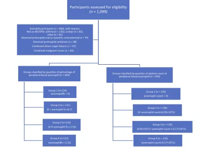
Overall, 1099 COPD patients who experienced acute exacer-bations were admitted during the study period, of whom 493
were analyzed (Figure 1). Medians of percentage and
absolute count of peripheral blood eosinophils were 0.7%
(interquartile range [IQR] 0–2.55) and 0.05 ×10⁹/L (IQR
0–0.17) in total. Patients were classified according to per-centage of eosinophil quartile as follows: Group 1 (n = 124), Group 2 (n = 131), Group 3 (n = 115), and Group 4 (n = 123). Similarly, patients were classified according to eosinophil count quartile as follows: Group 1 (n = 129), Group 2 (n = 120), Group 3 (n = 129) and Group 4 (n = 115). The proportion of males in the present study was 69.2%. The median (IQR) age, BMI, course of disease, and
length of hospital stay were 76 (68–83) years, 21.224
(18.5–24.315) kg/m2, 10 (5–20) days and 11(9–14) days, respectively. The clinical characteristics and laboratoryfi nd-ings on admission of the patients are summarized in Tables 1–4. AECOPD was treated with oxygen therapy, atomization, antibiotics, or systemic steroids. Antibiotics and systemic steroids were prescribed at the discretion of the attending physician.
The primary outcome measure was the length of the hospital stay. The length of hospital stays was found to be different in the two types of groups (p = 0.01, p = 0.002) (Tables 5and 6). In pairwise comparison, a significantly longer hospital stay was found in Group 1 than in Group 4
Participants assessed for eligibility (n = 1,099)
Groups classified by quartiles of percentage of peripheral blood eosinophil (n = 493)
Group 1 (n=124) (eosinophil% = 0)
Group 2 (n = 131)
(0 < eosinophil % ≤0.7)
Group 3 (n=115)
(0.7< eosinophil % ≤ 2.55)
Group 4 (n=113)
(eosinophil% > 2.55)
Groups classified by quartiles of abolute count of peripheral blood eosinophil (n = 493)
Group 1 (n = 129)
(eosinophil count = 0)
Group 2 (n = 120)
(0 <eosinophil count≤0.05×10⁹/L)
Group 3 (n = 129)
(0.05×10⁹/L< eosinophil count ≤ 0.17×10⁹/L)
Group 4 (n = 115)
(eosinophil count>0.17×10⁹/L)
Excluded participants (n = 606), with reasons: Not an AECOPD: asthma (n = 135); cardiac (n = 82);
other (n = 81)
Received prehospital oral or systemic corticosteroid (n = 79) Received prehospital antibiotic (n = 98)
Combined others organ failure ( n = 67)
Combined malignant tumor (n = 64)
Figure 1Flow chart of subjects.
International Journal of Chronic Obstructive Pulmonary Disease downloaded from https://www.dovepress.com/ by 118.70.13.36 on 22-Aug-2020
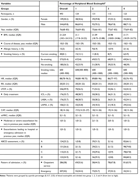
Table 1Patients’Characteristics on Admission of Quartile-Percentage of Eosinophil Cohorts
Variables Percentage of Peripheral Blood Eosinophila
Groups Overall 1 2 3 4
Participants, n 493 124 131 115 123
Gender, n (%) Female 149(30.2) 38(30.6) 39(29.8) 37(32.2) 35(28.5)
Male 344(69.8) 86(69.4) 92(70.2) 78(67.8) 88(71.5)
Year, median (IQR) 76(68–83) 76(69–83) 75(66–81) 77(67–83) 77(69–82)
● BMI, median (IQR) 21.224
(18.5–24.315)
21.1
(17.8–24.22)
21.09
(18.75–23.88)
20.98
(18.83–24.86)
21.71
(18.59–24.73)
● Course of disease, year, median (IQR) 10(5–20) 10(5–29) 10(5–20) 10(5–15) 10(5–10)
● Allergic history, n (%) 15(3) 3(2.4) 9(6.9) 1(0.9) 2(1.6)
● Smoking history, n (%) Current smoking 40(8.1) 15(12.1) 11(8.4) 8(7) 6(4.9)
Ex-smoking 273(55.4) 67(54) 69(52.7) 68(59.1) 69(56.1)
No-smoking 180(36.5) 42(33.9) 51(38.9) 39(33.9) 48(39)
Smoking index, median
600
(400–900)
600(300–800) 600
(400–1000)
600
(300–1000)
600
(300–900)
HR, median (IQR) 88(78–96.5) 90(80–98.75) 89(80–96) 86(77–97) 82(76–93)
RR, median (IQR) 20(20–21) 20(20–22) 20(20–21) 20(20–21) 20(20–21)
LTOT, n (%) 246(49.9) 70(56.5) 71(54.2) 53(46.1) 52(42.3)
● Daily treatment ICS, n (%) 176(35.7) 48(38.7) 50(38.2) 36(31.3) 42(34.1)
LABA, n (%) 176(35.7) 48(38.7) 50(38.2) 36(31.3) 42(34.1)
LAMA, n (%) 106(21.5) 32(25.8) 24(18.3) 21(18.3) 29(23.6)
CAT, median (IQR) 18(14–26) 19.5(14.25–27) 18(14–26) 19(14–26) 18(13–26)
mMRC, median (IQR) 2(1–3) 2(1–3) 2(1–3) 2(1–3) 2(1–3)
● Moderate or severe exacerbation
his-tory in previous year, median (IQR)
1(0–2) 1(0–2) 2(1–3) 2(0–3) 1(0–2)
● Exacerbations leading to hospital or
emergency admission in previous year, median (IQR)
1(0–2) 1(0–2) 1(0–3) 1(0–3) 1(0–2)
ABCD assessment, n (%) A 124(25.2) 1(0.8) 39(31.5) 2(1.6) 82(66.1)
B 131(26.6) 2(1.5) 29(22.1) 2(1.5) 98(74.8)
C 115(23.3) 2(1.7) 34(29.6) 1(0.9) 78(67.8)
D 123(24.9) 2(1.6) 36(29.3) 1(0.8) 84(68.3)
Pattern of admission, n (%) ● Outpatient
service
286(58) 69(55.6) 58(44.3) 78(67.8) 81(65.9)
Emergency 207(42) 55(44.4) 73(55.7) 37(32.2) 42(34.1)
Notes:aPatients were grouped by quartile percentage (0, 0.7, 2.55) of blood eosinophils and divided into group 1, 2, 3 and 4 (from low to high).
Abbreviations:IQR, interquartile range: 25%–75%; n, number; BMI, body mass index; ICS, inhaled corticosteroids; LAMA, acting muscarinic antagonist; LABA, long-acting beta agonist; HR, heart rate; RR, respiratory rate; LTOT, long-term oxygen therapy; CAT, COPD assessment test; mMRC, modified British medical research council.
International Journal of Chronic Obstructive Pulmonary Disease downloaded from https://www.dovepress.com/ by 118.70.13.36 on 22-Aug-2020
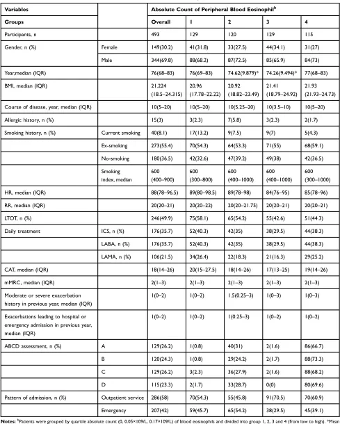
Table 2Patients’Characteristics on Admission of Quartile-Count of Eosinophil Cohorts
Variables Absolute Count of Peripheral Blood Eosinophilb
Groups Overall 1 2 3 4
Participants, n 493 129 120 129 115
Gender, n (%) Female 149(30.2) 41(31.8) 33(27.5) 44(34.1) 31(27)
Male 344(69.8) 88(68.2) 87(72.5) 85(65.9) 84(73)
Year,median (IQR) 76(68–83) 76(69–83) 74.62(9.879)* 74.26(9.494)* 77(68–83)
BMI, median (IQR) 21.224
(18.5–24.315)
20.96
(17.78–22.22)
20.92
(18.82–23.49)
21.41
(18.79–24.92)
21.93
(21.93–24.73)
Course of disease, year, median (IQR) 10(5–20) 10(5–20) 10(5.25–20) 10(3.5–10) 10(5–20)
Allergic history, n (%) 15(3) 3(2.3) 7(5.8) 3(2.3) 2(1.7)
Smoking history, n (%) Current smoking 40(8.1) 17(13.2) 9(7.5) 9(7) 5(4.3)
Ex-smoking 273(55.4) 70(54.3) 64(53.3) 71(55) 68(59.1)
No-smoking 180(36.5) 42(32.6) 47(39.2) 49(38) 42(36.5)
Smoking index, median
600
(400–900)
600
(300–800)
600
(400–1000)
600
(400–1000)
600
(300–1000)
HR, median (IQR) 88(78–96.5) 89(80–98.5) 89(78–98) 84(76–95) 85(78–96)
RR, median (IQR) 20(20–21) 20(20–22) 20(20–21.75) 20(20–21) 20(20–21)
LTOT, n (%) 246(49.9) 75(58.1) 65(54.2) 55(42.6) 51(44.3)
Daily treatment ICS, n (%) 176(35.7) 52(40.3) 42(35) 38(29.5) 44(38.3)
LABA, n (%) 176(35.7) 52(40.3) 42(35) 38(29.5) 44(38.3)
LAMA, n (%) 106(21.5) 34(26.4) 22(18.3) 21(16.3) 29(25.2)
CAT, median (IQR) 18(14–26) 20(15–27.5) 18(14–26) 17(13–25) 19(14–26)
mMRC, median (IQR) 2(1–3) 2(1–3) 2(1–3) 2(1–3) 2(1–3)
Moderate or severe exacerbation history in previous year, median (IQR)
1(0–2) 1(0–2) 1.5(0.25–3) 1(0–3) 1(0–3)
Exacerbations leading to hospital or emergency admission in previous year, median (IQR)
1(0–2) 1(0–2) 1(0.25–3) 1(0–2) 1(0–2)
ABCD assessment, n (%) A 129(26.2) 1(0.8) 40(31) 2(1.6) 86(66.7)
B 120(24.3) 1(0.8) 29(24.2) 2(1.7) 88(73.3)
C 129(26.2) 3(2.3) 36(27.9) 2(1.6) 88(68.2)
D 115(23.3) 2(1.7) 33(28.7) 0(0) 80(69.6)
Pattern of admission, n (%) Outpatient service 286(58) 70(54.3) 55(45.8) 91(70.5) 70(60.9)
Emergency 207(42) 59(45.7) 65(54.2) 38(29.5) 45(39.1)
Notes:bPatients were grouped by quartile absolute count (0, 0.05×10⁹/L, 0.17×10⁹/L) of blood eosinophils and divided into group 1, 2, 3 and 4 (from low to high). *Mean (SD, standard deviation).
Abbreviations:IQR, interquartile range: 25%–75%; n, number; BMI, body mass index; ICS, inhaled corticosteroids; LAMA, acting muscarinic antagonist; LABA, long-acting beta agonist; HR, heart rate; RR, respiratory rate; LTOT, long-term oxygen therapy; CAT, COPD assessment test; mMRC, modified British medical research council.
International Journal of Chronic Obstructive Pulmonary Disease downloaded from https://www.dovepress.com/ by 118.70.13.36 on 22-Aug-2020
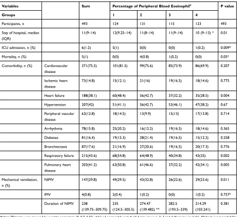
in the quartile-percentage eosinophil cohorts (12 vs 10 days; p = 0.005).
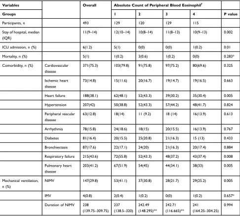
The secondary outcomes were the rate and duration of NIMV, comorbidities, mortality, and ICU admission. The frequencies of heart failure, respiratory failure, and pulmon-ary heart disease were found to be different between the two classified cohorts (p = 0.004, p = 0.005; p = 0.002, p = 0.008;
p = 0.005, p = 0.005, respectively) (Tables 5 and6). In the quartile-percentage eosinophil cohorts, patients in Group 1 experienced significantly higher rates of heart failure than those in Group 4 (48.4% vs 28.5%, p = 0.001), had higher frequencies of respiratory failure and pulmonary heart dis-ease compared with those in Groups 3 and 4 in a pairwise comparison (54.8% vs 34.8%, p = 0.002; 54.8% vs 35%, p =
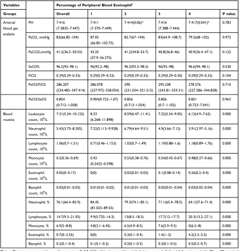
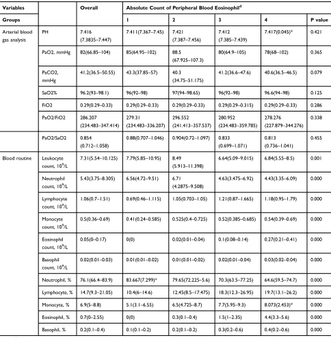
Table 3Patients’Laboratory Findings on Admission of Quartile-Percentage of Eosinophil Cohorts
Variables Percentage of Peripheral Blood Eosinophilc
Groups Overall 1 2 3 4 P value
Arterial blood gas analysis
PH 7.416
(7.3835–7.447)
7.411
(7.370–7.449)
7.414(0.06)* 7.416
(7.388–7.444)
7.417(0.041)* 0.783
PaO2, mmHg 82(66.85–104) 87.05
(66.85–103.75)
82.7(67–104) 83(64.9–108.7) 79.2(68–102) 0.972
PaCO2,mmHg 41.2(36.5–50.55) 43.25
(37.9–56.275)
41.2(34.8–53.7) 40.8(36.8–46) 40.9(36.4–47.1) 0.122
SaO2% 96.2(93–98.1) 96(92.2–98) 96.3(93.3–98.3) 96(92–98) 96.6(94–98.1) 0.530
FiO2 0.29(0.29–0.33) 0.29(0.29–0.33) 0.29(0.29–0.33) 0.29(0.29–0.30) 0.29(0.29–0.33) 0.104
PaO2/FiO2 286.207
(234.483–347.414)
286.078
(237.972–338.054)
290
(231.034–351.515)
295.238
(243.81–359.31)
278.276
(227.586–344.828)
0.710
PaO2/SaO2 0.854
(0.712–1.058)
0.904(0.722–1.07) 0.856
(0.713–1.054)
0.856 (0.7–1.102)
0.821
(0.723–7.041)
0.963
Blood routine
Leukocyte count, 109/L
7.31(5.54–10.125) 8.33
(6.268–11.898)
8.59(6.47–11.41) 7.22(5.54–9.05) 6.12(4.9–7.62) 0.000
Neutrophil count, 109/L
5.43(3.75–8.305) 7.32(5.113–9.928) 6.79(4.64–9.51) 4.9(3.66–7.12) 3.91(2.97–5.16) 0.000
Lymphocyte count, 109/L
1.06(0.7–1.51) 0.71(0.46–1.153) 1.03(0.7–1.49) 1.19(0.88–1.6) 1.18(0.89–1.76) 0.000
Monocyte count, 109/L
0.5(0.36–0.69) 0.43
(0.2425–0.598)
0.55(0.38–0.76) 0.54(0.43–0.67) 0.48(0.37–0.66) 0.000
Eosinophil count, 109/L
0.05(0–0.17) 0(0) 0.02(0.01–0.03) 0.1(0.08–0.14) 0.26(0.2–0.4) 0.000
Basophil count, 109/L
0.02(0.01–0.03) 0.01(0.01–0.02) 0.01(0.01–0.03) 0.02(0.01–0.04) 0.03(0.02–0.04) 0.000
Neutrophil, % 76.1(66.4–83.9) 84.45
(81.025–89.55)
79.3(74.1–85.1) 71.1(65.4–78.5) 64.1(57.6–71.4) 0.000
Lymphocyte, % 14.7(9.3–21.05) 9.9(5.725–14.3) 13(8.5–18.3) 17.7(12–17.7) 20.3(15.2–27.1) 0.000
Monocyte, % 6.9(5–8.8) 4.8(3.1–6.45) 6.5(4.9–8.5) 7.6(5.9–9.5) 0(6.5–8) 0.000
Eosinophil, % 0.7(0–2.55) 0(0) 0.3(0.1–0.4) 1.4(1–2) 4.2(3.3–5.5) 0.000
Basophil, % 0.2(0.1–0.4) 0.1(0.1–0.2) 0.2(0.1–0.3) 0.3(0.1–0.5) 0.5(0.2–0.7) 0.000
Notes:c
Patients were grouped by quartile percentage (0, 0.7, 2.55) of blood eosinophils and divided into group 1, 2, 3 and 4 (from low to high). *Mean (SD, standard deviation).
Abbreviations:IQR, interquartile range: 25%–75%; E%, percent of blood eosinophil in white blood cell; E, absolute count of peripheral blood eosinophil.
International Journal of Chronic Obstructive Pulmonary Disease downloaded from https://www.dovepress.com/ by 118.70.13.36 on 22-Aug-2020
0.003; 50.8% vs 32.2%, p = 0.004; 50.8% vs 34.1%, p = 0.008). In the eosinophil count cohorts, patients in Group 1 had significantly higher rates of heart failure, respiratory failure, and pulmonary heart disease than those in Groups 3 and 4 in a pairwise comparison (48.1% vs 30.2%, p = 0.003; 48.1% vs 30.4%, p = 0.005; 55.8% vs 37.2%, p=0.003; 55.8% vs 37.4%, p=0.004; 51.9% vs 34.1%, p = 0.004; 51.9% vs 33%, p = 0.003).
Except for the duration of NIMV, the proportion of ICU admission, NIMV, and mortality were found to vary among groups (p = 0.009, p = 0.01; p = 0.011, p = 0.005;
p = 0.05, p = 0.283) (Tables 5 and 6). There was
a significantly higher rate of NIMV in Group 1 than in
Group 4 in the percentage of eosinophil cohorts (29.5% vs 23.6%, p = 0.007). Comparable results were found in Group 1 compared to Group 3 in the eosinophil count
Table 4Patients’Laboratory Findings on Admission of Quartile-Count of Eosinophil Cohorts
Variables Overall Absolute Count of Peripheral Blood Eosinophild
Groups 1 2 3 4 P value
Arterial blood
gas analysis
PH 7.416
(7.3835–7.447)
7.411(7.367–7.45) 7.421
(7.387–7.456)
7.412
(7.385–7.439)
7.417(0.045)* 0.421
PaO2, mmHg 82(66.85–104) 85(64.95–102) 88.5
(67.925–107.3)
80(64.9–105) 78(68–102) 0.365
PaCO2,
mmHg
41.2(36.5–50.55) 43.3(37.85–57) 40.3
(34.75–51.175)
41.2(36.6–47.6) 40.6(36.5–46.5) 0.079
SaO2% 96.2(93–98.1) 96(92–98) 97(94–98.65) 96(92–98) 96.6(94–98) 0.125
FiO2 0.29(0.29–0.33) 0.29(0.29–0.33) 0.29(0.29–0.33) 0.29(0.29–0.315) 0.29(0.29–0.33) 0.286
PaO2/FiO2 286.207
(234.483–347.414)
279.31
(234.483–336.207)
296.552
(241.413–357.537)
280.952
(234.483–359.785)
278.276
(227.879–344.276)
0.338
PaO2/SaO2 0.854
(0.712–1.058)
0.88(0.707–1.046) 0.904(0.72–1.097) 0.833
(0.699–1.071)
0.813
(0.736–1.041)
0.455
Blood routine Leukocyte
count, 109/L
7.31(5.54–10.125) 7.79(5.85–10.95) 8.49
(5.913–11.398)
6.64(5.09–9.015) 6.84(5.55–8.5) 0.001
Neutrophil
count, 109/L
5.43(3.75–8.305) 6.56(4.72–9.51) 6.71
(4.2875–9.508)
4.63(3.475–6.92) 4.43(3.35–6.09) 0.000
Lymphocyte
count, 109/L
1.06(0.7–1.51) 0.69(0.46–1.115) 1.05(0.703–1.05) 1.21(0.87–1.665) 1.18(0.95–1.79) 0.000
Monocyte
count, 109/L
0.5(0.36–0.69) 0.41(0.24–0.585) 0.525(0.4–0.725) 0.52(0.385–0.685) 0.54(0.39–0.69) 0.000
Eosinophil
count, 109/L
0.05(0–0.17) 0(0) 0.02(0.01–0.04) 0.1(0.08–0.14) 0.27(0.21–0.41) 0.000
Basophil
count, 109/L
0.02(0.01–0.03) 0.01(0.01–0.02) 0.01(0.01–0.02) 0.02(0.01–0.04) 0.03(0.02–0.04) 0.000
Neutrophil, % 76.1(66.4–83.9) 83.667(7.299)* 79.65(72.225–5.6) 70.3(63.5–77.25) 64.6(59.5–74.7) 0.000
Lymphocyte, % 14.7(9.3–21.05) 10.4(6–14.6) 12.45(8.5–17.475) 18.3(12.3–26.95) 19.7(13.1–26.2) 0.000
Monocyte, % 6.9(5–8.8) 5.1(3.1–6.55) 6.5(4.725–8.7) 7.7(5.95–9.3) 8.073(2.453)* 0.000
Eosinophil, % 0.7(0–2.55) 0(0) 0.3(0.1–0.4) 1.5(1–2.35) 4.4(3.3–5.6) 0.000
Basophil, % 0.2(0.1–0.4) 0.1(0.1–0.2) 0.2(0.1–0.2) 0.3(0.2–0.6) 0.4(0.2–0.6) 0.000
Notes:d
Patients were grouped by quartile absolute count (0, 0.05×10⁹/L, 0.17×10⁹/L) of blood eosinophils and divided into group 1, 2, 3 and 4 (from low to high). *Mean (SD, standard deviation).
Abbreviations:IQR, interquartile range: 25%–75%; E%, percent of blood eosinophil in white blood cell; E, absolute count of peripheral blood eosinophil.
International Journal of Chronic Obstructive Pulmonary Disease downloaded from https://www.dovepress.com/ by 118.70.13.36 on 22-Aug-2020
cohorts (41.1% vs 21.7%, p = 0.001). No difference was found in mortality or ICU admission in a pairwise com-parison (p>0.008).
Kaplan-Meier analyses identified a significant difference between eosinophil groups in length of hospital stay in both quartile-percentage and absolute count of eosinophil groups (P < 0.023; P < 0.035) (Figure S1 andS2,Tables S1–S4). The median hospital stays were both 11 days for the abso-lute count and quartile-percentage eosinophil groups. Using the median hospital stay (11 days) as the cutoff value, ROC analysis of the cutoff values of blood eosinophil for longer
hospital stay at ≥ 11 days were as follows: percentage of
eosinophil < 0.45 was associated with a longer hospital stay
(AUC: 0.585, sensitivity: 0.534, specificity: 0.613, P =
0.001), while an eosinophil count of < 0.025×10⁹/L
(AUC: 0.579, sensitivity: 0.336, specificity: 0.805, P =
0.003) was associated with a longer hospital stay. Details
are shown inFigure S3andTable S5.
A sensitivity analysis was performed to address potential bias from analytical methods. Associations between eosino-phil classification and clinical outcomes were further
sur-veyed using different cutoffs to define eosinophilia.
Table 5Comparison of Clinical Outcomes of Quartile-Percentage of Eosinophil Cohorts
Variables Sum Percentage of Peripheral Blood Eosinophile P value
Groups 1 2 3 4
Participants, n 493 124 131 115 123 493
Stay of hospital, median (IQR)
11(9–14) 12(9.25–14) 11(8–14) 11(9–14) 10 (9–13) * 0.01
ICU admission, n (%) 6(1.2) 5(1) 0(0) 0(0) 1(0.2) 0.009*
Mortality, n (%) 5(1) 0(0) 4(0.8) 1(0.2) 0(0) 0.05*
Comorbidity, n (%) Cardiovascular
disease
371(75.3) 101(81.5) 99(75.6) 85(73.9) 86(69.9) 0.207
Ischemic heart disease
73(14.8) 15(12.1) 21(16) 19(16.5) 18(14.6) 0.775
Heart failure 188(38.1) 60(48.4) 56(42.7) 37(32.2) 35(28.5) 0.004
Hypertension 207(42) 51(41.1) 56(42.7) 53(46.1) 47(38.2) 0.67
Peripheral vascular disease
63(12.8) 18(14.5) 13(9.9) 15(13) 17(13.8) 0.714
Arrhythmia 78(15.8) 25(20.2) 16(12.2) 19(16.5) 18(14.6) 0.365
Diabetes 81(16.4) 19(15.3) 28(21.4) 19(16.5) 15(12.2) 0.258
Bronchiectasis 87(17.6) 21(16.9) 27(20.6) 19(16.5) 20(17.3) 0.776
Respiratory failure 215(43.6) 68(54.8) 64(48.9) 40(34.8) 43(35) 0.002
Pulmonary heart disease
203(41.2) 63(50.8) 61(46.6) 37(32.2) 42(34.1) 0.005
Mechanical ventilation, n (%)
NIMV 147(29.8) 49(29.5) 43(32.8) 26(22.6) 29(23.6) 0.011
IMV 4(0.8) 2(0.4) 1(0.2) 0(0) 1(0.2) 0.757*
Duration of NIMV 238
(139.75–309.75)
235
(124.5–305.5)
274.47 (139.482) **
282.5
(193.5–339)
214.29 (103.241)
0.381
Notes:e
Patients were grouped by quartile percentage (0, 0.7, 2.55) of blood eosinophils and divided into group 1, 2, 3 and 4 (from low to high). *Fisher’s exact probability method; **Mean (SD, standard deviation).
Abbreviations:IQR, interquartile range: 25%–75%; N, number; E%, percent of blood eosinophil in white blood cell; E, absolute count of peripheral blood eosinophil; ICU, intensive care unit; NIMV, noninvasive mechanical ventilation; IMV, invasive mechanical ventilation.
International Journal of Chronic Obstructive Pulmonary Disease downloaded from https://www.dovepress.com/ by 118.70.13.36 on 22-Aug-2020
Alternative cutoffs of 2%, 100/µL, and 300/µL were used to verify the difference in the hospital stay length, ICU admis-sion, rate, and duration of noninvasive ventilation, comorbid-ities, and mortality. Analysis of the 2% cutoff showed there
are 151 patients (30.63%) with eosinophil ≥ 2%. Patients
with eosinophil < 2% experience longer hospital stays and more respiratory failures (Table S6). There are 177 patients
(35.9%) with eosinophil≥100/µL. Patients with eosinophil
counts < 100/µL were associated with longer hospital stays and higher proportions of NIMV, respiratory failure, heart
failure, and pulmonary heart disease (Table S7). Only 43
patients (8.7%) had eosinophil≥300/µL. Patients with eosi-nophil counts < 300/µL were associated with longer dura-tions of NIMV, and higher propordura-tions of heart failure and pulmonary heart disease (Table S8). Different ways of group-ing showed comparable results; patients with lower eosino-phil experience poorer clinical outcomes in patients with AECOPD.
Discussion
We analyzed the clinical characteristics and outcomes of AECOPD patients according to the percent and absolute
Table 6Comparison of Clinical Outcomes of Quartile-Count of Eosinophil Cohorts
Variables Overall Absolute Count of Peripheral Blood Eosinophilf
Groups 1 2 3 4 P value
Participants, n 493 129 120 129 115
Stay of hospital, median (IQR)
11(9–14) 12(10–14) 10(8–14) 11(8–13) 10(9–13) 0.002
ICU admission, n (%) 6(1.2) 5(1) 0(0) 0(0) 1(0.2) 0.01
Mortality, n (%) 5(1) 1(0.2) 3(0.6) 1(0.2) 0(0) 0.283*
Comorbidity, n (%) Cardiovascular
disease
371(75.3) 103(79.8) 91(75.8) 97(75.2) 80(69.6) 0.325
Ischemic heart disease
73(14.8) 15(11.6) 20(16.7) 19(14.7) 19(16.5) 0.663
Heart failure 188(38.1) 62(48.1) 52(43.3) 39(30.2) 35(30.4) 0.005
Hypertension 207(42) 50(38.8) 52(43.3) 57(44.2) 48(41.7) 0.824
Peripheral vascular disease
63(12.8) 18(14) 11 (9.2) 18 (14) 16(13.9) 0.613
Arrhythmia 78(15.8) 24(18.6) 18(15) 20(15.5) 16(13.9) 0.767
Diabetes 81(16.4) 20(15.5) 25(20.8) 21(16.3) 15 (13) 0.433
Bronchiectasis 87(17.6) 22(17.1) 24(20) 21(16.3) 20(17.4) 0.884
Respiratory failure 215(43.6) 72(55.8) 52(43.3) 48(37.2) 43(37.4) 0.008
Pulmonary heart disease
203(41.2) 67(51.9) 54(45) 44(34.1) 38(33) 0.005
Mechanical ventilation, n (%)
NIMV 147(29.8) 53(41.1) 37(30.8) 28(21.7) 29(25.2) 0.005
IMV 4(0.8) 2(0.4) 1(0.2) 0(0) 1(0.2) 0.657*
Duration of NIMV 238
(139.75–309.75)
237
(138.5–320)
242.49 (148.295)**
242.71 (116.665)**
241
(164.25–304.25)
0.994
Notes:f
Patients were grouped by quartile absolute count (0, 0.05×10⁹/L, 0.17×10⁹/L) of blood eosinophils and divided into group 1, 2, 3 and 4 (from low to high).IQR, interquartile range: 25%–75%; *Fisher’s exact probability method; **Mean (SD, standard deviation).
Abbreviations:N, number; E%, percent of blood eosinophil in white blood cell; E, absolute count of peripheral blood eosinophil; ICU, intensive care unit; NIMV, noninvasive mechanical ventilation; IMV, invasive mechanical ventilation.
International Journal of Chronic Obstructive Pulmonary Disease downloaded from https://www.dovepress.com/ by 118.70.13.36 on 22-Aug-2020
count of eosinophils based on routine blood counts. We found that patients with higher eosinophil levels experi-enced better clinical outcomes. A significantly higher pro-portion of COPD patients with lower eosinophil counts required a longer hospital stay, NIMV, and experienced more complications. Patients with lower eosinophilic COPD exhibited a higher rate of heart failure, respiratory failure, and pulmonary heart disease than those in the higher eosinophilic COPD group.
There are several explanations for why COPD patients with lower eosinophil counts experienced poorer out-comes. First, lower eosinophilic COPD patients had higher neutrophil counts in our analysis. Neutrophilia is known to be a marker of bacterial infection, which is a common cause of exacerbation. COPD exacerbations caused by bacterial infection are also associated with longer hospital
stays.4,24,27 Collinearity diagnosis was performed on the
logistic regression model to verify the correlation between eosinophil and neutrophil. There was no collinearity between the eosinophil count, neutrophil count, percentage
of eosinophil, and neutrophil variation (Table S9).
Therefore, we believe that neutrophil and eosinophil are independent factors. Second, an appropriate treatment with antibiotics and systemic corticosteroids can shorten recov-ery time and hospital stay. The decision for antibiotics and systemic corticosteroids used in our study was based on
white blood cell counts, neutrophil levels, inflammatory
biomarkers, patient signs and symptoms, chest imaging, and general clinical practice, without controlled peripheral eosinophils. Most patients in our study were prescribed antibiotics. However, lower eosinophilic COPD patients
had a significantly higher frequency of systemic
corticos-teroid treatment. It has been reported that exacerbations associated with an increase in sputum or blood eosinophil
levels may be more responsive to systemic steroids.28
Additionally, two recent studies reported that glucocor-ticoids might be less effective in AECOPD patients with lower levels of blood eosinophils.4,29In this study, higher
eosinophilic COPD patients may have benefitted from
receiving more systemic steroids. More prospective trials are needed to verify this assertion.
Although the proportion of ICU admissions and mor-tality were different among the groups, no difference was found in the pairwise comparison. The number of ICU admissions and mortalities in our study was only 6 and 5, respectively. Moreover, there were no participants in some groups. As such, the validity and reliability of these
two analyses are limited. Thus, a study with larger sample size is needed.
In an analysis of the SubPopulations and InteRmediate Outcome Measures In COPD Study (SPIROMICS),
sig-nificant differences were found in age, sex, BMI,
percen-tage, predicted forced expiratory volume in one second
(ppFEV1), FEV1/FVC ratio and smoking. However, there
is no evidence of a GOLD stage between lower eosinophil (< 200/μL) and higher eosinophil (≥ 200/μL) groups.14 SPIROMICS was a retrospective observational cohort study that enrolled patients with a smoking history of at least 20 packs of cigarettes per year. Patients exhibited symptoms, exacerbations, activity limitations, and radiolo-gical evidence of airway disease. However, preserved lung functions not meeting the criteria for COPD diagnosis were included. These early COPD participants may have
influenced the results. In a retrospective, observational
study conducted in the ICU, patients with
non-eosinophilic COPD had a higher rate of NIMV on admis-sion, NIMV failure, ICU mortality, arrhythmia, and
a longer ICU stay than those with eosinophilic COPD.18
In this study, COPD patients were classified according to
eosinophil levels (eosinophilic > 2% or non-eosinophilic≤ 2%). However, some patients were treated with antibiotics or steroids before ICU admission, which may have
affected the results. Singh et al5 reported on COPD
sub-jects with eosinophils ≥ 2%, who were characterized by
older age, a higher proportion of males, higher ppFEV1,
fewer current smokers, better scores on the St. George’s
Respiratory Questionnaire (SGRQ) and mMRC than non-eosinophil COPD and healthy control groups. Data from this analysis were from the Evaluation of COPD Longitudinally to Identify Predictive Surrogate Endpoints
(ECLIPSE) study, which enrolled GOLD stage II–IV
COPD patients with a smoking history≥10 pack of
cigar-ettes per year. The ECLIPSE study was a 3-year investiga-tion, involving subjects > 75 years old with severe complications, who might not complete the study and were excluded. Data from those with mild COPD, older age, and those with severe complications were absent in this study.
However, Couillard et al30 reported that there was no
significant difference in sex, age, smoking, home oxygen
use, comorbidity, lung function, GOLD stage, and hospi-talization for COPD in the previous year between the two COPD phenotypes. Moreover, there was no difference in length of hospital stay. That was a retrospective observa-tional study that enrolled patients who were hospitalized
International Journal of Chronic Obstructive Pulmonary Disease downloaded from https://www.dovepress.com/ by 118.70.13.36 on 22-Aug-2020
for AECOPD. Eosinophils≥200/μL or≥2% was consid-ered as the cutoff for group allocation. Participants with a history of asthma and bronchiectasis, admission for pneumonia, therapy for systemic corticosteroids between 1 hr and 48 hrs before admission were excluded. However, the use of antibiotics was not detailed. Although the use of antibiotics would not directly affect the absolute eosino-phil count, it would influence the eosinophil percentage of total white blood cells.
Recently, Ko et al found that an eosinophil value of < 0.144×10⁹/L or < 2% on admission was associated with longer hospital stays for AECOPD independent of age,
lung function and previous hospital admissions.22 The
median of the absolute eosinophil count, percent
eosino-phil, and hospital stay were 0.11×10⁹/L, 1% and 5 days,
respectively. That was a single-center study, and not all subjects had eosinophil count data, but the results were
similar to our research. MacDonald et al21 found no
sig-nificant difference in baseline characteristics between
patients with low (< 50/mL), normal (50–150/mL), or
high (> 150/mL) blood eosinophils in two cohorts. Patients with low eosinophil counts were associated with infection (91% vs 51.9%, P < 0.001), longer hospital stay (7 vs 4 days, P < 0.001), and lower 12-month survival (82.4% vs 90.7%, P < 0.028) than those in high eosinophil counts group.
Our study had some limitations. First, for low eosino-phil levels in our study, we did not group patients accord-ing to 2% blood eosinophils as in previous investigations. Grouping according to the 2% cutoff could have led to several differences in the number of participants and induced an imbalance in the results. However, we found comparable results in a sensitivity analysis using the 2% cutoff. Second, the use of steroids was not according to eosinophil levels but determined by the physician accord-ing to patient signs and symptoms. The rate of steroid use was different among the groups. Third, the diagnosis of COPD was based on medical history records of spirome-try; patients with asthma were excluded. Spirometry results were not recorded on admission because some patients were not able to take the test. Fourth, most patients enrolled in our study had poor symptom scores
and were in stage B or D of the refined ABCD assessment.
As such, our results cannot be applied to all stages of COPD. Finally, this was a prospective observational study of patients with AECOPD, and we used the data to assess the effect of peripheral blood eosinophil on their hospital stays during acute admissions.
Our study also had several advantages. First, it was a prospective multicenter study, with a large sample of
AECOPD patients recruited from three teaching
hospitals. Second, our research excluded patients with histories of steroid use. Corticosteroids affect eosinophil levels and induce eosinopenia. Thus, we excluded patients who possibly had taken steroids before enrollment. Third, although several comparative studies investigating eosino-philic COPD have been published, all have been
retro-spective analyses18,20,31,32 and included patients taking
steroids before enrollment. Fourth, we did sensitivity ana-lysis using alternativity cutoffs at 2%, 100/µL, and 300/µL to avoid bias. Finally, our results suggest that lower per-ipheral eosinophil levels are associated with poor clinical outcomes. This information will aid clinicians who must evaluate and predict the clinical course of patients hospi-talized for AECOPD.
Conclusion
Identifying biomarkers of AECOPD could be useful in classifying exacerbation phenotypes. Lower-eosinophilic COPD inpatients can be more severely ill, experience longer hospital stays, a higher rate of NIMV, and more complications. A lower eosinophilic state can be a helpful indicator to predict outcomes of COPD and may be useful for the management of patients who experience AECOPD. More studies are needed to evaluate if peripheral blood
eosinophil can guide the use of antibiotics and
corticosteroids.
Abbreviations
COPD, chronic obstructive pulmonary disease; BMI,
body-mass index; ppFEV1, percent of predicted forced
expiratory volume in 1s; GOLD, Global Initiative for Chronic Obstructive Lung Disease.
Data Sharing Statement
The datasets used and/or analyzed during the current study are available from the corresponding author on reasonable request.
Acknowledgements
This study was partially funded by the Science and
Technology Support Project of Sichuan Province
(2015SZ0234-3). We thank all the people who participated in this study.
International Journal of Chronic Obstructive Pulmonary Disease downloaded from https://www.dovepress.com/ by 118.70.13.36 on 22-Aug-2020
Author Contributions
All authors contributed to data analysis, drafting or revising the article, gavefinal approval of the version to be published, and agree to be accountable for all aspects of the work. H-XW and D-YC initiated and coordinated the study. K-QZ were responsible for the data collection and data analysis. Studies
were reviewed by D-YC. H-XW wrote thefirst draft of the
manuscript. All the authors were involved in the interpretation of the analyses and gave input to thefinal manuscript.
Funding
This study was partially funded by the Science and
Technology Support Project of Sichuan Province
(2015SZ0234-3).
Disclosure
The authors declare that they have no conflicts of interest regarding this manuscript.
References
1. The Global Initiative for Chronic Obstructive Lung Disease (GOLD). Global strategy for diagnosis, management and prevention of COPD
2019 UPDATE+. https://goldcopd.org/wp-content/uploads/2018/11/
GOLD-2019-v1.7-FINAL-14Nov2018-WMS.pdf. Accessed December 19,2019.
2. Wedzicha JA, Seemungal TA. COPD exacerbations: defining their
cause and prevention.Lancet.2007;370(9589):786–796. doi:10.1016/
S0140-6736(07)61382-8
3. Seemungal TA, Donaldson GC, Paul EA, Bestall JC, Jeffries DJ, Wedzicha JA. Effect of exacerbation on quality of life in patients with
chronic obstructive pulmonary disease.Am J Respir Crit Care Med.
1998;157(5 Pt 1):1418–1422. doi:10.1164/ajrccm.157.5.9709032
4. Bafadhel M, McKenna S, Terry S, et al. Acute exacerbations of
chronic obstructive pulmonary disease: identification of biologic
clus-ters and their biomarkers. Am J Respir Crit Care Med. 2011;184
(6):662–671. doi:10.1164/rccm.201104-0597OC
5. Singh D, Kolsum U, Brightling CE, et al. Eosinophilic inflammation in
COPD: prevalence and clinical characteristics. Eur Respir J.
2014;44:1697–1700. doi:10.1183/09031936.00162414
6. Brightling CE, McKenna S, Hargadon B, et al. Sputum eosinophilia and the short term response to inhaled mometasone in chronic
obstruc-tive pulmonary disease. Thorax. 2005;60:193–198. doi:10.1136/
thx.2004.032516
7. Brightling CE, Monteiro W, Ward R, et al. Sputum eosinophilia and short-term response to prednisolone in chronic obstructive pulmonary
disease: a randomised controlled trial. Lancet.2000;356:1480–1485.
doi:10.1016/S0140-6736(00)02872-5
8. Leigh R, Pizzichini MM, Morris MM, Maltais F, Hargreave FE,
Pizzichini E. Stable COPD: predicting benefit from high-dose inhaled
corticosteroid treatment.Eur Respir J.2006;27:964–971. doi:10.1183/
09031936.06.00072105
9. Pizzichini E, Pizzichini MM, Gibson P, et al. Sputum eosinophilia
predicts benefit from prednisone in smokers with chronic obstructive
bronchitis. Am J Respir Crit Care Med. 1998;158:1511–1517.
doi:10.1164/ajrccm.158.5.9804028
10. Pascoe S, Locantore N, Dransfield MT, Barnes NC, Pavord ID. Blood
eosinophil counts, exacerbations, and response to the addition of
inhaled fluticasone furoate to vilanterol in patients with chronic
obstructive pulmonary disease: a secondary analysis of data from
two parallel randomised controlled trials. Lancet Respir Med.
2015;3:435–442. doi:10.1016/S2213-2600(15)00106-X
11. Pavord ID, Lettis S, Locantore N, et al. Blood eosinophils and
inhaled corticosteroid/long-acting β-2 agonist efficacy in COPD.
Thorax.2016;71:118–125. doi:10.1136/thoraxjnl-2015-207021 12. Watz H, Tetzla K, Wouters EF, et al. Blood eosinophil count and
exacerbations in severe chronic obstructive pulmonary disease after withdrawal of inhaled corticosteroids: a post-hoc analysis of the
WISDOM trial.Lancet Respir Med.2016;4:390–398. doi:10.1016/
S2213-2600(16)00100-4
13. Barnes NC, Sharma R, Lettis S, Calverley PM. Blood eosinophils as
a marker of response to inhaled corticosteroids in COPD.Eur Respir
J.2016;47:1374–1382. doi:10.1183/13993003.01370-2015
14. Hastie AT, Martinez FJ, Curtis JL, et al. Association of sputum and blood eosinophil concentrations with clinical measures of COPD
severity: an analysis of the SPIROMICS cohort. Lancet Respir
Med.2017;5(12):956–967. doi:10.1016/S2213-2600(17)30432-0
15. Regan EA, Hokanson JE, Murphy JR, et al. Genetic epidemiology of
COPD (COPDGene) study design.COPD J Chronic Obstr Pulm Dis.
2011;7:32–43. doi:10.3109/15412550903499522
16. Vestbo J, Anderson W, Coxson HO, et al. Evaluation of COPD
long-itudinally to identify predictive surrogate end-points (ECLIPSE). Eur
Respir J.2008;31:869–873. doi:10.1183/09031936.00111707
17. Bafadhel M, Peterson S, De Blas MA, et al. Predictors of exacerbation risk and response to budesonide in patients with chronic obstructive pulmonary disease: a post-hoc analysis of three randomised trials.Lancet Respir Med.
2018;6(2):117–126. doi:10.1016/S2213-2600(18)30006-7
18. Saltürk C, Karakurt Z, Adiguzel N, et al. Does eosinophilic COPD exacerbation have a better patient outcome than non-eosinophilic in
the intensive care unit? Int J Chron Obstruct Pulmon Dis.
2015;10:1837–1846. doi:10.2147/COPD.S88058
19. Kang HS, Rhee CK, Kim SK, et al. Comparison of the clinical characteristics and treatment outcomes of patients requiring hospital admission to treat eosinophilic and neutrophilic exacerbations of
COPD. Int J Chron Obstruct Pulmon Dis. 2016;11:2467–2473.
doi:10.2147/COPD.S116072
20. Duman D, Aksoy E, Agca MC, et al. The utility of inflammatory
markers to predict readmissions and mortality in COPD cases with or
without eosinophilia. Int J Chron Obstruct Pulmon Dis.
2015;10:2469–2478. doi:10.2147/COPD.S90330
21. MacDonald MI, Osadnik CR, Bulfin L, et al. Low and high blood
eosinophil counts as biomarkers in hospitalized acute exacerbations of
COPD.CHEST.2019;156(1):92–100. doi:10.1016/j.chest.2019.02.406
22. Ko FWS, Chan KP, Ngai J, et al. Blood eosinophil count as a predictor of hospital length of stay in COPD exacerbations. Respirology.2019. doi:10.1111/resp.13660
23. Rahimi-Rad MH, Asgari B, Hosseinzadeh N, Eishi A. Eosinopenia as a marker of outcome in acute exacerbations of chronic obstructive
pulmonary disease.Maedica (Buchar).2015;10(1):10–13.
24. Holland M, Alkhalil M, Chandromouli S, Janjua A, Babores M. Eosinopenia as a marker of mortality and length of stay in patients admitted with exacerbations of chronic obstructive pulmonary disease. Respirology.2010;15(1):165–167. doi:10.1111/resp.2010.15.issue-1 25. Hasegawa K, Camargo CA Jr. Prevalence of blood eosinophilia in
hospitalized patients with acute exacerbation of COPD.Respirology.
2016;21(4):761–764. doi:10.1111/resp.12724
26. Price D, Rigazio A, Postma D, et al. Blood eosinophilia and the
number of exacerbations in COPD patients [abstract].Eur Respir J.
2014;44(Suppl. 58):4416. doi:10.1183/09031936.00003814
International Journal of Chronic Obstructive Pulmonary Disease downloaded from https://www.dovepress.com/ by 118.70.13.36 on 22-Aug-2020
27. Chang C, Zhu H, Shen N, et al. Bacterial infection, airway and
systemic inflammation and clinical outcomes before and after
treat-ment of AECOPD, a longitudinal and cross-sectional study.COPD.
2015;12(1):19–30. doi:10.3109/15412555.2014.898043
28. Bafadhel M, McKenna S, Terry S, et al. Blood eosinophils to direct corticosteroid treatment of exacerbations of chronic
obstruc-tive pulmonary disease: a randomized placebo controlled trial.Am
J Respir Crit Care Med. 2012;186:48–55. doi:10.1164/rccm. 201108-1553OC
29. Hurst JR, Vestbo J, Anzueto A, et al. Susceptibility to exacerbation in
chronic obstructive pulmonary disease. N Engl J Med. 2010;363
(12):1128–1138. doi:10.1056/NEJMoa0909883
30. Couillard S, Larivée P, Courteau J, Vanasse A. Eosinophils in COPD exacerbations are associated with increased readmissions. Chest.2017;151(2):366–373. doi:10.1016/j.chest.2016.10.003 31. Bafadhel M, Greening NJ, Harvey-Dunstan TC, et al. Blood
eosino-phils and outcomes in severe hospitalized exacerbations of COPD. Chest.2016;150(2):320–328. doi:10.1016/j.chest.2016.01.026
32. Serafino-Agrusa L, Scichilone N, Spatafora M, Battaglia S. Blood
eosinophils and treatment response in hospitalized exacerbations of
chronic obstructive pulmonary disease: a case-control study.Pulm
Pharmacol Ther.2016;37:89–94. doi:10.1016/j.pupt.2016.03.004
International Journal of Chronic Obstructive Pulmonary Disease
Dove
press
Publish your work in this journal
The International Journal of COPD is an international, peer-reviewed journal of therapeutics and pharmacology focusing on concise rapid reporting of clinical studies and reviews in COPD. Special focus is given to the pathophysiological processes underlying the disease, inter-vention programs, patient focused education, and self management
protocols. This journal is indexed on PubMed Central, MedLine and CAS. The manuscript management system is completely online and includes a very quick and fair peer-review system, which is all easy to use. Visit http://www.dovepress.com/testimonials.php to read real quotes from published authors.
Submit your manuscript here:https://www.dovepress.com/international-journal-of-chronic-obstructive-pulmonary-disease-journal
International Journal of Chronic Obstructive Pulmonary Disease downloaded from https://www.dovepress.com/ by 118.70.13.36 on 22-Aug-2020