Laser treatment of epistaxis in hereditary haemorrhagic telangiectasia
D. Yaniv,1 L. Wolfson,2 T. Hadar,1 E. Yaniv3Departments of 1Otorhinolaryngology-Head and Neck Surgery, and 2Pathology, Rabin Medical Center, Beilinson Hospital, Petach Tikva, and Sackler Faculty of Medicine, Tel Aviv University, Tel Aviv; and
3Assuta Medical Center, Tel Aviv; Israel
INTRODUCTION
Hereditary Haemorrhagic Telangiectasia (HHT), also known as Osler-Weber-Rendu syndrome, is a relatively common albeit under-recognized autosomal-dominant multisystemic vascular disorder. It affects all ethnic and racial groups and has a wide geographic distribution, with an overall incidence of 1 per 5000 to 10,000 persons (1-4). The initial diagnosis in a family is made on the basis of the clinical examination, medical history, and vigilant family history (3). De novo mutations are rare. Penetrance approaches 100% by age 40 years.
The underlying vascular dysplasia is responsible for the two main manifestations of the disease: telangiectasia and arteriovenous malformations (AVMs)(1-3). Telangiectases are small arteriovenous shunts involving dilated arterioles and venules. They appear as red spots of 1-2 mm diameter on the skin. In an electron microscopy study of HHT, Braveman et al. hypothesised that the earliest clinically detectable lesion is a focal dilatation of postcapillary venules. Over time, the venules continue to enlarge and eventually connect with dilated arterioles through capillaries(5). As the vascular lesion increases in size, the capillary
segments disappear, and a direct arterio-venous communication is formed. In fully developed telangiectases, the venules are markedly dilated and convoluted, extend through the entire dermis, and have excessive layers of smooth muscle without elastic fibers. However, few reports have provided direct evidence of the actual morphogenic evolution of cutaneous telangiectatic lesions. Ultrastructural studies have so far been performed only on skin samples, and there are no available histological data on mucosal (oral and nasal) telangiectases, despite their high clinical significance. AVMs, the second most prominent lesion of HHT, lack capillaries. They consist of direct connections between arteries and veins and are much larger than telangiectases. It is these vascular abnormalities that lead to the common presenting symptoms of patients with HHT.
Most telangiectases in HHT are found in the oral, nasal, and gastrointestinal mucosa and the fingertips, whereas AVMs occur most commonly in the lungs, liver, and central nervous system(6,7). Epistaxis due to telangiectases in the nasal mucosa is the most common and often the earliest symptom of HHT. As many as 95% of affected individuals eventually experience recurrent epistaxis, with a mean frequency of 18 episodes per month. Some patients have severe epistaxis which can lead to chronic anemia, and others have mild nosebleeds that do not require treatment. In general, the frequency and severity of the epistaxis increases
with advancing age, although there are patients who report no particular change in episodes over time, and some even show improvement (8,9).
The treatment of epistaxis consists of local measures, including application of local pressure and ice, combined with high-dose antifibrinolytic agents, such as tranexamic acid, administered either orally (1–2 g 3x daily) or intravenously at the time of epistaxis(10). Systemic antiestrogen drugs such as tamoxifen have also proven very effective in most patients, although they are limited to women of reproductive age and no other contraindications(11). In the presence of anemia, patients may require oral iron supplementation or, rarely, parenteral iron therapy with or without blood transfusion, depending on the level of anemia and clinical symptoms. If conservative measures are insufficient and the frequency and duration of episodes impair the patient’s quality of life, chemical and electro-coagulation may be recommended. However, while these modalities are tolerable as a single treatment or for a restricted area inside the nasal cavity, more extensive use poses a risk of widespread scarring and damage to the cilia and mucous glands with crust formation, leading to compromised function. Embolisation of the external carotid artery branches is effective for short-term relief of symptoms, but when used for long-term management, it can lead to ischaemia-related complications(12).Chemical cauterisation should always be avoided because it can harm nasal structures.
Prompted by the limitations of invasive treatment, researchers have directed attention to laser photocoagulation as an alternative nonsurgical treatment modality with promising results (13, 14). The aim of
the present study was to describe our experience with the state-of-the-art 980 nm diode laser for the treatment of epistaxis in patients with HHT.
MATERIALS AND METHODS
Patients and setting
The study group consisted of patients with a diagnosis of HHT with intractable bleeding who attended a single university-affiliated, tertiary, medical center from June 2013 to October 2014. All were treated with coagulation using the neoV980 nm diode laser (neoLaser, Caesarea, Israel).
Laser procedure
Haemoglobin levels were measured before and after treatment. Laser photocoagulation was performed as an office procedure under local anesthesia. For nasal lesions, anesthesia was achieved with a nasal spray of lidocaine 2% and adrenaline 1:20,000; for lesions of the skin and oral cavity mucosa, anaesthesia was achieved with infiltration with lidocaine 2% and adrenaline 1:100,000. Patients were treated in continuous wave (CW) mode, with power levels starting from 12W. The optic fiber was maintained at a distance of 3-5
mm from the lesion. Laser shots were controlled by activating a foot pedal. Protective goggles were provided to both patient and treating physician.
Self-report questionnaire
Patients were asked to complete the Rhinosinusitis Disability Index (RDI)(15) at the clinic visits just before and after treatment. The instrument measures the severity of epistaxis in the previous month by 3 items graded on a rising scale, as follows: intensity of bleeding (0=none, 1=slight/few drops; 2=mild/significant; 3=severe/hard to control); frequency of bleeding (0=none; 1=1-5 episodes; 2=6-10 episodes; 3=11-29 episodes; 4=daily episodes); and number of blood transfusions (0=none, 1=one, 2=more than one).
Outcome measures
The effect of laser photocoagulation was measured by the change in haemoglobin levels and RDI scores from before to after treatment.
Statistical analysis
Data are presented as mean ± standard deviation. The Wilcoxon signed ranks sum test was used to analyze differences in the objective and subjective measure before and after treatment. All reported p values are two-sided; p<0.05 was considered statistically significant. For the statistical analyses, we used SPSS 15.01.1 software (SPSS Inc. Chicago, IL, USA).
RESULTS
Sixteen patients (10 female, 6 male) aged 29 to 76 years (average 56.6) met the inclusion criteria. Lesion sites were the nose in 9 patients, the nose and facial skin in 2, and the oral cavity in 5. All patients were haemodynamically stable at the time of the laser procedure. Ten patients required one session of laser photocoagulation and 6 patients required two sessions, 2-6 weeks apart. The duration of follow-up ranged from 2 to 12 months.
Seven patients had a single bleeding lesion at the time of treatment. All of these patients showed complete cessation of bleeding after treatment. Their mean haemoglobin level was 8.9±1.8 g/dL before treatment and 12.1±1 g/dL after (p=0.018). Corresponding mean severity scores were 4.0±1.8 and 0 (p=0.018).
Nine patients had multiple lesions at the time of treatment. In this group, we treated the lesions with the most bleeding (1-4 at each session) Patients reported a marked improvement in the intensity of bleeding and in the frequency and severity of epistaxis, with longer bleeding-free periods. Mean haemoglobin level in this group was 8.4±3.8 g/dL before treatment and 11.2±1 g/dL after (p=0.008), and mean severity scores, 5.33±1.6 and 2.8±1.1 (p=0.007), respectively.
There were no recurrences in the treated lesions in either group during follow-up.
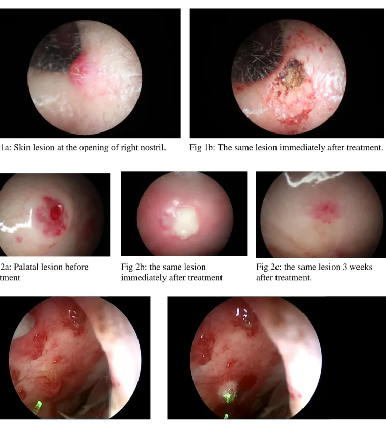
Fig 1a: Skin lesion at the opening of right nostril. Fig 1b: The same lesion immediately after treatment.
Fig 2a: Palatal lesion before treatment
Fig 2b: the same lesion immediately after treatment
Fig 2c: the same lesion 3 weeks after treatment.
Fig 3a: Multiple nasal lesions before treatment Fig 3b: The nasal vascular lesion at the end of the treatment
DISCUSSION
The most common symptom of HHT is recurrent epistaxis (up to 90% of patients), which may lead to a decrease in quality of life, hospitalizations, and need for multiple blood transfusions. Ideally, epistaxis should be treated locally with minimum side effects and without mucosal and glandular damage. Laser
photocoagulation is a potentially better method of treatment than chemical and electrocoagulation because it avoids the risk of large areas of scar tissue and nasal structure or vascular damage. Popular devices include the KTP laser (532 nm), the pulse dye laser (585 nm), and various diode lasers at the near infrared range (780-1064 nm)(16).
The present study evaluated the effect of the neoV980-nm diode laser. The 980 nm wavelength is selectively absorbed by haemoglobin and minimally absorbed in water, allowing for its transmission through the epithelium and coagulation of telangiectasis on the nasal mucosa, without damaging the vessel wall or mucosal-coating epithelium(17). Because it penetrates more deeply into tissue, the neoV980 laser may yield better results than lower-wavelength lasers when larger vessels located a few millimeters below the mucosal surface are involved(18-20). The laser transmits up to 28 watts of power, either continuous or pulsed, through a flexible optical fiber consisting of a 400-micron silica core, 440-micron silica cladding with 465-micron polymide coating, and a bare tip measuring 2.6 meters, with a numerical aperture of 0.22 at the tip. The laser may be applied with and without contact of the fiber with the tissue. The thin optical fibers are highly amenable for use inside the nasal cavity.
The good long-term clinical outcome of the laser-treated patients with HHT in the present study suggests the absence of a clinically significant adverse effect of the laser treatment on mucosal or glandular tissue. As the mucosa is undamaged, repeated laser sessions at minimal intervals to optimize nasal function are feasible and safe.
Division of the patients in our study by number of lesions affected (one vs multiple on endoscopic examination) yielded interesting findings. Patients with one lesion had an excellent outcome, with complete cessation of bleeding after the procedure and throughout follow-up (2-12 months). Their mean haemoglobin level increased by 4.2 g/dL, and their severity score improved by 2.2 points. These patients appear to be ideal candidates for local treatment with the 980 nm diode laser and can be spared systemic treatment with its possible side effects.
In the patients with multiple lesions, results were satisfactory. All showed a marked improvement in quality of life, with a decrease in bleeding. Mean haemoglobin increased by 2.8 g/dL and severity score improved by 2.5 points.
In conclusion, the neoV 980-mm laser is an excellent treatment option for the treatment of HHT-associated epistaxis, with good results and no side effects. The procedure can be done in the office. Its use allows physicians to reserve systemic treatment for more complicated cases or cases.
REFERENCES
1. Peery WH. Clinical spectrum of hereditary hemorrhagic telangiectasia (Osler-Weber-Rendu disease). Am J Med 1987; 82: 989-997.
2. Guttmacher AE, Marchuk DA, White RI Jr. Hereditary hemorrhagic telangiectasia. N Engl J Med 1995; 333: 918-924.
3. Shovlin CL, Guttmacher AE, Buscarini E, et al. Diagnostic criteria for hereditary hemorrhagic telangiectasia (Rendu-Osler-Weber syndrome). Am J Med Genet 2000; 91: 66-67.
4. Kjeldsen AD, Vase P, Green A. Hereditary haemorrhagic telangiectasia: a population-based study of prevalence and mortality in Danish patients. J Intern Med 1999; 245: 31-39.
5. Braverman IM, Keh A, Jacobson BS. Ultrastructure and three-dimensional organization of the telangiectases of hereditary hemorrhagic telangiectasia. J Invest Dermatol 1990; 95: 422-427. 6. Haitjema T, Disch F, Overtoom TT, Westermann CJ, Lammers JW. Screening family members of
patients with hereditary hemorrhagic telangiectasia. Am J Med 1995; 99: 519-524.
7. Kjeldsen AD, Oxhoj H, Andersen PE, Elle B, Jacobsen JP, Vase P. Pulmonary arteriovenous malformations: screening procedures and pulmonary angiography in patients with hereditary hemorrhagic telangiectasia. Chest 1999; 116: 432-439.
8. Assar O, Friedman CM, White RI Jr. The natural history of epistaxis in hereditary hemorrhagic telangiectasia. Laryngoscope 1991; 101: 977-980.
9. Plauchu H, De Chadarevian JP, Bideau A, Robert JM. Age-related clinical profile of hereditary hemorrhagic telangiectasia in an epidemiologically recruited population. Am J Med Genet 1989; 32: 291-297.
10. Sabba C, Gallitelli M, Palasciano G. Efficacy of unusually high doses of tranexamic acid for the treatment of epistaxis in hereditary hemorrhagic telangiectasia. N Engl J Med 2001; 345: 926. 11. Yaniv E, Preis M, Hadar T, Shvero J, Haddad M. Antiestrogen therapy for hereditary hemorrhagic
telangiectasia: a double-blind placebo-controlled clinical trial. Laryngoscope 2009; 119: 284-288. 12. Strother CM, Newton TH. Percutaneous embolization to control epistaxis in Rendu-Osler-Weber
disease. Arch Otolaryngol 1986; 102: 58-60.
13. Pau H, Carney AS, Murty GE. Hereditary haemorrhagic telangiectasia (Osler-Weber-Rendu syndrome): otorhinolaryngological manifestations. Clin Otolaryngol Allied Sci 2001; 26: 93-98. 14. Sabba C, Pasculli G, Cirulli A, et al. Hereditary hemorrhagic teleangiectasia (Rendu-Osler-Weber
disease). Minerva Cardioangiol 2002; 50: 221-238.
15. Al-Deen S, Bachmann-Harilstrad G. A grading scale for epistaxis in hereditary hemorrhagic teleangiectasia. Laryngoscope 2004; 114: 750-759.
16. Yaniv E, Hadar T, Shvero J, Tamir R, Nageris B. KTP\532 YAG laser treatment for allergic rhinitis. Am J Rhinol Allergy 2009; 23: 527-530.
17. Fiorella ML, Ross DA, White RI, et al. Hereditary haemorrhagic telangiectasia: state of the art. Acta Otorhinolaryngol Ital 2004; 24: 330-336.
18. Desiate A, Cantore S, Tullo D, Profeta G, Grassi FR, Ballini A. 980 nm diode lasers in oral and facial practice: current state of the science and art. Int J Med Sci 2009; 6: 358-364.
19. Levy JL, Berwald C. Treatment of vascular abnormalities with a long-pulse diode at 980 nm. J Cosmet Laser Ther 2004; 6: 217-221.
20. Kunishige JH, Goldberg LH, Friedman PM. Laser therapy for leg veins. Clin Dermatol 2007; 25: 454-461.