DISEASE PATHOGENS ASSOCIATED WITH PETRI DISEASE AND
ESCA
PROVIDENCE MOYO
Thesis presented in partial fulfilment of the requirements for the degree of Master of Science in Agriculture at the University of Stellenbosch
Supervisor: Dr. L. Mostert Co-supervisor: Dr. F. Halleen
DECLARATION
By submitting this thesis/dissertation electronically, I declare that the entirety of the work contained therein is my own, original work, that I am the sole author thereof (save to the extent explicitly otherwise stated), that reproduction and publication thereof by Stellenbosch University will not infringe any third party rights and that I have not previously in its entirety or in part submitted it for obtaining any qualification.
Date: March 2013 &RS\ULJKW6WHOOHQERVFK8QLYHUVLW\ $OOULJKWVUHVHUYHG
ABSTRACT
Petri disease and esca are devastating grapevine trunk diseases and compromise the sustainability of viticulture world-wide. Despite being extensively studied, knowledge of inoculum sources and mechanisms of spread of the causal pathogens is limited. Arthropods have been suspected to play a role in the spread of Petri disease and esca pathogens. However, little information is known about the extent to which arthropods are associated with these pathogens. This study aimed to determine whether arthropods occurring within or on declining grapevines, are associated with trunk disease pathogens and to identify arthropods associated with pruning wounds. The potential of selected arthropods to act as vectors of trunk disease pathogens was also investigated.
Two vineyards exhibiting grapevine trunk disease infections were sampled weekly for two years for collection of arthropods. Arthropods were collected using pruning wound traps, visual searches as well as trunk and cordon traps. Fungal spores from surfaces of arthropods were collected in water. Samples were subjected to nested PCR using primers Pm1/Pm2 and Pch1/Pch2 to verify the presence of Phaeoacremonium spp. and Phaeomoniella chlamydospora, respectively. Water samples were also cultured and
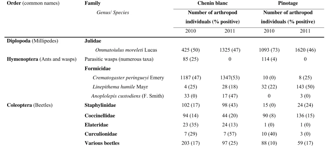
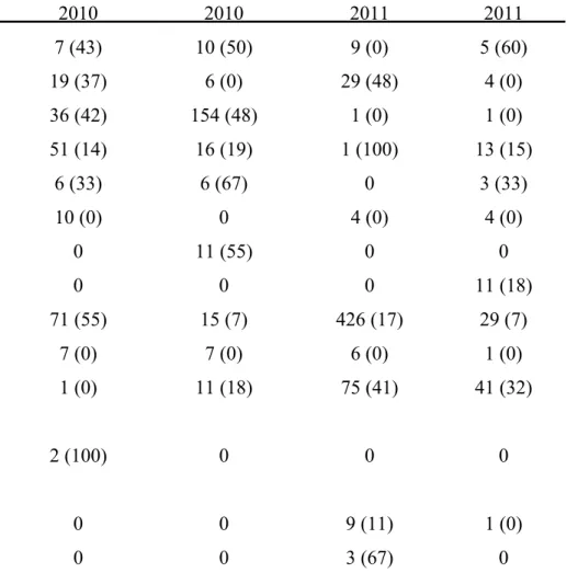
grapevine trunk disease pathogens obtained were identified by sequencing the internal transcribed spacers 1 and 2 and the 5.8S rRNA gene or the partial beta-tubulin gene. A total of 10 875 arthropod individuals, belonging to more than 31 families, were collected from declining grapevines. The most abundant arthropods included millipedes, ants, spiders and beetles. Portuguese millipedes and cocktail ants were associated with fresh grapevine pruning wounds. Thirty-three percent of the 5677 water samples analysed, contained propagules of pathogens associated with Petri disease and esca. Of these, 37 % were recovered from millipedes, 22 % from cocktail ants, 15 % from spiders and 10 % from beetles. All the major groups of grapevine trunk diseases were detected on the arthropods.
Phaeoacremonium species were detected in 1242 samples while Phaeomoniella chlamydospora was identified from 855 samples. Other fungi isolated included members of
the Botryosphaeriaceae, Diatrypaceae and Diaporthales.
The potential of grapevine sap as a food source for Portuguese millipedes and cocktail ants was investigated, in vitro. Millipede individuals were offered a choice between
water and grapevine sap while ants in nests were presented with grapevine sap, tuna and water and monitored for ingestion of sap. Both taxa preferred grapevine sap over the other
food items, indicating close association with pruning wounds. Subsequently, the ability of both taxa to transmit a DsRed-transformed Phaeomoniella chlamydospora isolate to fresh
pruning wounds of canes in polystyrene strips, floating in water, and potted vines was tested. Arthropods were exposed to the fungus for 24 hours and transferred to the base of the plants and canes and were removed after three days. Isolations after a month revealed that millipedes and ants were capable of transmitting the fungus onto wounds and cause infection. Millipede faecal pellets were also evaluated as potential sources of inoculum. Millipedes were fed on Phaeomoniella chlamydospora for 24 hours, surface sterilised and
allowed to defaecate in sterile Petri dishes overnight. Faecal material was collected, macerated in water and plated onto potato dextrose agar. Propagules of Phaeomoniella chlamydospora survived passage through the gut of millipedes and were passed out in a
viable state to form colonies of Phaeomoniellachlamydospora.
This study concludes that a wide variety of arthropods can be a source of inoculum of trunk diseases in vineyards. The results of the dissemination trial provides evidence that millipedes and ants are able to disseminate and infect vines with Phaeomoniella chlamydospora. It is therefore, highly likely that other grapevine trunk disease pathogens
are transmitted in the same manner. This knowledge highlights the need for control of certain arthropods to be taken into consideration when managing grapevine trunk disease pathogens.
OPSOMMING
Petri siekte en esca is verwoestende wingerd stamsiektes en verhinder die volhoubaarheid van wingerdproduksie wêreldwyd. Hierdie siektes is al intensief bestudeer, maar kennis rakende die inokulum bronne en meganismes van verspreiding van die veroorsakende patogene is beperk. Arthropoda is al vermoed om ‘n rol te speel in die verspreiding van Petri siekte en esca patogene, maar weinig informasie is bekend oor die mate waartoe arthropoda geassosieer is met die patogene. Hierdie studie het ten doel gestel om die arthropoda wat op of in wingerdstokke wat terugsterf voorkom te identifiseer en te bepaal watter van die arthropoda geassosieer is met stamsiekte patogene. Daar is ook ten doel gestel om die arthropoda wat geassosieer is met vars snoeiwonde te identifiseer en ook die moontlike vektor status van die stamsiekte patogene deur arthropoda.
Arthropoda is weekliks vir twee jaar gekollekteer vanaf twee wingerde met stamsiekte infeksies. Snoeiwond lokvalle, visuele soektogte en stam- en kordon lokvalle was gebruik om arthropoda te vang. Swamspore van die oppervlak van die arthropoda is afgewas met water. Van hierdie water monsters is gebruik om dubbelvoudige polimerase ketting reaksies (PKR) te doen met die inleiers Pm1/Pm2 en Pch1/Pch2 om vir die teenwoordigheid van Phaeoacremonium spp. en Phaeomoniella chlamydospora
onderskeidelik te toets. Die oorblywende water monster is gekweek op medium om die swamme teenwoordig te bepaal. Die wingerd stamsiekte patogene is verder geidentifiseer deur die DNS volgordes te bepaal van die interne getranskribeerde spasies 1 en 2 en die 5.8S rRNS geen of ‘n gedeelte van die beta-tubulien geen. In totaal is 10 875 arthropoda, wat behoort tot 31 families, gekollekteer vanaf wingerde wat terugsterf. Die mees algemene arthropoda was duisendpote, miere, spinnekoppe en kewers. Die Portugese duisendpote en die wipstert mier is geassosieer met vars wingerd snoeiwonde. Van die 5677 water monsters wat geanaliseer is, het 33% propagules van die Petri siekte of esca patogene gehad. Van hierdie was 37 % afkomstig vanaf duisendpote, 22 % van wipstert miere, 15 % van spinnekoppe en 10 % van kewers. Al die hoofgroepe van wingerd stampatogene is opgespoor op die arthropoda. Phaeoacremonium species is opgespoor in 1242 monsters en Phaeomoniellachlamydospora is gevind in 855 monsters. Ander swamme wat ook geisoleer
is sluit lede van die Botryosphaeriaceae, Diatrypaceae en Diaporthales in.
Die potensiaal van wingerdsap as ‘n bron van voedsel vir Portugese duisendpote en wipstert miere is in vitro ondersoek. Duisendpoot invidue is ‘n keuse gegee tussen water en
wingerd sap terwyl mierneste ‘n keuse gehad het tussen water, wingerd sap en tuna. Die duisendpote en miere is gemonitor vir die inname van wingerdsap in die teenwoordigheid van die ander bronne. Beide die duisendpote en miere het wingerdsap verkies wat aandui dat hulle ‘n noue assosiasie met wingerd snoeiwonde het. Vervolgens is beide taksons getoets vir hul vermoë om ‘n DsRooi-getransformeerde Phaeomoniella chlamydospora isolaat te
vektor na vars snoeiwonde op lote gemonteer op polistireen stroke wat in water dryf en op wingerd plante in potte. Die duisendpote en miere is blootgestel aan die swam vir 24 uur en oorgedra na die basis van die plante en lote en is weer verwyder na drie dae. Na ‘n maand is isolasies gedoen wat gewys het dat die duisendpote en miere die swam suksesvol kon oordra na die snoeiwonde en infeksie veroorsaak. Duisendpoot uitwerpsels is geëvalueer vir die potensiaal as inokulum bron. Duisendpote het gevoed op Phaeomoniellachlamydospora vir
24 uur, daarna oppervlakkig gesteriliseer en toegelaat om oornag uitwerpsels te maak in steriele Petri bakkies. Uitwerpsels was gekollekteer, fyngemaak in water en op aartappel dekstrose agar uitgeplaat. Propagules van Phaeomoniella chlamydospora het die
verteringskanaal van die duisendpote oorleef en het tipiese kolonies op die agar gevorm. Hierdie studie het vasgestel dat ‘n verskeidenheid van arthropoda ‘n bron van inokulum van stamsiektes in wingerd kan wees. Die resultate van die vektor proewe het gewys dat duisendpote en miere die vermoë het om Phaeomoniella chlamydospora te
versprei na snoeiwonde wat die swam dan suksesvol geinfekteer het. Dit is daarom hoogs waarskynlik dat van die ander wingerd stamsiekte patogene ook versprei kan word op dieselfde manier. Hierdie kennis demonstreer dat die beheer van spesifieke arthropoda in ag geneem moet word in die bestuur van wingerd stamsiektes.
ACKNOWLEDGEMENTS
First and foremost, I would like to thank the Lord Almighty. I could never have done this without the love, grace and strength He gave me each day.
I wish to express my sincere gratitude to my supervisors Dr. Lizel Mostert and Dr. Francois Halleen for guidance and mentoring. Thank you for remaining patient when I made mistakes and learnt from them.
Sincere thanks to Dr. Francois Roets for sharing his abundant knowledge on arthropods, statistical analysis, identification of arthropods and editing of this manuscript.
To Cheusi Mutawila, thank you very much for the encouragement, advice, and the friendship we built. I am very grateful for all that you taught and made me aware of during my study.
Many thanks to Gugu for the plants and the love we shared. To Annabella, thanks girl for the love we shared together and also for the time we spent watching millipedes at night. I wish to thank all the people in the department for their kindness and friendship throughout the course of my study.
Iam very grateful to Winetech, Agricultural Research Council of South Africa and NRF for financial support. Many thanks to Marieta van de Rijst for statistical analyses.
Iam very grateful to Palesa Lesuthu, Muriel Knipe, Danie, Adoration Shubane, Carine, Julia, Lydia, Levocia and other marvellous people from the Agricultural Research Council of South Africa for their willingness to help with the field work and isolations.
To my daughter Faith and niece Hope, I would like to dedicate this thesis to you and thank you for bringing a smile on my face always, your understanding and patience when I could not be with you. You gave me reason to continue doing this even when it was hardest. To my parents, thank you for being the best parents and I can never thank you enough for helping me take care of your granddaughter. To my brothers, Praise and Nomatter, thank you for being good uncles to Faith when I was away.
Many thanks to my good friend Masauso for prayers and support and lending an ear each time I needed to mourn. I would also like to thank everyone who was involved in this project at any level and I offer my sincere apologies to those I did not mention by name.
CONTENTS
CHAPTER 1: A REVIEW OF PETRI DISEASE AND ESCA AND THE ROLE OF ARTHROPODS IN THE DISPERSAL OF PLANT PATHOGENIC FUNGI 1
1.1 Importance of Petri disease and esca 1
1.2 Etiology of Petri disease and esca 1
1.3 Symptoms of Petri disease and esca 2
1.4 Sources and dispersal of Petri disease and esca pathogens 3
1.5 Disease management 6
1.5.1 Chemical control 6
1.5.2 Biological control 7
1.5.3 Cultural control practices 8
1.6 Dissemination of fungal pathogens by arthropods and development of
plant disease 9
1.7 Fungal adaptations for arthropod dispersal 11
1.8 Potential dissemination of grapevine trunk disease pathogens by arthropods 12
1.9 Aims of this study 14
1.10 References 16
CHAPTER 2: ARTHROPODS AS CARRIERS OF PATHOGENS ASSOCIATED WITH PETRI DISEASE AND ESCA IN VINEYARDS 29
2.1 Abstract 29
2.2 Introduction 30
2.3 Materials and methods 33
2.3.1 Selection of vineyards 33
2.3.2 Arthropod collection 33
2.3.2.1 Trunk and cordon traps 33
2.3.2.2 Visual surveys 34
2.3.2.3 Pruning wound traps 34
2.3.3 Arthropod identification 35
2.3.4 Screening of arthropods for presence of Petri and other grapevine
2.3.4.1 Washing of arthropods and identification of fungal pathogens
using plating techniques 35
2.3.4.2 Identification of fungal pathogens obtained from water washings
using molecular techniques 36
2.3.4.2.1 DNA extraction from water 36
2.3.4.2.2 DNA extraction from pure cultures 36
2.3.4.2.3 Detection of Petri disease fungi using nested PCR 37 2.3.4.2.4 PCR from DNA isolated from fungal cultures 38
2.3.4.2.5 Sequencing of PCR products 38
2.3.4.2.6 Restriction enzyme identification of Phaeoacremonium spp.
from PCR products 39
2.3.4.3 Detection of grapevine trunk pathogens from pruning wounds on which
pruning wound traps were placed 39
2.3.5 The influence of environmental factors on the presence of trunk disease
pathogens on arthropods 40
2.4 Results 40
2.4.1 Arthropod collection 40
2.4.1.1 Arthropods associated with declining grapevines 40 2.4.1.2 Arthropods collected from pruning wound traps 41 2.4.2 Screening of arthropods for presence of Petri and other grapevine trunk
disease pathogens 42
2.4.2.1 Identification of fungal pathogens obtained from water washings using
plating techniques 42
2.4.2.2 Identification of fungal pathogens obtained from water washings using
molecular techniques 43
2.4.2.2.1 Detection of Petri disease fungi using nested PCR 43 2.4.2.2.2 Restriction enzyme identification of Phaeoacremonium spp.
from PCR products 43
2.4.2.3 Detection of grapevine trunk pathogens from pruning wounds on which
pruning wound traps were placed 44
2.4.3 The influence of environmental factors on the presence of trunk disease
pathogens on arthropods 44
2.5 Discussion 45
CHAPTER 3: ANTS AND MILLIPEDES AS VECTORS OF PHAEOMONIELLA
CHLAMYDOSPORA TO GRAPEVINE PRUNING WOUNDS 83
3.1 Abstract 83
3.2 Introduction 84
3.3 Materials and methods 86
3.3.1 Grapevine sap as food source for millipedes and ants 86
3.3.1.1 Millipedes 86
3.3.1.2 Cocktail ants 87
3.3.2 Transmission of fungal propagules from external surfaces of arthropods to
pruning wounds 88
3.3.2.1 Fungal isolate used 88
3.3.2.2 Preparation of grapevine plants 88
3.3.2.2.1 Potted plants 88
3.3.2.2.2 Floating canes 89
3.3.2.3 Arthropod collection, treatment and placement on grapevine plants 89 3.3.2.4 Inoculum viability and spore load of Pa. chlamydospora on
millipedes and ants 90
3.3.2.5 Transmission of inoculum onto pruning wounds 90
3.3.3 Millipede faecal pellets as sources of inoculum on grapevine
pruning wounds 91
3.4 Results 92
3.4.1 Grapevine sap as food source for millipedes and ants 92 3.4.2 Transmission of fungal propagules from external surfaces of arthropods
to pruning wounds 92
3.4.3 Millipede faeces as sources of inoculum on grapevine pruning wounds 93
3.5 Discussion 93
3.6 References 97
CHAPTER 4: GENERAL DISCUSSION 109
4.1 Arthropods as carriers of pathogens associated with Petri disease and esca
in vineyards 109
4.2 Ants and millipedes as vectors of Phaeomoniella chlamydospora to grapevine pruning
4.3 Conclusion and future research 111
CHAPTER 1
A REVIEW OF PETRI DISEASE AND ESCA AND THE ROLE OF ARTHROPODS IN THE DISPERSAL OF PLANT PATHOGENIC FUNGI
1.1 Importance of Petri disease and esca
Petri disease and esca are important grapevine trunk diseases as they have become a limiting factor for grapevine production. Their incidence and severity have increased in the last two decades and they have been reported in all the major viticultural areas around the world (Mugnai et al., 1999; Chicau et al., 2000; Larignon and Dubos, 2000; Edwards et al.,
2001b; Eskalen and Gubler, 2001; Fourie and Halleen, 2004a; Zanzotto et al., 2007;
Gramaje etal., 2008). It has been suggested that the increase in the incidence and severity is
a result of factors such as the changes in the nursery and vineyard management practices, lack of pruning wound protection and the deregistration of effective fungicides due to toxicity (Graniti et al., 2000). Both Petri disease and esca are an impediment to profitability
and sustained production because they can shorten the productive life of vineyards dramatically (Rolshausen et al., 2010). They are responsible for graft failure, loss of vigor
and productivity in established vines (Mugnai et al., 1999; Rumbos and Rumbou, 2001;
Whiting et al., 2001). Vineyard managements costs can increase as a result of labour
involved in remedial pruning and/or replanting in vineyards where there is poor vine establishment (Rumbos and Rumbou, 2001; Whiting et al., 2001; Rolshausen et al., 2010).
The clusters of berries borne on esca-infected vines display dark spots on the epidermis, known as black measles (Reisenzein et al., 2000) and this can be a problem to table grape
growers because such clusters are unmarketable (Rooney-Latham etal., 2005b). In addition,
the wine produced from affected clusters can be of poor quality (Calzarano etal., 2001).
1.2 Etiology of Petri disease and esca
Petri disease is caused by a combination of Phaeomoniella(Pa.)chlamydospora (W.
Gams, Crous and M.J. Wingf. and L. Mugnai) Crous & W. Gams and several
Phaeoacremonium (Pm.) W. Gams, Crous & M.J. Wingf. species (Scheck et al., 1998;
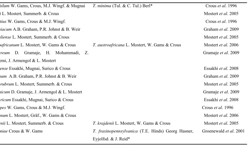
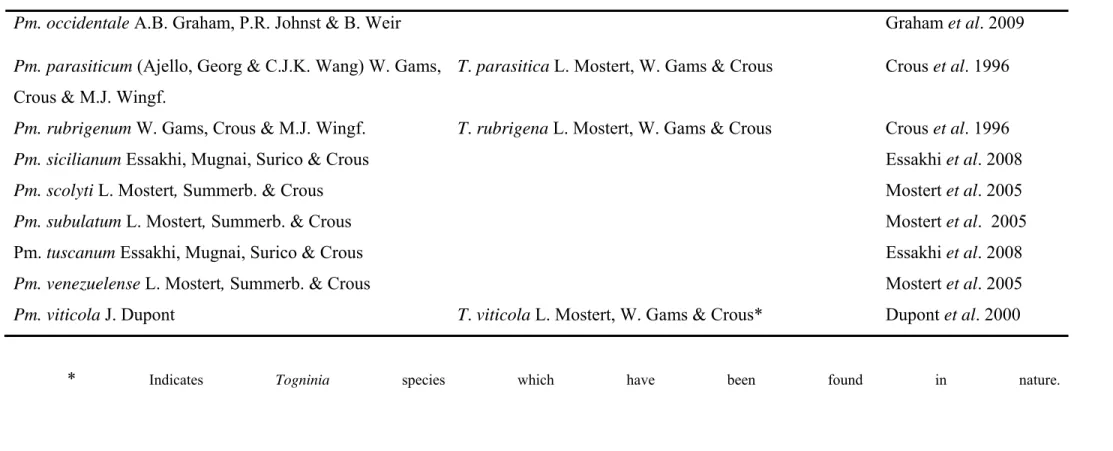
Mugnai et al., 1999; Groenewald et al., 2001). Twenty-five species of Phaeoacremonium
have been isolated from declining grapevines (Table 1), but Pm. aleophilum W. Gams,
1996; Mugnai et al., 1999; Mostert et al., 2006; Essakhi et al., 2008). Togninia Berl.
teleomorphs have been described for seven of the 25 Phaeoacremonium species (Mostert et al., 2006). Perithecia of three of the Togninia species, have been found in nature namely, T. minima (Tul. & C. Tul.) Berl. (Rooney-Latham etal., 2005a), T.fraxinopennsylvanica (T.E.
Hinds) Georg Hasner, Eyjolfsd. & J. Reid and T. viticola L. Mostert, W. Gams & Crous
(Eskalen etal., 2005a, 2005b).
A new definition of esca has been proposed and the term ‘esca’ is to be used when referring to wood decay (its original meaning) caused by basidiomycete fungi and the term ‘grapevine leaf stripe’ (previously known as ‘young esca’) is to be used for the vascular disease caused by Pa. chlamydospora and Pm. aleophilum. The term ‘esca-proper’ will then
be used when both esca and grapevine leaf stripe occur together in a vine (Surico, 2009). However, several other fungi, which are involved in other grapevine declines, have been isolated from esca-affected vines. Such fungi include species of the Botryosphaeriaceae, Diatrypaceace as well as Diaporthales (Fischer and Kassemeyer, 2003; Edwards and Pascoe, 2004; Calzarano and Di Marco, 2007; Péros etal., 2008; White etal., 2011). However, for
the purpose of this study, the biology of the pathogens that cause grapevine leaf stripe will be discussed with regards to esca.
1.3 Symptoms of Petri disease and esca
External symptoms of Petri disease include stunted growth, shortened internodes, reduced vigor, shoot dieback as well as a general decline of young grapevines resulting in plant death (Fourie and Halleen, 2004a; Retief et al., 2005; Gramaje et al., 2008). The
common internal symptom is the black/brown wood streaking with the presence of gummy masses in the xylem vessels which have been found to originate from wounds or the graft union. The gummy masses, also known as black goo (Fig. 1A), appear as minute black/brown spots in cross section and as black/brown streaks when viewed longitudinally.
Phaeomoniella chlamydospora and species of Phaeoacremonium have been isolated from
these black spots of declining grapevines (Mugnai etal., 1999; Del Rio etal., 2001; Whiting etal., 2001).
The term ‘esca’, in this context, will be used to include the occurrence of both the external and/or internal symptoms on vines. The leaves of esca-affected vines develop an interveinal foliar chlorosis or reddening resulting in a ‘tiger stripe’ pattern (Fig. 1B).
However, these leaf symptoms are discontinuous, not occurring every year on diseased vines (Redondo et al., 2001; Marchi et al., 2006). The berries on affected vines become
shrivelled and discoloured with minute black spots known as ‘black measles’(Fig. 1C) and dieback of the shoot tips occur (Mugnai et al., 1999; Reisenzein et al., 2000; Edwards and
Pascoe, 2004). The severe form of esca, also known as apoplexy, is a sudden wilting of the entire plant including the clusters of berries. Apoplexy is thought to be favoured by hot summers, in particular when rainfall is followed by dry, hot weather (Mugnai et al., 1999;
Peros etal., 2008). Internal symptoms of esca are similar to those of Petri disease but can be
distinguished by white rot (Fig. 1D) in cross sections of trunks or cordons. The white rot is often surrounded by a thick black/brown margin (Mugnai et al., 1999; Koklu, 2000;
Pollastro et al., 2000; White et al., 2011). White rot has been found in the trunk next to
pruning wounds and extending into the basal parts of the plant, usually within a section of the internal tissue and can spread to the surface of the trunk (Fischer and Kassemeyer, 2003).
1.4 Sources and dispersal of Petri disease and esca pathogens
Pathogens that cause Petri disease and esca are thought to occur as latent infections in the tissues of grapevines and that they probably become pathogenic after vines are subjected to stress. Such stress include water deficit and improper planting procedures (Mugnai etal., 1999; Whiting etal., 2001; Retief etal., 2005; Rooney-Latham etal., 2005c;
Di Marco and Osti, 2009).
Several inoculum sources for Pa. chlamydospora and Phaeoacremonium spp. have
been identified. Rootstock mother vines and various propagation processes have been shown to be primary inoculum sources for these pathogens (Mugnai et al., 1999; Halleen et al.,
2003; Edwards and Pascoe, 2004; Aroca et al., 2010). Isolations have shown that these
pathogens are present in apparently healthy rootstock mother vines (Fourie and Halleen, 2004b) and cuttings (Halleen etal., 2003) as well as grafted nursery plants (Zanzotto etal.,
2001). Mycelial growth of Pa. chlamydospora (Pascoe and Cottral, 2000) and spores of Pm. aleophilum and Pm. inflatipes W. Gams, Crous & M.J. Wingf. (Feliciano and Gubler, 2001)
have been observed within xylem vessels of grapevines. These findings led to the hypothesis that infection by these pathogens occur via spores or hyphae from mother vines into canes (Fourie and Halleen, 2002; Edwards et al., 2003). Different propagation processes and
of inoculum for Petri disease pathogens. In 2001, researchers in Italy investigated the occurrence of Petri disease fungi on grafted vines and reported detecting Petri disease pathogens and suggested that contamination occurred after grafting (Zanzotto et al., 2001). Phaeomoniella chlamydospora was later found to be present in hydration tanks, grafting
tools, callusing media and soil in South African nurseries (Retief et al., 2006). Phaeomoniella chlamydospora and some species of Phaeoacremonium were also detected
in hydration tanks, scissors, grafting machines as well as in peat used for root development in Spanish nurseries, using PCR-based detection techniques (Aroca et al., 2010). Retief et al. (2006) suggested that mycelium and conidia present on the surfaces of grapevine cuttings
probably wash off into the water during hydration or it oozes from the xylem vessels into the water.
The potential of soil as a source of inoculum is shown through the recovery of Petri disease pathogens from soil in vineyards. Phaeoacremoniuminflatipes was recovered from soil and standing water in Californian vineyards (Rooney et al., 2001). Phaeomoniella
chlamydospora was detected, using molecular techniques, from soil samples collected from
beneath rootstock mother vines known to be infected with the pathogen in South African (Damm and Fourie, 2005; Retief et al., 2006) and New Zealand (Whiteman et al., 2005) vineyards. Phaeomoniellachlamydospora has been suggested to be present in vineyard soils as mycelium, conidia, chlamydospores or fruiting structures (Retief et al., 2006). It has also been suggested that Pa. chlamydospora could be a soil-borne pathogen because of its ability to form chlamydospores in culture (Bertelli et al., 1998). The chlamydospores are also thought to form conidia that can germinate and penetrate roots of vines in nurseries and vineyards (Bertelli etal., 1998; Mugnai etal., 1999).
Diseased grapevine wood act as inoculum sources for Petri disease pathogens in vineyards. Phaeomoniella chlamydospora was recovered from plant sap and on the outer bark of diseased grapevines (Rooney et al., 2001). Pycnidia of Pa. chlamydospora have been found to survive on grapevine bark tissue (Edwards and Pascoe, 2001) and are thought to be the sources of Pa. chlamydospora spores in the vineyards (Eskalen and Gubler, 2001).
Phaeomoniella chlamydospora was found sporulating inside cracks of diseased vines
(Edwards et al., 2001a; Edwards and Pascoe, 2001). Perithecia of Togninia species have
been found on dead vascular tissue in deep cracks of trunks and cordons and on decaying pruning wounds of affected grapevines in vineyards (Eskalen etal., 2005a, 2005b;
on ash trees located close to the vineyards in California (Eskalen etal., 2005a, 2005b). The
presence of perithecia on ash trees illustrates the possibility of infected trees surrounding vineyards of being sources of inoculum (Eskalen etal., 2007a).
In vitro studies conducted in California showed that ascospores of T. minima are
released from perithecia after precipitation (Rooney-Latham et al., 2005b). The asci
emerged through the ostiole and either accumulated at the ostiole or contracted and forcibly discharged the ascospores. It was then speculated that the same mechanism of spore release occurs in vineyards and the ascospores that are forcibly discharged are then aerially dispersed and could land on fresh grapevine pruning wounds and cause infection. Spore trapping studies conducted in Californian vineyards showed that Pa. chlamydospora and Pm. aleophilum can be spread as airborne inoculum (Eskalen and Gubler, 2001). Spore
release for Pa. chlamydospora occurred during and after rainfall in late winter and early
spring and coincided with pruning and pruning wounds, but spore release for Pm. aleophilum was not always correlated with rainfall. Larignon and Dubos (2000) also found
that the occurrence of Pa.chlamydospora was correlated with rainfall and rainfall plays an
important role in the release of aerial inoculum in French vineyards.
The presence of spores of T. minima in the vineyards, in the absence of rainfall,
could be explained by the ascospores that accumulate at the ostiole of perithecia of T. minima (Rooney-Latham et al., 2005a). These ascospores are then spread by irrigation
practices or insects (Rooney-Latham et al., 2005a). Spores of T. minima were successfully
isolated from drip irrigation puddles under grapevines in California, but were not isolated directly from irrigation water as it passed through the emitter (Rooney et al., 2001). This
indicates that the water was contaminated after splashing over the vine. Insects may also contribute to the dispersal of ascospores because they are produced in a slimy droplet (Rooney-Latham etal., 2005a) which is ideal to stick or smear onto insects moving over the
diseased wood. Evidence that aerial inoculum might not be the only mechanism of pathogen dispersal in the field is shown in recent studies in Italy (Michelon et al., 2007) and South
Africa (Van Niekerk et al., 2010), which failed to trap spores of pathogens in the air using
volumetric spore traps. The different spatial patterns of disease symptoms observed in vineyards affected by esca further supports the hypothesis that several mechanisms may be involved in the dispersal of pathogens. The different spatial patterns observed included a tendency for infected vines to be aggregated along rows (Mugnai et al., 1999; Pollastro et al., 2000), a random spatial pattern of infected vines (Reisenzein et al., 2000; Redondo et
al., 2001; Marchi et al., 2006) as well as both aggregation and random spatial patterns
(Surico et al., 2000; Edwards et al., 2001b). These patterns can be attributed to different
modes of dispersal which may include insects, propagation material, rain splash as well as air currents (Reisenzein etal., 2000; Surico etal., 2000).
1.5 Disease management
The eradication of Petri disease and esca fungi once they have colonised the grapevines is difficult (Mugnai et al., 1999; Di Marco et al., 2004). Several studies have
shown that grapevines are infected during nursery propagation stages and therefore, propagation material and different nursery stages act as a source of inoculum for Petri disease and esca fungi (Zanzotto etal., 2001; Halleen etal., 2003; Aroca etal., 2010). In the
vineyards, pathogens infect vines through pruning wounds (Eskalen and Gubler, 2001; Rolshausen et al., 2010) and therefore, these need to be protected. Various chemical,
biological and cultural strategies have been studied to control Petri disease and esca of grapevines during the grapevine propagation process as well as in the vineyards. Disease management options are mostly preventative and limited to minimising infection risk (Hunt, 2004).
1.5.1 Chemical control
Chemical strategies in the control of Petri disease and esca in nurseries mainly involve drenches and dips of propagation material in fungicides at the various propagation stages (Fourie and Halleen, 2004a, 2006; Gramaje et al., 2009). A number of chemical
products have been tested to prevent or reduce Petri disease and esca infection of woody tissues of grapevine propagation material but no chemical product has been registered yet (Jaspers, 2001; Gramaje et al., 2009). Soaking propagation material prior to cold storage or
grafting in fungicides such as benomyl, carbendazim and captan has been shown to be effective in reducing Petri disease pathogens in nursery plants (Fourie and Halleen, 2004a, 2006). In 2007, California researchers reported that soaking dormant rootstocks and scions in ziram, thiram, thiophanate-methyl and lime sulphur, before grafting reduced incidence of
Pa. chlamydospora and Pm. aleophilum in vines (Eskalen etal., 2007b). However, although
some fungicides were found to be effective in controlling Petri disease fungi, some such as iprodione have been found to be poorly effective in reducing the germination and growth of Petri disease fungi (Jaspers, 2001; Gramaje et al., 2009). Petri disease and esca pathogens
are vascular pathogens that inhabit xylem vessels and the success of fungicide treatment in nurseries are therefore, limited by the inability of the fungicides to penetrate the wood tissue leading to poor efficacy (Waite and May, 2005).
Pruning wound protection with chemicals has also been studied. Eskalen et al.
(2007b) examined the potential of fungicides such as thiophanate-methyl, cyproconazole, boron and pyraclostrobin in protecting pruning wounds in the field against Pa. chlamydospora and Pm. aleophilum and found that these fungicides were effective.
Rolshausen et al. (2010) later also found boron to be effective in controlling Petri disease
fungi when the fungicide was applied directly on pruning wounds. Sodium arsenite has been used as a preventative application with great effect (Mugnai etal., 1999). The chemical was
applied as a foliar spray or painted onto the trunk or arms of infected vines. However, due to its toxicity and negative impact on the environment, it has been banned from most countries (Mugnai etal., 1999; Di Marco etal., 2000).
1.5.2 Biological control
Studies have been carried out on the potential application of Trichoderma Pers.
(Schumach) spp. in bio-control of Petri disease and esca in nurseries and the field.
Trichoderma formulations have been found to be suitable agents in the protection of pruning
wounds against infection by Petri disease and esca fungi (Hunt etal., 2001; Di Marco etal.,
2004; Kotze et al., 2011). Di Marco et al. (2004) reported that Trichoderma strains were
able to reduce infection of grapevine cuttings and pruning wounds of potted vines by Pa. chlamydospora. The incidence of Pa. chlamydospora and Phaeacremonium species in
grapevine rootstock material was reduced by soaking the rootstocks in Trichoderma
formulations (Fourie and Halleen, 2004a). Kotze et al. (2011) reported that a Trichoderma
isolate, USPP-T1, was effective in reducing the incidence of Pa. chlamydospora when
tested on pruning wounds of field grapevines. The mechanisms used by Trichoderma
include production of antibiotics, mycoparasitism, competition for nutrients and space with pathogenic fungi as well as stimulation of host resistance (Di Marco et al., 2004; Kotze et al., 2011).
1.5.3 Cultural control practices
Implementing traditional cultural practices remains essential in reducing the inoculum load and the spread of esca and Petri disease. Sanitary measures reduce inoculum originating from the vineyard and these include remedial pruning, which is cutting off dead arms below the diseased and discoloured wood, as well as uprooting dead or dying vines and removal of pruned material from the vineyard floor (Mugnai etal., 1999). However, the
effectiveness of remedial pruning is dependent on whether all infected wood is removed ensuring that shoots used for training are not infected.
Pathogens have been isolated from apparently healthy vines and hence occur as latent pathogens in the host becoming pathogenic when vines are stressed (Mugnai et al.,
1999; Whiting et al., 2001; Retief et al., 2005; Rooney-Latham et al., 2005c). Stress
conditions such as improper soil preparation, fertilisation and irrigation, in vineyards can result in symptom expression of infected plants. Maintaining optimal fertilisation and irrigation, therefore, results in vines which are healthy. Vines that are grown in conditions where cultural practices are carried out according to the best recommendations are less susceptible to disease since they are not predisposed to stress conditions that favour pathogenic infections (Fourie etal., 2000).
Treating propagation material with hot water for 30 minutes at 50 °C followed by 30 minutes in cold water has been found to be effective in disinfecting shoots during the propagation process (Crous et al., 2001; Fourie and Halleen 2004a; Waite and May, 2005).
However, there are conflicting reports on the effectiveness of hot water treatment. In vitro
tests carried out by Whiting et al. (2001) found that Pa. chlamydospora and Pm. inflatipes
were not killed by hot water treatment at 51 °C for 30 minutes and suggested that the treatment can not eliminate the pathogens in dormant canes. These findings were further confirmed by Rooney and Gubler (2001) who found hot water treatment at 51 °C for 30 minutes to be ineffective in the control of Petri disease pathogens in inoculated dormant wood. A study carried out by Gramaje et al. (2008) evaluated the effect of hot water
treatment in vitro on mycelial growth and germination of Petri disease pathogens. In this
study, the authors did not inoculate dormant grapevine wood. This was done in the study by Gramaje et al. (2009) which showed that hot water treatment at 53 °C for 30 minutes was
infected planting material at 50 °C for 45 minutes reduced the frequency of isolation of Pa. chlamydospora compared to untreated controls.
1.6 Dissemination of fungal pathogens by arthropods and development of plant disease
Arthropods facilitate the development of plant diseases in several ways. They can serve as agents of dispersal of the pathogens, be responsible for inoculation and ingression as well as allowing pathogens to over-season in and/or on their bodies (Leach, 1940; Agrios, 2005). A number of arthropods facilitate the entry of pathogens into plants through the wounds they make on plant parts, which can be either above or below ground. Arthropods feeding on plant parts predispose them to attack by pathogenic fungi by weakening the plant and creating wounds which can serve as ports of entry for fungi. Arthropods play a major role in the dispersal of fungal pathogens, although most transmission of fungi by arthropods is unintentional (Agrios, 2005). Spore dissemination occurs as a result of arthropods becoming contaminated with fungal reproductive propagules, either externally and/or internally, as they visit infected tissues. These can then transport the spores to uncolonised plants or plant parts (Agrios, 2005). Various arthropods have been implicated in the dispersal of different fungal pathogens including flies and springtails (Abbott, 2002; Lilleskov and Bruns, 2005), mites (Roets et al., 2011), beetles (Lilleskov and Bruns, 2005)
and ants (El-Hamalawi and Menge, 1996). Arthropod morphology often help in spore acquisition as they have cuticular processes such as setae and microtrichia (fixed hairs consisting of very small pointed extensions of the cuticula) and body appendages such as legs, wings and antennae that serve as structures that can hook and distribute spores (Leach, 1940).
Ambrosia fungi are fungi which have obligate associations with arthropods (especially beetles in the Platypodidae and Scolytidae) and they are a source of food for these beetles (Batra, 1963; Cassar and Blackwell, 1996; Henriques etal., 2006). These fungi
flourish inside tunnels made by the beetles, in dead trees, and are introduced into the plant when the beetles bore into xylem vessels and deposit the ectosymbiotic fungal spores (Cassar and Blackwell, 1996). In some instances, the fungi are transmitted in specialised glandular pouches called mycangia and usually only one fungal species is transmitted in the mycangium of female beetles (Cassar and Blackwell, 1996).
Oak wilt is caused by the fungal pathogen, Ceratocystis fagacearum (Bretz) Hunt.,
which enters xylem vessels of oak trees through fresh wounds (Juzwik etal., 2004; Agrios,
2005). Sap beetles (Coleoptera: Nitidulidae) are the primary spore vectors and are attracted to the aromatic volatiles produced by the growing fungus on the diseased trees (Juzwik et al., 2004). The beetles crawl over, and may tunnel into the mats as they feed on the fungal
tissues, acquiring viable fungal propagules on their external surfaces, and internally as they ingest fungal material (Juzwik et al., 2004; Agrios, 2005). Successful disease transmission
occurs when C. fagacearum infested sap beetles visit susceptible fresh wounds and
fermented sap from older wounds on healthy oaks (Juzwik et al., 2004). The spores of the
fungus have been shown to be able to survive for the entire period of hibernation of the beetles (Carter, 1973; Agrios, 2005).
Dutch elm disease kills elm trees by clogging their xylem vessels thereby, blocking movement of water from the roots to other parts of the tree (Agrios, 2005). It is caused by
Ophiostomaulmi (Buisman) Melin & Nannf. and O. novo-ulmi Brasier which are spread by
bark beetles (Scolytidae) (Jacobi etal., 2007). The fungi colonise the wood of dying or dead
elm trees which is also where the bark beetle larvae develop. The fungi sporulate in the larval tunnels and the newly emerged adult beetles leave the tunnels carrying thousands of sticky spores on their bodies (Agrios, 2005). New infections occur when the spores from beetles are deposited on the moist, freshly wounded tissues of a susceptible tree, usually as a result of burrowing activities of beetles (Agrios, 2005). Fungal spores produced in infected wood can be carried upward in the tree via xylem sap and may cause additional infections
(Agrios, 2005; Purcell and Almeida, 2005).
Similar to beetles, fungal reproductive propagules can also be transported by other arthropods. Botrytis cinerea Pers.:Fr., a fungus which causes bunch rot of grapes, is
vectored by larvae of the grape berry moth, Lobesiabotrana Den. & Schiff. from infected to
healthy grape berries (Fermaud and Le Menn, 1992). Argentine ants, Iridomyrmex humilis
(Mayr), have been reported to vector Phytophthora citricola Sawada to healthy lauraceous
trees, Perseaindica (L.) Spreng and cause 73 % infection (El-Hamalawi and Menge, 1996).
A wide variety of arthropods including beetles, springtails, oribatid mites and centipedes have also been found to carry the ectomycorrhizal fungus, Tomentella sublilacina (Ellis &
Holw.), on their exoskeleton and internally (Lilleskov and Bruns, 2005). The corn earworm moth, Helicoverpa zea Boddie is a pest of sorghum and has also been shown to transmit Clavicepsafricana Frederickson, Mantle & de Millano and cause ergot infection on healthy
sorghum plants (Prom et al., 2003). Mites in the genus Trichouropoda Berlese were found
to carry spores of the fungus Gondwanamyces proteae (M.J.Wingf., P.S.van Wyk &
Marasas) G.J.Marais & M.J.Wingf. within pit mycangia at the base of their legs, vectoring spores in the flower heads of Protea repens (L.) L (Roets etal., 2011).
1.7 Fungal adaptations for arthropod dispersal
Fungi produce three general types of inoculum and these are vegetative mycelium, sclerotia and spores. Sclerotia and mycelium are poorly adapted to arthropod dissemination although the mycelium in plant parts eaten by arthropods can act as inoculum (Leach, 1940). The most common type of reproductive structure for fungi is spores, including asexual and/or sexual spores. Spore masses are often well adapted for arthropod dispersal (Leach, 1940). Several fungal pathogens produce their spores in sticky exudates that become hard when dry and can easily be dispersed by the wind. However, before the spore droplets are dry, arthropods can play an important role in their dissemination (Leach, 1940). Any arthropod that comes into contact with moist, sticky spores can potentially act as a vector of these. Such sticky spores readily adhere to the legs, wings, bristles and other body parts and could easily be brushed off (Leach, 1940; Abbott, 2002) when the arthropods move about. Ascospores which are wet and produced in sticky masses and are associated with arthropods for dispersal have a better chance of reaching an infection court than those that are dispersed by wind and/or water because they are usually transported directly to possible infection courts (Leach, 1940; Carter, 1973) to initiate infection. It is also suspected that characteristics of ascomycetes such as evanescent asci, loss of forcible ascospore discharge, as well as long necked perithecia are a result of selection for arthropod dispersal (Cassar and Blackwell, 1996).
The production of slimy droplets is common among hyphomycetes and ascomycetes, with the latter also often producing long necked perithecia (Abbott, 2002). For example, spores of the blue-stain fungi, Ceratostomella Sacc. species, are produced in sticky solutions
under the bark making wind dissemination impossible (Leach, 1940). Sporulation of the blue-stain fungi occurs in tunnels and pupal chambers of bark beetles and the sporophores and perithecia point toward the centre of the tunnels such that the spore masses are in a perfect position for contaminating emerging arthropods (Leach, 1940). Hyphomycetous species such as Graphium Corda and Leptographium R.W. Davidson often produce large
arthropods moving over the surface of the substratum they colonise (Abbott, 2002). Other species (Trichoderma Pers. and Acremonium Fr.) produce large numbers of small droplets at
the apex of simple conidiophores whereas genera such as Microascus Zukal and Chaetomium Corda, extrude ascospores from the ascocarp neck in a droplet or cirrhi (spore
masses) from where these can easily adhere to arthropods (Abbott, 2002).
Spores of several fungal species are resistant to digestive enzymes found in the guts of insects since they stay viable after ingestion. For example, spores of the cotton wilt pathogen, Fusarium vasinfectum G.F.Atk), were disseminated in the faecal pellets of
grasshoppers that ingested infected plant material (Leach, 1940). Some ambrosia fungi are transmitted in the crop of the female beetle, Xyleborusdispar Fabricius, which regurgitates
them to initiate a culture in a new brood channel (Carter, 1973). Spores of the fungus,
Clavicepspaspali Stev., can survive the passage through the intestinal tract of the green fly Pyrellia coerulea (Wied.) (Carter, 1973). Other fungal genera such as Ceratocystis Ellis &
Halst., can also produce conidia coated with mucous to prevent them from being digested from within the guts of the beetles which disseminate them (Carter, 1973). El-Hamalawi and Menge (1996) found that faeces of garden snails (Helixaspera Muller), when fed avocado
plants (Persea americana Mill.) infected with Phytophthora citricola, contained viable
propagules of the fungus. Inoculation of wounds of healthy avocado plants with these faeces resulted in 77 % infection (El-Hamalawi and Menge, 1996). Researchers in Spain were able to isolate Phytophthora citrophthora (R.E.Sm. & E.H.Sm.) Leonian, a pathogen that causes
Phytophthora branch canker in citrus, from faeces of naturally infested Helix aspersa snails
(Alvarez et al., 2009). Faecal pellets of the millipede, Harpaphehaydeniana (Wood), were
also found to contain the fungus, Tomentellasublilacina. Inoculation of healthy seedlings of
the Bishop Pine, Pinusmuricata D. Don., with the millipede faeces revealed that the spores
of Tomentella sublilacina were still viable after passing through the gut of the millipede
(Lilleskov and Bruns, 2005).
1.8 Potential dissemination of grapevine trunk disease pathogens by arthropods
An ideal vector should be able to acquire the pathogen and transport it to uncolonised tissues for initiation of the disease in plants (Leach, 1940; Purcell and Almeida, 2005). Many arthropods may carry plant pathogens but cannot transmit them to a particular part where infection may result and therefore are not vectors (Leach, 1940; Purcell and Almeida, 2005).
Limited information is known about the role arthropods play in the dissemination of Petri disease and esca pathogens. Edwards etal. (2001a) speculated about the possible role
of arthropods in the dispersal of Pa. chlamydospora. Rooney-Latham et al. (2005a) stated
that wood-boring insects might play a role in the dispersal of T. minima. To date only Diplodia seriata De Not. (‘Botryosphaeria obtusa (Schwein.) Shoemaker’), which is
sometimes isolated from esca-affected vines, has been found on rove beetles collected from pruning wounds in a vineyard (Epstein etal., 2008).
Collembolans and mites were observed in the cracks and crevices on the bark of diseased grapevines (Edwards et al., 2001a) where pycnidia and sporulating hyphae of Pa. chlamydospora were found (Edwards et al., 2001a; Edwards and Pascoe, 2001). The
sporulation of this pathogen in the same niche as the arthropods were found could promote effective, though incidental, dispersal of the pathogen to healthy vines. It is suspected that the collembolans and mites that carry fungal spores on their exoskeletons can deposit them onto open xylem vessels of pruned shoots (Edwards et al., 2001a). Furthermore, the
sheltered nature of the cracks and crevices provides limited scope for other dispersal methods such as rain or wind and both the phialidic conidial heads and the pycnidial cirrhi of Pa. chlamydospora are presented in a way that they can readily come into contact with
small arthropods (Edwards etal., 2001a).
The perithecia of Togninia have long necks (Rooney-Latham et al., 2005a; Mostert etal., 2003) and have been found within trunks and cordons of diseased vines. Perithecia of Togninia are phototropic, thus their necks were found oriented towards openings of cracks,
pruning wounds and insect tunnels (Eskalen et al., 2005a, 2005b; Rooney-Latham et al.,
2005a). Invitro tests to determine how ascospores of T. minima are released from perithecia
showed that when perithecia were moistened, asci exude from the perithecial ostioles and accumulated in groups around the perithecial neck. This suggests that a similar mechanism of ascospore release occurs in infested vineyards following precipitation (Rooney-Latham et al., 2005b). The presence of perithecia clustered inside cracks and insect tunnels in
grapevines supports the possibility of an association between insects and T. minima
(Rooney-Latham et al., 2005a), and probable transmission of ascospores by insects.
Similarly, the production of ascospores in slimy droplets favours the involvement of insects in their dispersal rather than aerial dispersal (Cassar and Blackwell, 1996).
In an investigation of Botryosphaeriaceae-related dieback of grapevines, Epstein et al. (2008) postulated that although pruning wounds were primarily infected with D. seriata
conidia that are disseminated by windblown rain, some inoculum may be disseminated by other mechanisms. To test their theory, duct tape covered with a sticky substance, was placed over pruning wounds and from it, rove beetles (Staphilinidae) infested with D. seriata were recovered. Beetles have been found to vector numerous plant pathogens
(Leach, 1940; Carter, 1973; Juzwik etal., 2004; Agrios, 2005) and therefore, the possibility
of rove beetles acting as vectors of Petri disease and esca pathogens cannot be ruled out. Furthermore, Pm. rubrigenum W. Gams, Crous & M.J. Wingf. (a Petri disease fungus)was
isolated from larvae of the oak bark beetles, Scolytus intricatus (Ratz.) (Scolytinae), and
their galleries on oak trees, Querus robur L. and also from ash bark beetles, Leperisinus fraxini Panzer (Scolytinae) found under the bark of ash trees, Fraxinus excelsior L.
(Kubatova etal., 2004).
The mere association of an arthropod with diseased plants or the presence of inoculum on or in the arthropod’s body, however, does not establish that the arthropod is a vector of a pathogen. Any evidence that presents a certain arthropod as a vector of a plant pathogen should always meet Leach’s rules for proof of arthropod transmission (Leach, 1940). These rules state that the: i) arthropod should have a close association with diseased plants, ii) arthropod should regularly visit healthy plants under conditions suitable for transmission of the disease, iii) arthropod should carry the pathogen in nature or following visitation to a diseased plant and iv) disease should be produced experimentally by arthropod visitation under controlled conditions.
1.9 Aims of this study
From the literature reviewed in this chapter it is evident that arthropods could be involved in the dissemination of Petri disease and esca pathogens on grapevines. The sporulation structures of Petri disease and esca fungi, which produce spore droplets either on conidiophores or on perithecial necks, hold the potential to be dispersed by arthropods. Spore droplets are ideal to stick onto the exoskeletons of arthropods crawling over and into the crevices or cracks of diseased tissues and possibly be deposited onto the exposed xylem vessels of healthy vines as the arthropods explore fresh pruning wounds.
The role possibly played by arthropods in the dissemination of these pathogens needs to be investigated and possible vectors identified. The objectives of this study, therefore, were to:
i) determine which arthropods are associated with declining grapevines and freshly made pruning wounds,
ii) determine whether pathogens associated with Petri disease and esca can be detected on the arthropods and
iii) determine whether arthropods can vector Petri disease pathogens to pruning wounds.
1.10 REFERENCES
Abbott, S.P. 2002. Insects and other arthropods as agents of vector-dispersal of fungi. www.precisionenv.com./PDFS/Abbott/Insectdispersal.pdf.
Agrios, G.N. 2005. Insect involvement in the transmission of fungal pathogens. In: Harris KF, Maramorosch K (eds) Vectors of plant pathogens. Academic Press, New York: 293-323.
Alvarez, L.A., Gramaje, D., Abad-Campos, P. and García-Jiménez, J. 2009. Role of the
Helix aspersa snail as a vector of Phytopthora citrophthora causing branch cankers
on clementine trees in Spain. Plant Pathology 58: 956-963.
Aroca, A., Gramaje, D., Armengol, J., García-Jiménez, J. and Raposo, R. 2010. Evaluation of the grapevine nursery propagation process as a source of Phaeoacremonium spp.
and Phaeomoniella chlamydospora and occurrence of trunk disease pathogens in
rootstock mother vines in Spain. European Journal of Plant Pathology 126: 165-174. Batra, L.R. 1963. Ecology of ambrosia fungi and their dissemination by beetles.
Transactions of the Kansas Academy of Science 66: 213-236.
Bertelli E., Mugnai, L. and Surico, G. 1998. Presence of Phaeoacremonium chlamydosporum in apparently healthy rooted grapevine cuttings. Phytopathologia
Mediterranea 37: 79-82.
Calzarano, F., Cichelli, A. and Odoardi, M. 2001. Preliminary evaluations in composition induced by esca on cv. Trebbiano d'Abruzzo grapes and wines. Phytopathologia Mediterranea 40 (Supplement): S443-S448.
Calzarano, F. and Di Marco, S. 2007. Wood discoloration and decay in grapevines with esca proper and their relationship with foliar symptoms. Phytopathologia Mediterranea 46: 96-101.
Carter, W. 1973. Insects in Relation to Plant Disease. J. Wiley & Sons: New York.
Cassar, S. and Blackwell, M. 1996. Convergent origins of ambrosia fungi. Mycologia 88: 596-601.
Chicau, G., Aboim-Inglezi, M., Cabral, S. and Cabral, J.P.S. 2000. Phaeoacremonium chlamydosporum and Phaeoacremonium angustius associated with esca and
grapevine decline in Vinho Verde grapevines in northwest Portugal. Phytopathologia
Mediterrenea 39: 80-86.
Crous, P.W., Gams, W., Wingfield, M.J. and van Wyk, P.S. 1996. Phaeoacremonium gen.
nov. associated with wilt and decline diseases of woody hosts and human infections. Mycologia 88: 786-796.
Crous, P.W., Swart, L. and Coertze, S. 2001. The effect of hot water treatment on fungi occuring in apparently healthy grapevine cuttings. Phytopathologia Mediterranea 40 (Supplement): S464-S466.
Damm, U. and Fourie, P.H. 2005. Development of a cost-effective protocol for molecular detection of fungal pathogens in soil. South African Journal of Science 101: 135-139.
Del Rio, J., Gonzalez, A., Fuster, M.D., Botia, J.M., Gomez, P., Frias, V. and Ortuno, A. 2001. Tylose formation and changes in phenolic compounds of grape roots infected with Phaeomoniella chlamydospora and Phaeoacremonium species.
Phytopathologia Mediterranea 40 (Supplement): S394-S399.
Di Marco, S., Mazzullo, A., Calzarano, F. and Cesari, A. 2000. The control of esca: status and perspectives. Phytopathologia Mediterranea 39: 232-240.
Di Marco, S., Osti, F. and Cesari, A. 2004. Experiments on the control of esca by
Trichoderma. Phytopathologia Mediterranea 43: 108-115.
Di Marco, S. and Osti, F. 2009. Effect of biostimulant sprays on Phaeomoniella chlamydospora and esca proper infected vines under greenhouse and field
conditions. Phytopathologia Mediterranea 48: 47-58.
Dupont, J., Magnin, S., Paronnaud, J. and Roquebert, M-F. 2000. The genus
Phaeoacremonium from a molecular point of view. Phytopathologia Mediterranea
39: 119-124.
Edwards, J. and Pascoe, I.G. 2001. Pcynidial state of Phaeomoniella chlamydospora found
Edwards, J., Laukart, N. and Pascoe, I.G. 2001a. In situ sporulation of Phaeomoniella chlamydospora in the yard. Phytopathologia Mediterranea 40 (Supplement):
S61-S66.
Edwards, J., Marchi, G. and Pascoe, I.G. 2001b. Young esca in Australia. Phytopathologia 40 (Supplement): S303-S310.
Edwards, J., Pascoe, I.G., Salib, S. and Laukart, N. 2003. Phaeomoniella chlamydospora
and Phaeoacremonium aleophilum can spread into grapevine canes from trunks of
infected mother vines. In Proceedings, 3rd International Workshop Grapevine Trunk Disease, Lincoln University, Canterbury, New Zealand: 29.
Edwards, J. and Pascoe, I.G. 2004. Occurrence of Phaeomoniella chlamydospora and Phaeoacremonium aleophilum associated with Petri disease and esca in Australian
grapevines. Australasian Plant Pathology 33: 273-279.
El-Hamalawi, Z.A. and Menge, J.A. 1996. The role of snails and ants in transmitting the avocado stem canker pathogen, Phytophthora citricola. Journal of American Society
of Horticultural Science 121: 973-977.
Epstein, L., Kaur, S. and VanderGheyst, J.S. 2008. Botryosphaeria-related dieback and
control investigated in noncoastal California grapevines. California Agriculture 62: 160-166.
Eskalen, A. and Gubler, W.D. 2001. Association of spores of Phaeomoniella chlamydospora, Phaeoacremonium inflatipes, and Pm. aleophilum with grapevine
cordons in California. Phytopathologia Mediterranea 40 (Supplement): S429-S432. Eskalen, A., Rooney, S.N. and Gubler, W.D. 2005a. Occurrence of Togninia
fraxinopennsylvanica on esca-diseased grapevines (Vitis vinifera) and declining ash
trees (Fraxinus latifolia) in California. Plant Disease 89: 528.
Eskalen, A., Rooney, S.N. and Gubler, W.D. 2005b. First report of perithecia of
Phaeoacremonium viticola on grapevine (Vitis vinifera) and ash trees (Fraxinus latifolia) in California. Plant Disease 89: 528.
Eskalen, A., Feliciano, A.J. and Gubler, W.D. 2007a. Susceptibility of grapevine pruning wounds and symptom development in response to infection by Phaeoacremonium aleophilum and Phaeomoniella chlamydospora. Plant Disease 91: 1100-1104.
Eskalen, A., Rooney-Latham, S. and Gubler, W.D. 2007b. Identifying effective management strategies for esca and Petri disease. Phytopathologia Mediterranea 46: 125-126.
Essakhi, S., Mugnai, L., Crous, P.W., Groenewald, J.Z. and Surico, G. 2008. Molecular and phenotypic characterization of novel Phaeoacremonium species isolated from esca
diseased grapevines. Persoonia 21: 119-34.
Feliciano, A.J. and Gubler, W.D. 2001. Histological investigations on infection of grape roots and shoots by Phaeoacremonium spp. Phytopathologia Mediterranea
(Supplement) 40: S387-S393.
Fermaud, M. and Le Menn, R. 1992. Transmission of Botrytis cinerea to grapes by grape
berry moth larvae. Phytopathology 82: 1393-1398.
Fischer, M. and Kassemeyer, H.H. 2003. Fungi associated with esca disease of grapevine in Germany. Vitis 42: 109-116.
Fourie, P.H., Halleen, F., Groenewald, M. and Crous, P.W. 2000. Black goo decline of grapevine: Current understanding of this mysterious disease. Winelands 2000: 93-96. Fourie, P.H. and Halleen, F. 2002. Investigation on the occurence of Phaeomoniella
chlamydospora in canes of rootstock mother vines. Australasian Plant Pathology 31:
425-426.
Fourie, P.H. and Halleen, F. 2004a. Proactive control of Petri disease of grapevine through treatment of propagation material. Plant Disease 88: 1241-1245.
Fourie, P.H. and Halleen, F. 2004b. Occurrence of grapevine trunk disease pathogens in rootstock mother plants in South Africa. Australasian Plant Pathology 33: 313-315. Fourie, P.H. and Halleen, F. 2006. Chemical and biological protection of grapevine
propagation material from trunk disease pathogens. European Journal of Plant Pathology 116: 255-265.
Graham, A.B., Johnston, P.R. and Weir, B.S. 2009. Three new species of Phaeoacremonium
on grapevines in New Zealand. Australasian Plant Pathology 38: 505-513.
Gramaje, D., García-Jiménez, J. and Armengol, J. 2008. Sensitivity of Petri disease pathogens to hot water treatments invitro. Annals of Applied Biology 153:95-103.
Gramaje, D., Aroca, A., Raposo, R., Garcia-Jimenez, J. and Armengol, J. 2009. Evaluation of fungicides to control Petri disease pathogens in the grapevine propagation process. Crop Protection 28: 1091-1097.
Graniti, A., Surico, G. and Mugnai, L. 2000. Esca of grapevine: a disease complex or a complex of diseases. Phytopathologia Mediterranea 39: 16-20.
Groenewald, M., Kang, J-C., Crous, P.W. and Gams, W. 2001. ITS and beta-tubulin phylogeny of Phaeoacremonium and Phaeomoniella species. Mycological Research
105: 651-657.
Habib, W., Pichierri, A., Masiello, N., Pollastro, S. and Faretra, F. 2009. Application of hot water treatment to control Phaeomoniella chlamydospora in grapevine plant
propagation materials. Phytopathologia Mediterranea 48: 186.
Halleen, F., Crous, P.W. and Petrini, O. 2003. Fungi associated with healthy grapevine cuttings in nurseries, with special reference to pathogens involved in the decline of young vines. Australasian Plant Pathology 32: 47-52.
Henriques, J., Macio, M.L. and Sousa, E. 2006. Ambrosia fungi in the insect-fungi symbiosis in relation to cork oak decline. Revista Iberoamericana de Micologia 23: 185-188.
Hunt, J.S., Gale, D.S.J. and Harvey, I.C. 2001. Evaluation of Trichoderma as bio-control for
protection against wood-inhabiting fungi implicated in grapevine trunk diseases. Phytopathologia Mediterranea 40 (Supplement): S485.
Hunt, J.S. 2004. Trichoderma and trunk disease fungi: prospects for new protective
management options. The Australian & New Zealand Grapegrower & Winemaker: 17-20.
Jacobi, W.R., Koski, R.D., Harrington, T.C. and Witcosky, J.J. 2007. Association of
Ophiostoma novo-ulmi with Scolytus schevyrewi (Scolytidae) in Colorado. Plant
Disease 91: 245-247.
Jaspers, M.V. 2001. Effects of fungicides invitro on germination and growth of
Phaeomoniella chlamydospora. Phytopathologia Mediterranea 40 (Supplement):
S453-S458.
Juzwik, J., Skalbeck, T. and Neuman, M.F. 2004. Sap beetle species (Coleoptera: Nitidulidae) visiting fresh wounds on healthy oaks during spring in Minnesota. Forest Science 50: 757.
Koklu, G. 2000. Notes on esca disease on some grapevine varieties grown in Turkish Thrace. Phytopathologia Mediterranea 39: 38-40.
Kotze, C., Van Niekerk, J., Mostert, L., Halleen, F. and Fourie, P.H. 2011. Evaluation of biocontrol agents for grapevine pruning wound protection against trunk pathogen infection. Phytopathologia Mediterranea 50 (Supplement): S247-S263.
Kubatova, A., Kolarik, M. and Pazoutova, S. 2004. Phaeoacremonium
rubrigenum-Hyphomycete associated with bark beetles found in Czechia. Foilar Microbiology 49: 99-104.
Larignon, P. and Dubos, B. 2000. Preliminary studies on the biology of Phaeoacremonium.
Phytopathologia Mediterranea 39: 184-189.
Leach, J.G. 1940. Insect Transmission of Plant Diseases. McGraw-Hill Book Company, Inc. New York and London.
Lilleskov, A. and Bruns, T.D. 2005. Spore dispersal of a resupinate ectomycorrhizal fungus,
Tomentella sublilacina, via soil food webs. Mycologia 97: 762-769.
Marchi, G., Peduto, F., Mugnai, L., Di Marco, S., Calzarano, F. and Surico, G. 2006. Some observations on the relationship of manifest and hidden esca to rainfall. Phytopathologia Mediterranea 45 (Supplement): S117-S126.
Michelon, L., Pellegrini, C., and Pertot, I. 2007. First observations of esca disease in the Trentino area, northern Italy: Monitoring of spores, evolution of symptoms and evaluation of incidence. Phytopathologia Mediterranea 46: 105.
Mostert, L., Crous, P.W., Groenwald, J.Z, Gams, W. and Summerbell, R.C. 2003. Togninia
(Calosphaerials) is confirmed as teleomorph of Phaeoacremonium by means of
morphology, sexual compatibility and DNA phylogeny. Mycologia 95: 646-659. Mostert, L., Groenwald, J.Z., Summerbell, R.C., Robert, V., Sutton, D.A., Padhye, A.A. and
Crous, P.W. 2005. Species of Phaeoacremonium associated with infections in
Humans and environmental reservoirs in infected woody plants. Journal of Clinical Microbiology 43: 1752-1767.
Mostert, L., Groenwald, J.Z., Summerbell, R.C., Gams, W. and Crous, P.W. 2006. Taxonomy and Pathology of Togninia (Diaporthales) and its Phaeoacremonium
anamorphs. Studies in Mycology 54: 1-115.
Mugnai, L., Graniti, A. and Surico, G. 1999. Esca (Black measles) and brown wood streaking: Two old and elusive diseases of grapevines. Plant Disease 83: 404-418. Pascoe, I. and Cottral, E. 2000. Developments in grapevine trunk diseases research in
Australia. Phytopathologia Mediterranea 39: 68-75.
Péros, J.P., Berger, G. and Jamaux-Despréaux, I. 2008. Symptoms, wood lesions and fungi associated with esca in organic vineyards in Languedoc-Roussillon (France). Journal of Phytopathology 156: 297-303.
Pollastro, S., Dongiovanni, C., Abbatecola, A. and Faretra, F. 2000. Observations on the fungi associated with esca and on spatial distribution of esca-symptomatic plants in Apulian (Italy) vineyards. Phytopathologia Mediterranea 39: 206-210.
Prom, L.K., Lopez, J.D. and Latheef, M.A. 2003. Transmission of Claviceps africana spores
from diseased to non-infected sorghum by corn earworm moths, Helicoverpa zea.
Journal of Sustainable Agriculture 21: 49-58.
Purcell, A.H. and Almeida, R.P.P. 2005. Insects as vectors of disease agents. Encyclopedia of Plant and Crop Science DOI: 10.1081: 1-4.
Redondo, C., Tello, M.L., Avilla, A. and Sagasta, E.M. 2001. Spatial distribution of symptomatic grapevines with esca disease in the Madrid region (Spain). Phytopathologia Mediterranea 40 (Supplement): S439-S442.
Reisenzein, H., Berger, N. and Nieder, G. 2000. Esca in Austria. Phytopathologia Mediterranea 39: 26-34.
Retief, E., Damm, U., Van Niekerk, J.M., McLeod, A. and Fourie, P.H. 2005. A protocol for molecular detection of Phaeomoniella chlamydospora in grapevine wood. South
African Journal of Science 101: 139-142.
Retief, E., McLeod, A. and Fourie, P.H. 2006. Potential inoculum sources of Phaeomoniella chlamydospora in South African grapevine nurseries. European Journal of Plant
Pathology 115: 331-339.
Roets, F., Wingfield, M.J., Wingfield, B.D. and Dreyer, L.L. 2011. Mites are the most common vectors of the fungus Gondwanamyces proteae in Protea infructescences.
Fungal Biology 115: 343-350.
Rooney, S.N. and Gubler, W.D. 2001. Effect of hot water treatments on eradication of
Phaeomoniella chlamydospora and Phaeoacremonium inflatipes from dormant
grapevine wood. Phytopathologia Mediterranea 40 (Supplemement): S467-S472. Rooney, S.N., Eskalen, A. and Gubler, W.D. 2001. Recovery of Phaeomoniella
chlamydospora and Phaeoacremonium inflatipes from soil and grapevine tissues.
Phytopathologia Mediterranea 40 (Supplement): S351-S356.
Rooney-Latham, S., Eskalen, A. and Gubler, W.D. 2005a. Occurrence of Togninia minima
perithecia in esca affected vineyards in California. Plant Disease 89: 867-871. Rooney-Latham, S., Eskalen, A. and Gubler, W.D. 2005b. Ascospore release of Togninia
minima, cause of esca and grapevine decline in California. Online. Plant Health
Progress doi:10.1094/PHP-2005-0209-01-RS.
Rooney-Latham, S. Eskalen, A. and Gubler, W.D. 2005c. Teleomorph formation of
Phaeoacremonium aleophilum, cause of esca and grapevine decline in California.
Plant Disease 89: 177-184.
Rolshausen, P.E., Urbez-Torres, J.R., Rooney-Latham, S., Eskalen, A., Smith, R.J. and Gubler, W.D. 2010. Evaluation of Pruning wound Susceptibility and Protection against fungi associated with grapevine trunk diseases. American Journal of Enology and Viticulture 61: 113-119.