Received: 17 March 2016
Revised: 12 August 2016
Accepted:
17 October 2016 of the Creative Commons Attribution-NonCommercial 4.0 Unported Licensehttp://creativecommons.org/licenses/by-nc/4.0/, which permits unrestricted non-commercial reuse, provided the original author and source are credited.
Cite this article as:
Wang X, Joseph AA, Kalentev O, Merboldt K-D, Voit D, Roeloffs VB, et al. High-resolution myocardialT1mapping using single-shot inversion recovery fast low-angle shot MRI with radial undersampling and iterative reconstruction.Br J Radiol2016;89: 20160255.
FULL PAPER
High-resolution myocardial
T
1
mapping using single-shot
inversion recovery fast low-angle shot MRI with radial
undersampling and iterative reconstruction
1XIAOQING WANG,MSc,1,2ARUN A JOSEPH,PhD,1OLEKSANDR KALENTEV,PhD,1KLAUS-DIETMAR MERBOLDT,PhD, 1DIRK VOIT,PhD,1VOLKERT B ROELOFFS,PhD,1MAAIKE VAN ZALK,PhDand1,2JENS FRAHM,PhD
1Biomedizinische NMR Forschungs GmbH, Max-Planck Institut f¨ur Biophysikalische Chemie, G¨ottingen, Germany 2
DZHK (German Centre for Cardiovascular Research), partner site G¨ottingen, Germany Address correspondence to:Mr Xiaoqing Wang
E-mail:xwang1@gwdg.de
Objective:To develop a novel method for rapid myocar-dialT1mapping at high spatial resolution.
Methods: The proposed strategy represents a single-shot inversion recovery experiment triggered to early diastole during a brief breath-hold. The measurement combines an adiabatic inversion pulse with a real-time readout by highly undersampled radial FLASH, iterative image reconstruction and T1 fitting with automatic deletion of systolic frames. The method was imple-mented on a 3-T MRI system using a graphics processing unit-equipped bypass computer for online application. Validations employed aT1reference phantom including analyses at simulated heart rates from 40 to 100 beats per minute. In vivo applications involved myocardial
T1 mapping in short-axis views of healthy young volunteers.
Results: At 1-mm in-plane resolution and 6-mm section thickness, the inversion recovery measurement could be shortened to 3 s without compromising T1 quantitation. Phantom studies demonstrated T1 accuracy and high precision for values ranging from 300 to 1500 ms and up to a heart rate of 100 beats per minute. Similar results were obtainedin vivoyielding septalT1values of 1246624 ms (base), 1256633 ms (mid-ventricular) and 1288630 ms (apex), respectively (mean6standard deviation,n56). Conclusion:Diastolic myocardialT1mapping with use of single-shot inversion recovery FLASH offers high spatial resolution, T1 accuracy and precision, and practical robustness and speed.
Advances in knowledge:The proposed method will be beneficial for clinical applications relying on native and post-contrastT1quantitation.
INTRODUCTION
Tissue characterization by native myocardialT1 mapping as well as quantitation of perfusion and extracellular volume after contrast administration are essential ingredients of Cardio-vascular magnetic resonance imaging (CMR) investigations and commonly performed by inversion recovery (IR) meth-ods using Fast low-angle shot (FLASH),1,2 Echo planar im-aging (EPI)3or Steady-state free precession (SSFP) readouts4 according to the Look-Locker technique.5To date, respective applications commonly rely on a modified Look-Locker in-version (MOLLI) experiment6or manifold derivatives there-from (for a recent review of possibilities and limitations, see Kellman and Hansen7). In fact, despite widespread usage, most approaches still suffer from practical restrictions such as limited spatial resolution and/or compromisedT1accuracy, so that further technical improvements are warranted.
Following the recommendations of theT1mapping
Con-Magnetic Resonance and CMR Working Group of the European Society of Cardiology,8 the basic requirements and clinical needs for cardiac T1 mapping comprise (i) speed,i.e.single-shot applications with measuring times of a few seconds only, (ii)T1accuracy,i.e.validatedT1values with small standard deviations and without dependency on heart rate, (iii) sufficiently high spatial resolution,i.e.
about 1-mm in-plane resolution and (iv) practical ro-bustness,i.e.no motion sensitivity and no image artefacts due to susceptibility problems, SSFP bandings or radial streakings.
This work describes a novel method which effectively meets all aforementioned challenges. It is based on a single-slice acquisition during a brief breath-hold (typically 3 s only) which combines a single-shot IR-FLASH technique with pronounced radial undersampling of individual frames, iterative reconstruction by non-linear inversion
described10,11andfitting of a diastolicT1map after deletion of systolic (i.e., motion-affected) frames. The entire pro-cedure is fully automatic and only requires triggering of the initial inversion pulse to early diastole. The results comprise both an image series representing the entire IR experiment and a colour-coded T1 map where pixel intensities directly refer toT1values in milliseconds. The proposed T1mapping method complements previous real-time MRI acquisitions of cardiac function and flow12–15 which together bear the potential to develop a comprehensive real-time CMR examination.
METHODS AND MATERIALS
All measurements were performed on a human MRI system operating at 3 T (Magnetom® Prismafit; Siemens Healthcare, Erlangen, Germany). Phantom measurements employed the 64-channel head coil, whereas human heart studies were per-formed using the 18-element thorax coil in combination with 12 elements of the 32-element spine coil. Six young subjects (two female, four male, age range 24–27 years) with no known illness [heart rate about 50–55 beats per minute (bpm)] were recruited among the students of the local university. Written informed
consent, according to the recommendations of the local ethics committee, was obtained from all subjects prior to MRI. According to the T1 mapping Consensus Statement,8 experi-mental validations of the proposed technique were performed at different simulated heart rates with the use of a commercial reference phantom (Diagnostic Sonar Ltd, Scotland, UK) con-sisting of six compartments with definedT1values surrounded by water. As suggested, a long-repetition time (TR) IR fast spin echo (FSE) sequence with 13 logarithmically spaced inversion times between 50 and 2300 ms served for T1 determination [TR57.2 s, echo time (TE)512 ms, 6 echoes and measuring time550 min].
The procedures for cardiac T1 mapping described below (i.e.data acquisition, image reconstruction and T1fitting) were implemented as an easy-to-use protocol on the MRI system by taking advantage of a bypass computer (sysGen/TYAN Octuple-GPU; Sysgen, Bremen, Germany) previously developed for real-time MRI9,15and equipped with eight graphics processing units (NVIDIA® GeForce® GTX TITAN Black; NVIDIA, Santa Clara, CA). This bypass computer could be fully integrated into
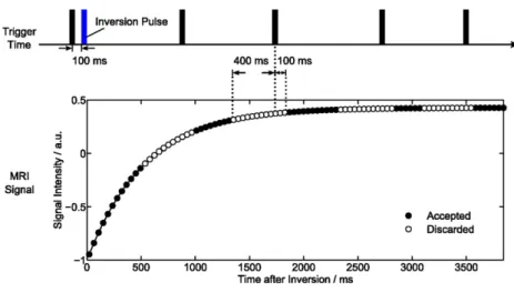
Figure 1. MyocardialT1mapping using single-shot inversion recovery FLASH with radial undersampling. Finger pulse trigger5
black bar.
Table 1.T1relaxation times for a reference phantom and simulated heart rates
T1, msa T2, msb Heart ratec 0 40 60 80 100 331611 10162 315613 315613 314613 316613 317616 494622 4662 476618 475618 475618 476620 479623 676619 8163 660625 659626 661627 663629 665631 857625 13265 850628 850628 852630 853630 853635 1225620 13864 1227634 1226634 1230636 1230637 1236638 1501623 16665 1511642 1513643 1513644 1516646 1517650
Single-shot inversion recovery FLASH was performed at 43-ms resolution (19 spokes) for a duration of 3 s.
aT
1values for a long-repetition time inversion recovery fast spin echo sequence.
b
T2values according to Sumpf et al.
29
cSimulated heart rates (in beats per minute) correspond to the deletion of a 500-ms period (
“systole”) in each cardiac cycle. No images are deleted for
the reconstruction pipeline of the commercial MRI system (Magnetom Prisma fit) by a single network connection. If the system software is compatible, the implementation takes less than an hour including installation of ready-to-use measuring protocols for cardiacT1mapping and other real-time CMR applications. MRI acquisition and reconstruction
The chosen acquisition scheme for cardiac T1 mapping is illustrated inFigure 1. In order to achieve maximum robustness andT1 accuracy, a previously developed IR FLASH sequence10 was applied as a single-slice technique using a non-selective adiabatic 180° inversion pulse triggered to early diastole. The present study employed a simple and robustfinger pulse trigger and a 100-ms delay to inversion. Although the method yields similar accuracy for a slice-selective inversion pulse when ap-plied to stationary tissue (data not shown), cardiacT1mapping exclusively used a non-selective inversion pulse to minimize the effects of through-plane motion and myocardial perfusion. Continuous image readout after inversion was based on a radial FLASH sequence with pronounced undersampling. Time-efficient spoiling of residual transverse magnetizations was accomplished by random radiofrequency (RF) phases.16CardiacT1maps were then acquired at a nominal in-plane resolution of 1.031.0 mm2 and 6-mm section thickness using afield of view52563256 mm2in combination with a resolution of 512 complex data points per radial spoke (using two-fold oversampling). All spokes were ho-mogeneously distributed over 360°, whereasfive successive frames used complementary sets of spokes in sequential order. Other parameters were TR52.26 ms, TE51.47 ms andflip angle of 4°.
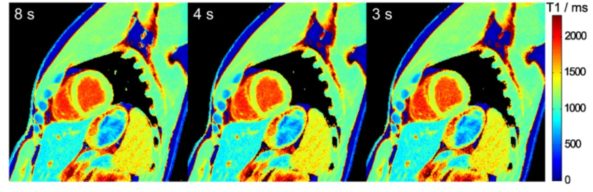
The number of spokes per frame varied from 27 to 23 andfinally 19 spokes yielding a temporal resolution of 61, 52 and 43 ms, respectively. The total acquisition time was initially chosen to be 8 s but later reduced to 4 and 3 s.
Image reconstruction has previously been described for the case of non-cardiacT1mapping10,11and employs the same iteratively reg-ularized NLINV algorithm as developed for real-time MRI; for details, see Uecker et al.9 Apart from an advanced gradient-delay correction14and data compression to 10 virtual channels based on a principal component analysis, the method takes advantage of some degree of spatial smoothness of coil sensitivities as well as of temporal regularization to the preceding frame. However, this latter term of the underlying cost function is downsized relative to the data consistency term by a factor of two during each iteration. The reconstruction ensures high temporalfidelity as demonstrated for a motion phan-tom rotating at defined speed17and therefore does not compromise the resolution of contrast changes during inversion recovery. The actual reconstruction process starts immediately after the end of data acquisition,first by a reverse NLINV reconstruction of the last 10 frames to obtain high-quality coil sensitivity maps using 6 iterations. Subsequently, the entire image series was reconstructed in the reverse order byfixing the coil sensitivities to those obtained by NLINV. The resulting linear inverse problem was solved by the iteratively regularized conjugate gradient method, again using six iterations.
Prior toT1fitting, the images were spatiallyfiltered by a recently developed modified non-local means algorithm.18 The filter
Figure 2. MyocardialT1maps for different temporal resolutions. Single-shot inversion recovery FLASH was performed with 27, 23
and 19 spokes per frame (i.e.acquisition times of 63, 52 and 43 ms) for a duration of 8 s.
Figure 3. MyocardialT1maps for different measurement durations. Single-shot inversion recovery FLASH was performed at 43 ms
preserves small isolated details and efficiently removes back-ground noise [corresponding to a 60% signal-to-noise (SNR) improvement] without introducing blur, smearing or patch artefacts. This is accomplished by extending the conventional non-local means algorithm to adapt the influence of the original pixel value according to a simple measure for patch regularity. Detail preservation is improved by a compactly supported weighting kernel which closely approximates the commonly used exponential weight.
Temporal median filtering was only used for the purpose of displaying image series, whereas no temporalfilter was used for
T1 mapping. The median filter extended over five frames to match the number of frames with different sets of spokes,e.g. in the studies of Uecker et al9and Frahm et al.17As illustrated in Figure 1, the influence of systolic motion on thefitting of a di-astolicT1map was minimized by automatically deleting images
over a period of 500 ms starting from 400 ms prior to eachfinger pulse trigger signal.
T1quantitation
After reconstruction, spatial filtering and systolic deletion, the remaining complex images were fitted to the complex sig-nal model2 MðtÞ5Mini g2ð11gÞexp 2t T1p (1) whereMiniis the initial complex signal after inversion; tis the central time point (i.e.radial spoke) of each frame during in-version recovery;gis the ratio between the steady-state signal
Mss and Mini; and T1p is the shortened apparent T1 due to multiple low flip-angle RF excitations. The same phase is as-sumed for Mss and Mini, which leads to four unknown real-valued parameters: Re{Mini}, Im{Mini},g and T1p. A pixelwise estimation was performed using the Trust-Region algorithm (Chapters 4.1 and 4.3 in the study of Nocedal and Wright19) based on the Dlib C11library.20 The algorithm performs an unconstrained minimization of the cost function defined by
1 2+t
ðRefMðtÞ2YðtÞgÞ21ðImfMðtÞ2YðtÞgÞ2 (2) where Y corresponds to the vector of pixel intensities during inversion recovery. The iterative optimization was stopped if the relative difference of the objective function values between successive iterations was ,1025. T
1 was then calculated according to:21
T15
T1p
g 12dt (3)
Table 2. MyocardialT1relaxation times for different temporal
resolutions and measuring times
Spokes per frame 27 23 19
Temporal resolutiona 61 ms 52 ms 43 ms IR-FLASH measurementb T1, ms c 8 s 1253629 1254629 1253631 4 s 1252630 1254630 1253631 3 s 1254632 1257633 1256633
IR, inversion recovery.
aNumber of spokes and acquisition time per frame.
b
Total measuring time.
cAveraged across subjects (mean
6standard deviation,n56).
Figure 4. Cardiac images andT1maps (top) without and (bottom) with spatial filtering. Single-shot inversion recovery FLASH was
performed at 43 ms temporal resolution (19 spokes) for a duration of 3 s. The images (magnified views) refer to an early time point after inversion, nulling of myocardial tissue and nulling of blood signal, respectively.
with dt the delay between inversion and the start of data ac-quisition. In the present implementation, this period covered half of the inversion pulse (5 ms) and a following spoiler gra-dient (10 ms). For the assessment of myocardial T1 values, the regions of interest were carefully selected to exclude con-tributions from the blood pool. These analyses were accom-plished using the arrayShow tool22in MATLAB® (MathWorks®, Natick, MA).
RESULTS
Table 1summarizesT1relaxation times for a reference phantom. The data were acquired with the radial IR-FLASH method proposed for cardiac T1 mapping (43-ms resolution and 3-s duration) at different simulated heart rates ranging from 40 to 100 bpm. Zero heart rate refers toT1fitting without deletion of any frames. A comparison withT1relaxation times obtained by a long-TR IR-FSE technique reveals excellent agreement for most (long) values, whereas two tubes with shorter values are slightly underestimated (maximum deviation 5%).
Figures 2 and 3 demonstrate cardiac T1 maps for different numbers of spokes per frame,i.e.different temporal resolution, and different durations of the IR-FLASH measurement, re-spectively. In all cases, visual inspection reveals no detectable difference. This qualitativefinding is confirmed by the quanti-tative analysis inTable 2.
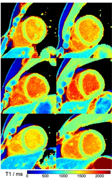
The effect of filtering prior to T1 fitting is demonstrated in Figure 4comparing raw images andT1maps with and without application of a spatialfilter.Figure 5shows threeT1maps for a single subject in a basal, mid-ventricular and apical short-axis section, whereas Figure 6 summarizes the mid-ventricular T1 maps of all six subjects. A full cardiac image series which cor-responds to the T1 map in the mid-ventricular section of Figure 5is available asSupplementary Video A. The quantitative results for all six subjects are summarized inTable 3(septalT1 values in a basal, mid-ventricular and apical section) andTable 4 (segmentalT1values in a mid-ventricular section), respectively. DISCUSSION
This work describes a novel method for myocardialT1mapping which offers accuracy, high spatial resolution, practical robust-ness and speed. The results indicate that myocardialT1mapping by IR-FLASH may be performed at a nominal resolution of
1.0 mm, a temporal resolution of 43 ms per frame and within a measuring time of only 3 s.
T1 accuracy was confirmed in a phantom study providing ref-erence T1 values for a long-TR IR-FSE sequence. A slight
Figure 5. MyocardialT1maps of a basal, mid-ventricular and apical section. Single-shot inversion recovery FLASH was performed at
43 ms resolution (19 spokes) for a duration of 3 s.
Figure 6. MyocardialT1maps of all six subjects (mid-ventricular
section, magnified view). Single-shot inversion recovery FLASH was performed at 43 ms resolution (19 spokes) for a duration of 3 s.
underestimation ofT1relaxation times for two tubes with values of about 500 and 675 ms coincides with the occurrence of the shortest T2 relaxation times of 46 and 81 ms, respectively (compare Table 1). The slight deviation of T1 values may therefore be due to a partial failure of the “reference”IR-FSE acquisition which extends to a maximum TE of 72 ms and thus may affect the image of compounds and tissues with shortT2 relaxation times. Similar effects are to be expected for IR methods with a SSFP readout module, because such sequences require relatively longT2relaxation times to build up sufficiently strong transverse coherences.
Apart fromT1accuracy, the results inTable 1confirm the in-dependence ofT1quantitation on the heart rate up to 100 bpm which effectively refers to the independence of T1 on the number offitted images after elimination of “systolic” frames. This advantageous behaviour reflects the fact that the highly undersampled radial FLASH readout ensures a sufficiently large number of frames for a proper sampling of the IR signal time course. T1 precision was also demonstrated to be high both
in vitroand in vivo. It is characterized by small standard de-viation values of 3–5% of the mean for phantom measurements and 4–8% for septalT1values (compareTables 1–4). Moreover, the achievedT1mapping quality not necessarily depends on the use offiltering as shown by the comparison inFigure 4. Nev-ertheless, although high-quality T1 maps may be obtained by
fitting unfiltered images, the use of a new modified non-local meansfilter18further improves the SNR ofT1maps without the expense of blurring.
Although myocardial T1 relaxation times found here were in general agreement with literature values, comparisons to pre-vious results are compromised by numerous technical differ-ences or even inadequacies. As an example, the present values are in the range of those reported in the study by Kawel et al23 but slightly higher than in that by von Knobelsdorff-Brenkenhoff et al24and lower than in the study by Lee et al,25 who all used similar MOLLI sequences. Of course, all techniques including the one proposed here suffer from some general limitations of the Lock-Locker approach which often are due to
Table 3.T1relaxation times of the septal wall
Subject T Basal 1, #framesa Mid-ventricular T1, #frames Apical T1, #frames #1 1250669/48 1256664/48 1298672/38 #2 1237660/48 1263654/47 1291658/47 #3 1270674/35 1287669/35 1332660/38 #4 1277671/47 1295661/49 1298650/47 #5 1215668/49 1209651/47 1256648/47 #6 1227661/49 1227651/47 1253646/48 Meanb 1246624 1256633 1288630 Kawel et al23 1286 von Knobelsdorff-Brenkenhoff et al24 1157 1159 1181 Lee et al25 1315 a
T1(in milliseconds, mean6standard deviation in a region of interest covering most of the septal wall) for single-shot inversion recovery-FLASH at 43
ms resolution (19 spokes) and 3 s duration. #Frames refers to the number of images retained after deletion of systolic frames.
bMean
6standard deviation across subjects.
Table 4. Regional myocardialT1relaxation times
Subject AnteriorT 1, ms Septal T1, ms Inferior T1, ms Lateral T1, ms #1 1295670 1261670 1223695 1218670 #2 1191660 1259656 1245691 1157664 #3 1212662 1291668 1270689 1238669 #4 1224664 1304664 1230692 1217668 #5 1169656 1206659 1155679 1166657 #6 1173656 1234658 1171666 1156669 Meana 1211647 1259636 1216644 1192636
T1 (in milliseconds, mean 6 standard deviation per standardized region of interest in a mid-ventricular section) for single-shot inversion
recovery-FLASH at 43-ms resolution (19 spokes) and 3-s duration.
aMean
the presence of residual motion both in plane and through plane. For example, diastolic circulation of blood within the ventricles leads to image intensities which violate the expected IR signal model and preclude a reliablefitting of bloodT1 re-laxation times. Even myocardial movements may play a role during early diastolic phases. However, this mainly becomes a problem for motion-sensitive readouts such as SSFP sequen-ces, whereas short-TE FLASH sequences as used here and re-cently proposed by others26,27 are much less affected. This is because SSFP sequences inherently rely on the establishment of phase coherence over multiple repetition intervals which is commonly precluded (i.e.spoiled) in the presence of motion, whereas FLASH sequences interrogate a pool of longitudinal magnetization with independent low-flip angle excitations that give rise to spin-density weighted images for the lowflip angles used for T1 mapping. In fact, when exploiting the additional motion robustness of radial encodings in the present implementation, preliminary trials of myocardial T1 mapping during free breathing showed little if any qualitative and quantitative difference to breath-hold scans (data not shown). Thus, the proposed method seems to be robust enough to even work in patients who are unable to perform any breathing protocol.
Another factor contributing to myocardialT1values is the dif-ferent access to high spatial resolution and the concomitant consideration of partial volume effects. Such problems have been reported for thin myocardial walls7 including the assess-ment offibrosis in the peri-infarct zone as well as for the right ventricle.28TheseT1mapping studies using MOLLI techniques were performed at 1.431.938.0 mm3for low heart rates and 1.932.338.0 mm3at high heart rates.7A higher resolution of 1.231.234 mm2 was only achieved with the use of a seg-mented readout module after inversion which therefore required repetitive acquisitions and very long measuring times of 2.5 min
perT1map.28Another recent work using IR-FLASH employed a sliding-window reconstruction27 at 1.1731.1738 mm3 res-olution, which was achieved by twofold zero-padding, i.e. an interpolation of the acquired resolution. To the best of our knowledge, the method proposed here is thefirst technique for myocardialT1mapping which offers 1.031.036.0 mm3 reso-lution within a measuring time of only 3 s.
The most important limitation of this study is the small sample size. This is because the work represents a new technical de-velopment which requires basic validation with use of aT1 refer-ence phantom and a group of normal subjects. Obviously, this precedes any evaluation of the clinical utility of the proposedT1 mapping in a large cohort of patients. Moreover, at this stage, widespread clinical applications are hampered by the fact that the technical solution requires dedicated software and hardware which so far is only available for MRI systems of the same manufacturer as used here. A remaining temporary restriction is the computa-tional time needed for image reconstruction andT1fitting which currently takes about 13 s perT1map. Nevertheless, this may not necessarily block the clinical workflow, because a delayed re-construction does not interfere with continuous acquisitions. CONCLUSION
MyocardialT1mapping based on single-shot IR-FLASH with ra-dial undersampling and iterative reconstruction as well asT1fitting with automated deletion of systolic frames meets most current clinical challenges. The proposed method warrants extensive clinical trials as it promises significant advantages for CMR studies which rely on native or post-contrastT1quantitation.
FUNDING
AAJ is supported by the DZHK (German Centre for Cardio-vascular Research) and by the BMBF (German Ministry of Ed-ucation and Research).
REFERENCES
1. Haase A. Snapshot FLASH MRI. Applications to T1, T2, and chemical-shift imaging.Magn Reson Med1990;13: 77–89. doi:https://doi. org/10.1002/mrm.1910130109
2. Deichmann R, Haase A. Quantification of T1 values by SNAPSHOT-FLASH NMR imag-ing.J Magn Reson1992;96: 608–12. 3. Ordidge RJ, Gibbs P, Chapman B, Stehling
MK, Mansfield P. High-speed multislice T1 mapping using inversion-recovery echo-planar imaging.Magn Reson Med1990;16: 238–45. doi:https://doi.org/10.1002/ mrm.1910160205
4. Scheffler K, Hennig J. T1 quantification with inversion recovery TrueFISP.Magn Reson Med2001;45: 720–3. doi:https://doi.org/ 10.1002/mrm.1097
5. Look DC, Locker DR. Time saving in measurement of NMR and EPR relaxation
times.Rev Sci Instrum1970;41: 250–1. doi:
https://doi.org/10.1063/1.1684482
6. Messroghli DR, Radjenovic A, Kozerke S, Higgins DM, Sivananthan MU, Ridgway JP. Modified look-locker inversion recovery (MOLLI) for high-resolution T1 mapping of the heart.Magn Reson Med2004;52: 141–6. doi:https://doi.org/10.1002/mrm.20110
7. Kellman P, Hansen MS. T1-mapping in the heart: accuracy and precision.J Cardiovasc Magn Reson2014;16: 2. doi:https://doi.org/ 10.1186/1532-429X-16-2
8. Moon JC, Messroghli DR, Kellman P, Piechnik SK, Robson MD, Ugander M, et al. Myocardial T1 mapping and extracellular volume quantification: a Society for Cardio-vascular Magnetic Resonance (SCMR) and CMR Working Group of the European Society of Cardiology consensus statement.J
Cardiovasc Magn Reson2013;15: 92. doi:
https://doi.org/10.1186/1532-429X-15-92
9. Uecker M, Zhang S, Voit D, Karaus A, Merboldt KD, Frahm J. Real-time MRI at a resolution of 20 ms.NMR Biomed2010;23: 986–94. doi:https://doi.org/10.1002/ nbm.1585
10. Wang X, Roeloffs V, Merboldt KD, Voit D, Sch¨atz S, Frahm J. Single-shot multi-slice T1 mapping at high spatial resolution— inversion-recovery FLASH with radial undersampling and iterative reconstruction.Open Med Imag-ing J2015;9: 1–8. doi:https://doi.org/10.2174/ 1874347101509010001
11. Hofer S, Wang X, Roeloffs V, Frahm J. Single-shot T1 mapping of the corpus callosum: a rapid characterization offiber bundle anatomy.Front Neuroanat2015;9: 57. doi:
12. Zhang S, Uecker M, Voit D, Merboldt KD, Frahm J. Real-time cardiovascular magnetic resonance at high temporal resolution: radial FLASH with nonlinear inverse reconstruction.
J Cardiovasc Magn Reson2010;12: 39. doi:
https://doi.org/10.1186/1532-429X-12-39
13. Voit D, Zhang S, Unterberg-Buchwald C, Sohns JM, Lotz J, Frahm J. Real-time cardiovascular magnetic resonance at 1.5 T using balanced SSFP and 40 ms resolution.J Cardiovasc Magn Reson2013;15: 79. doi:
https://doi.org/10.1186/1532-429X-15-79
14. Untenberger M, Tan Z, Voit D, Joseph AA, Roeloffs V, Merboldt KD, et al. Advances in real-time phase-contrastflow MRI using asymmetric radial gradient echoes.Magn Reson Med2016;75: 1901–8. doi:https://doi. org/10.1002/mrm.25696
15. Zhang S, Joseph AA, Voit D, Schaetz S, Merboldt KD, Unterberg-Buchwald C, et al. Real-time magnetic resonance imaging of cardiac function andflow-recent progress.
Quant Imaging Med Surg2014;4: 313–29. doi: https://doi.org/10.3978/j.issn.2223-4292.2014.06.03
16. Roeloffs V, Voit D, Frahm J. Spoiling without additional gradients: radial FLASH MRI with randomized radiofrequency phases.Magn Reson Med2016;75: 2094–9. doi:https://doi. org/10.1002/mrm.25809
17. Frahm J, Sch¨atz S, Untenberger M, Zhang S, Voit D, Merboldt KD, et al. On the temporal
fidelity of nonlinear inverse reconstructions for real-time MRI—the motion challenge.
Open Med Imaging J2014;8: 1–7. doi:https:// doi.org/10.2174/1874347101408010001
18. Klosowski J, Frahm J. Image denoising for real-time MRI.Magn Reson Med2016; doi:
https://doi.org/10.1002/mrm.26205
19. Nocedal J, Wright SJ. Numerical Optimiza-tion. In: Mikosch TV, Sidney S, Resnick I, Robinson SM, eds.Springer Series in Oper-ations Research. New York, NY: Springer Science1Business Media; 2006. pp. 66–98. 20. King D. Dlib-ml: A Machine Learning Toolkit.
J Mach Learn Res2009;10: 1755–1758. 21. Deichmann R. Fast high-resolution T1
mapping of the human brain.Magn Reson Med2005;54: 20–7. doi:https://doi.org/ 10.1002/mrm.20552
22. Sumpf T, Unterberger M. arrayShow: a guide to an open source matlab tool for complex MRI data analysis. In:ISMRM; 2013. p. 2719. 23. Kawel N, Nacif M, Zavodni A, Jones J, Liu S,
Sibley CT, et al. T1 mapping of the myocardium: intra-individual assessment of the effect offield strength, cardiac cycle and variation by myocardial region.J Cardiovasc Magn Reson2012;14: 27. doi:https://doi.org/ 10.1186/1532-429X-14-27
24. von Knobelsdorff-Brenkenhoff F, Proth-mann M, Dieringer MA, Wassmuth R, Greiser A, Schwenke C, et al. Myocardial T1 and T2 mapping at 3 T: reference values, influencing factors and implica-tions.J Cardiovasc Magn Reson2013;15: 53. doi: https://doi.org/10.1186/1532-429X-15-53
25. Lee JJ, Liu S, Nacif MS, Ugander M, Han J, Kawel N, et al. Myocardial T1 and extracel-lular volume fraction mapping at 3 Tesla.J Cardiovasc Magn Reson2011;13: 75. doi:
https://doi.org/10.1186/1532-429X-13-75
26. Shao J, Rapacchi S, Nguyen KL, Hu P. Myocardial T1 mapping at 3.0 Tesla using an inversion recovery spoiled gradient echo readout and Bloch equation simulation with slice profile correction (BLESSPC) T1 esti-mation algorithm.J Magn Reson Imaging
2016;43: 414–25. doi:https://doi.org/ 10.1002/jmri.24999
27. Gensler D, M¨orchel P, Fidler F, Ritter O, Quick HH, Ladd ME, et al. Myocardial T1: quantification by using an ECG-triggered radial single-shot inversion-recovery MR imaging sequence.Radiology2015;274: 879–87. doi:https://doi.org/10.1148/ radiol.14131295
28. Mehta BB, Chen X, Bilchick KC, Salerno M, Epstein FH. Accelerated and navigator-gated look-locker imaging for cardiac T1 estima-tion (ANGIE): development and applicaestima-tion to T1 mapping of the right ventricle.Magn Reson Med2015;73: 150–60. doi:https://doi. org/10.1002/mrm.25100
29. Sumpf TJ, Petrovic A, Uecker M, Knoll F, Frahm J. Fast T2 mapping with improved accuracy using undersampled spin-echo MRI and model-based reconstructions with a gen-erating function.IEEE Trans Med Imaging
2014;33: 2213–22. doi:https://doi.org/ 10.1109/TMI.2014.2333370