Correlations of Biomechanical Characteristics with Ball Speed in Penalty Corner Push-In
An Investigation of Segmentation Techniques in Image Processing for MRI Brain Tumor Images
V.Vishalini1, K.Radha2 & A.Saraswathi31Research Scholar, Thanthai Hans Roever College, Perambalur, Tamilnadu, India. 2Research Scholar, Thanthai Hans Roever College, Perambalur, Tamilnadu, India.
3Asst. Prof. in Comuter Science, Thanthai Hans Roever College, Perambalur, Tamilnadu, India.
Received 30th July 2017, Accepted 5th September 2017
Abstract
The main goal of image segmentation is partitioning of an image into a set of disjoint regions that are visually different, homogeneous and meaningful with respect to some characteristics or computed properties, such as grey level, texture or color to enable easy image analysis (object identification, classification and processing). Image segmentation can be classified three categories traditionally including Threshold Technique, Region-Based Image Segmentation, and Edge-Based Image Segmentation. Magnetic Resonance Imaging (MRI) is an advanced medical imaging technique used to produce high quality images of the parts contained in the human body MRI imaging is often used when treating brain tumors. MRI can also be used to measure the tumor’s size. Segmentation is a process of extracting information from an image and to group pixels together into regions of similarity. Image segmentation is typically used to locate objects in images.
Keywords: Threshold Technique, Region-Based Image Segmentation, Edge-Based Image Segmentation, MRI, Brain Tumor.
© Copy Right, IJRRAS, 2017. All Rights Reserved.
Introduction
Brain tumor is naturally serious and life intimidating because of its character in the limited space of the intracranial cavity (space formed inside the skull). Most Research in developed countries proves that the numbers of people who have brain tumors were died due to the fact of inaccurate detection. Magnetic resonance images are a very useful tool to detect the tumor growth in brain. MRI is directed into intracranial cavity produces a complete image of brain. This image is visually examined by the physician for detection and diagnosis of brain tumor. However this method of detection resists the accurate determination of stage and size of tumor. To avoid that, the computer aided analysis for brain tumor detection is proposed. The image processing techniques allows the detection of tumor tissue with accuracy and reproducibility comparable to manual detection. In addition, it also reduces the time for analysis. At the end of the process the tumor is extracted from the MR image and its exact position and the shape also determined [1]. The following figure shows the fundamental steps in MRI system.
Correspondence
A.Saraswathi
E-mail: [email protected], Ph. +9195007 84950
Figure 1. Block Diagram for MRI System Correct segmentation of MR images is very important because most of the time MR images are not highly contrast thereby these segments can be easily overlapped with each other. So, to develop high contrast MR images. A brain tumor is an abnormal growth of tissue in the brain. Unlike other tumors, brain tumors spread by local extension and rarely metastasize (spread) outside the brain. A benign brain tumor is composed of non-cancerous cells and does not metastasize beyond the part of the brain where it originates. A brain tumor is considered malignant if it contains cancer cells, or if it is composed of harmless cells located in an area where it suppresses one or more vital functions. Each year, more than 17,000 brain tumors are diagnosed in the United States. About half of all primary brain tumors are benign, but in life threatening locations. The rest are malignant and invasive [2].
International
Journal of Recent Research and Applied Studies
(Multidisciplinary Open Access Refereed e-Journal)Figure 2. MRI Brain Tumor images
The above figure shows the some samples of MRI Brain Tumor images.
Segmentation
All image processing operations normally represent at a better recognition of objects of interest, i.e., at finding suitable local features that can be distinguished from other objects and from the background. The next step is to check each individual pixel to see whether it belongs to an object of interest or not. This operation is called segmentation and produces a binary image. A pixel has the value one if it belongs to the object otherwise it is zero. Segmentation is the operation at the threshold between low-level image processing and image analysis. After segmentation, it is known that which pixel belongs to which object. The image is parted into regions and we know the discontinuities as the boundaries between the regions [3].
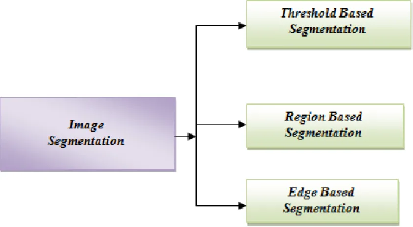
Segmentation is a process of extracting information from an image and to group pixels together into regions of similarity. Image segmentation is typically used to locate objects and boundaries in images. The main goal of image segmentation is partitioning of an image into a set of disjoint regions that are visually different, homogeneous and meaningful with respect to some characteristics or computed properties, such as grey level, texture or color to enable easy image analysis (object identification, classification and processing). Image segmentation can be classified three categories as follows,
1. Threshold Technique,
2. Region-Based Image Segmentation 3. Edge-Based Image Segmentation.
Figure 3. Image segmentation techniques
In terms of mathematical formulae, Image segmentation divides a digital image f(x, y) into continuous, disconnect and nonempty subsets, from these subsets higher level information can be easily extracted. Practical applications of image segmentation include object identification and recognition, facial recognition, medical image processing, criminal investigation, airport security system, satellite images, quality assurance in factories, etc [2]. Due to the importance of the image segmentation, large number of algorithms has been proposed but the selection of the algorithm purely
depends upon the image type and the nature of the problem [3].
Many image segmentation techniques have been developed by researchers and scientists, some of the most important and widely used image segmentation techniques are shown in Fig. 1.In recent years, a lot of research is done in the field of image segmentation process. There are currently thousands of algorithms, each doing the segmentation process slightly different from another, but still there is no particular algorithm that is applicable for all types of digital image, fulfilling
every objective. Thus, algorithm developed for a group of images may not always apply to images of another class [4].
Threshold segmentation
Thresholding is the simplest method of image segmentation. From a grayscale image, thresholding can be used to create binary images. During the thresholding process, individual pixels in an image are marked as "object" pixels if their value is greater than some threshold value (assuming an object to be brighter than the background) and as "background" pixels otherwise. This convention is known as threshold above. Variants include threshold below, which is opposite of threshold above. Typically, an object pixel is given a value of ―1‖ while a background pixel is given a value of ―0‖. Finally, a binary image is created by coloring each pixel white or black, depending on a pixel's labels.
Threshold selection
The key parameter in the thresholding process is the choice of the threshold value. Several different methods for choosing a threshold exist. Users can manually choose a threshold value or a thresholding algorithm can compute a value automatically, which is known as automatic thresholding. A simple method would be to choose the mean or median value, the basis being that if the object pixels are brighter than the background, they should also be brighter than the average. In a noiseless image with uniform background and object values, the mean or median will work well as the threshold, however, this will generally not be the case. A more sophisticated approach might be to create a histogram of the image pixel intensities and use the valley point as the threshold. The histogram approach assumes that there is some average values for both the background and object pixels, but that the actual pixel values have some variation around these average values. However, this may be computationally expensive, and image histograms may not have clearly defined valley points, often making the selection of an accurate threshold difficult. In such cases a unimodal threshold selection algorithm may be more appropriate. One method that is relatively simple, does not require much specific knowledge of the image, and is robust against image noise, is the following iterative method:
1. An initial threshold (T) is chosen, this can be done randomly or according to any other method desired.
2. The image is segmented into object and background pixels as described above, creating two sets:
1. = {f(m,n):f(m,n)>T} (object pixels)
2. = {f(m,n):f(m,n) T} (background pixels) (note: f(m,n) is the value of
the pixel located in the column, row)
3. The average of each set is computed. 1. = average value of 2. = average value of
4. A new threshold is created that is the average of and
1. T = ( + )/2
(Note: Threshold = (average
background + average objects)/2)
5. Go back to step two, now using the new threshold computed in step four, keep repeating until the new threshold matches the one before it (i.e. until convergence has been reached).
This iterative algorithm is a special one-dimensional case of the k-means clustering algorithm, which has been proven to converge at a local
minimum—meaning that a different initial threshold may
give a different final result.(Thresholding: All values above some threshold are set to 1 and those below the threshold are set to 0 or vice versa.)
Region-based segmentation
Region growing is a simple region-based image segmentation method. It is also classified as a pixel-based image segmentation method since it involves the selection of initial seed points. This approach to segmentation examines neighboring pixels of initial ―seed points‖ and determines whether the pixel neighboring pixel should be added to the region. The following steps are performed to find the initial seed point.
Convert the given color image into a gray image
Count the number of pixels whose intensities are greater than hundred and less than hundred and Store them in separate variables.
Find the difference between both the variables, if the difference is smaller (Less than 0.2) then go to image enhancement else convert the image into binary image.
Calculate the maximum length and breadth of the image.
Calculate the sum of all rows and columns and store in separate arrays.
Find the intersection of row and column having maximum sum. This is taken as seed Point.
Edge based segmentation
Edge based segmentation methods partition an image based on rapid changes in intensity near edges [4]. The result is a binary image. Based on theory there are two main edge based segmentation methods- gray histogram and gradient based method [5].
Gray Histogram Technique
The result of edge detection technique mainly depends upon selection of threshold T
Gradient Based Method
In gradient based method, the difference between two neighboring pixel values is considered. So, when there is an abrupt change in intensity near edge and there is little image noise then gradient based method works well. This method involves convolving gradient operators with the image. Common edge detection operators used in gradient based method are sobel operator, canny operator, Laplace operator, Laplacian of Gaussian (LOG) operator & so on, canny is most promising one [6], but takes more time as compared to sobel operator. Edge detection methods require a balance between detecting accuracy and noise immunity. If the level of detecting accuracy is too high, noise may bring in fake edges making the outline of images unreasonable and if the degree of noise immunity is too excessive [7], some parts of the image outline may get undetected and the position of objects may be mistaken. Thus, edge detection algorithms are suitable for images that are simple and noise-free as well often produce missing edges or extra edges on complex and noisy images. To detect boundaries between 2 distinct regions, edge based segmentation is used.
Algorithm:
Step1: Apply the derivative operator to detect edges of the image
Step2: Find the magnitude at the edges
Step3: Retain all edge having magnitude greater than threshold value T
Step4: Find the position of crack edge, the crack edge is either retained or rejected.
Step5: Repeat step 3 and step 4 with different values of threshold so as to find out the closed boundaries, segmentation of an image is obtained.
Review of Literature
The purpose of image segmentation is to partition an image into regions (spatially connected groups of pixels called classes. or subsets) and objects with respect to one or more characteristics or features.Image segmentation plays a significant role in image processing as it helps in the extraction of suspicious regions from the medical images. The idea behind segmentation is to segment an image into several clusters. The results will be such that, it is possible to identify regions of interest and objects in the original image.
R. Rajeswari. G. Gunasekaran proposed Watershed segmentation algorithm for brain tumor segmentation and this is to transform the gradient of a grey level image in a topographic surface. Maker based watershed segmentation solves image segmentation
problems .In this internal markers are produced from gray scale image and external markers are used to find the pixels between the internal markers. This is done by watershed transform. Computation of this transform along with modified gradient image produces watershed ridge lines and this lines are superimposed on the original image and produce the segmentation of tumor region from MRI. Tumor cells are clustered using hierarchical clustering algorithm. [8]
Color based segmentation using k-means clustering identifies the tumor region significantly form the pre-processed MR image as a clustering feature. Here the pre-processed gray-level brain MR image is converted into RGB color image. Histogram equalization technique is performed and it takes advantages of the neglected pixel values. The RGB color image is then been coarsely represented using 25 bins. Coarse representation uses the spatial information from a histogram based windowing process. K-means is been used to cluster the coarse image data. This shows better result when compared with other edge detection algorithm and enhance the tumor detection accuracy in less time. This was developed by Sarbani Datta. Dr. Monisha Chakraborty [9].
Easha Noureen. Dr. Md. Kamrul Hassan proposed Histogram Thresholding segmentation method for detecting the brain tumor in MRI image. This is based on thresholding of histogram features and gray level thresholding .It is suitable for an image with region or object of uniform brightness placed against a background of different gray level. A threshold must be applied to segment the object and background. Histogram presents the intensity values of an image and the thresholding is a technique for converting the grayscale or color image in to a binary image based on threshold values. MRI image of the brain is divided and the histogram of each part is drawn. Threshold point of the histogram is calculated and the segmentation is done using the threshold point for both the halves. Plot the histogram and it is between number of pixel and pixel intensity .Bar graph can be used to plot the histogram. Difference of the two histogram is calculated and the resultant difference is plotted using bar graph to select the threshold point. This results gives the great importance in detecting the brain tumor in MRI image [10].
Seeded Region Growing method is an approach to segmentation where it examines neighboring pixels of initial ‗seed points‘ and determines pixel neighbors should be added to the region. It is a technique for determining the regions directly. Formulation of the region –based segmentation is, it must be complete and every pixel in the region must be disjoint so that clear separation from each other can be identified. It satisfies the condition that the gray level of pixel is in the range of region. This segmentation is used to find the abnormality is present in the image or not. Fast and fully automatic algorithm, both the homogenous texture features and spatial features of the MRI are used to find the seed
points and segmentation results obtained are accurate. This was developed by Mukesh Kumar, Kamal Mehta in 2011[11].
Segmentation Results for Brain Tumor MRI Images
Images are first pre-processing by converting into gray image and it is denoised using various techniques. This pre-processed image is segmented using
the combined features of single seed region growing and threshold segmentation methods. The obtained results are analyzed by the quality measures [12]. The result of segmentation techniques are compared in following figure. In figure 4, 1st row images are the output of Threshold Technique, 2nd row represents the result of Region-Based Image Segmentation, and 3rd row shows the result of Edge-Based Image Segmentation.
Figure 4. Segmentation Results
Above figure clearly shows the new Region-Based segmentation method can able to generate the expected results by experimentally.
Table 1. Comparison of Segmentation Results
Segmentation Method
PSNR MSE Elapsed time Accuracy
Threshold 34.13 1.547 7.25 3.0234 Edge-Based 33.27 30.59 3.58 0.0025 Region-Based 41.908 1.357 2.980 3.262
Conclusion
Many image segmentation methods have been developed in the past several decades for segmenting MRI brain images, but still it remains a challenging task. A segmentation method may perform well for one MRI brain image but not for the other images of same type. Thus it is very hard to achieve a generic segmentation method that can be commonly used for all MRI brain images. In this paper, Threshold Technique, Region-Based Image Segmentation, Edge-Based Image Segmentation methods are analyzed in detail for MRI brain tumor images.
References
1. Deeplai Kelkar and Surendra Gupta (2008): ―Improved Quadtree method for split merge image segmentation‖, Emerging Trends in Engineering and Technology, 2008 ICETET.
2. Easha Noureen, Dr, Md, Kamrul Hassan ‗ Brain Tumor Detection Using Histogram Thresholding to Get the Threshold point‘ IOSR Journal of Electrical and Electronics Engineering (IOSR-JEEE) Volume 9, Issue 5 Ver, III (Sep – Oct, 2014), PP 14-19.
3. Gonzalez, Rafael C., Richard E.Woods (2007): ―Digital Image Processing‖, Pearson Education., 3rd Edition.
4. H.P. Narkhede, (2013) ―Review of Image Segmentation Techniques‖, International Journal of Science and Modern Engineering (IJISME), Vol. 1, No. 8, pp: 54-61.
http://medicaldictionary.
thefreedictionary.com/Brain+Tumor
5. Image Segmentation Techniques Rajeshwar Dass, priyanka, Swapna Devi IJECT Vol. 3, Issue 1, Jan. -March 2012
6. Kang, W.X., Yang, Q.Q., Liang, R.R (2009): ―The Comparative Research on Image Segmentation Algorithms‖, IEEE Conference on ETCS, pp. 703-707.
7. Mukesh Kumar, Kamal K,Mehta ‗A Texture based Tumor detection and automatic Segmentation using Seeded Region Growing Method‘ Int, J, Comp, Tech, Appl,, Vol 2 (4), 855-859 august 2011. 8. Pal, N.R., Pal, S.K (1993): ―A Review on Image
Segmentation Techniques‖, Pattern Recognition, Vol. 26, No. 9, pp. 1277-1294.
9. R, Rajeswari, G, Gunasekaran ‗Tumor Detection and Segmentation Using Watershed and
Threshold Technique
Edge-Based
Hierarchical Clustering Algorithms‘, IJIRCCE Vol,2, Special Issue 5, October 2014.
10. Rohini Paul Joseph, C. Senthil Singh, M.Manikandan ―brain tumor mri image segmentation and detection in image processing‖ IJRET, eISSN: 2319-1163 | pISSN: 2321-7308.
11. Sarbani Datta, Dr, Monisha Chakraborty ‗Brain Tumor Detection from Pre-Processed MR Images using Segmentation Techniques‘, IJCA Special Issue on ‗2nd National Conference- Computing, Communication and Sensor Network‘ CCSN, 2011.