Dcx reexpression reduces subcortical band heterotopia
and seizure threshold in an animal model of neuronal
migration disorder
Jean-Bernard Manent
1, Yu Wang
1, YoonJeung Chang
1, Murugan Paramasivam
1& Joseph J LoTurco
1 Disorders of neuronal migration can lead to malformations of the cerebral neocortex that greatly increase the risk of seizures. It remains untested whether malformations caused by disorders in neuronal migration can be reduced by reactivating cellular migration and whether such repair can decrease seizure risk. Here we show, in a rat model of subcortical band heterotopia (SBH) generated byin uteroRNA interference of theDcxgene, that aberrantly positioned neurons can be stimulated to migrate by reexpressingDcxafter birth. Restarting migration in this way both reduces neocortical malformations and restores neuronal patterning. We further find that the capacity to reduce SBH continues into early postnatal development. Moreover, intervention after birth reduces the convulsant-induced seizure threshold to a level similar to that in malformation-free controls. These results suggest that disorders of neuronal migration may be eventually treatable by reengaging developmental programs both to reduce the size of cortical malformations and to reduce seizure risk.A causal connection between disruptions in neuronal migration during fetal development and altered neocortical excitability is well established1–5. Foci of abnormally migrated neurons are particularly prevalent in individuals with pharmacologically intractable epilepsies, and surgical resection of malformed cortex can often effectively treat such drug-resistant epilepsy6,7. Many cases, however, remain untrea-table by surgery because of the location and/or widespread distribu-tion of malformadistribu-tion(s). One such malformadistribu-tion occurs in double cortex syndrome, in which a band of heterotopic gray matter composed of abnormally migrated neurons is located between the ventricular wall and the cortical mantle and is separated from both by a band of white matter8,9. Focal resection of epileptogenic tissue in double cortex syndrome shows poor clinical outcome10. Double cortex syndrome or SBH is also associated with mild to moderate mental retardation11and with intractable epilepsy in about 65% of affected individuals12, and it is most often caused in females by mutations in the X-linked geneDCX, a microtubule-binding protein essential to neuronal migration13–16.DCXmutations in males usually cause anterior lissencephaly15, but SBH associated with DCX muta-tions have also been described in males17.
Studies in animal models have revealed that several types of migration disruptions and malformations increase neuronal excitabil-ity and seizure risk. For example, spontaneous seizures are observed in the tish (telencephalic internal structure heterotopia) mutant rat18, and significantly reduced thresholds to convulsant agents are observed in rats with cortical migration anomalies caused by prenatal exposure to teratogens such as methylazoxymethanol19,20, cocaine21
or irradiation22. Similarly, in a freeze-lesion model of microgyria, epileptiform discharges are reliably evoked in brain slices containing malformations, and the threshold dose of convulsants to induce seizures is reduced23,24. A recent study also reported that spontaneous convulsive seizures can occasionally be observed in a subset of Dcx-knockout mice showing discrete hippocampal malformations but no cortical abnormalities25. Together, results from animal models and studies on surgically removed human tissue indicate that malformed neocortex is associated with reorganized neuronal networks and altered cellular physiologies that create hyperexcitable tissue. It is currently unknown whether there is a time in development that interventions to reverse or reduce formed or forming malformations would also prevent neuronal hyperexcitability and seizure risk.
We previously developed a rat model of SBH by decreasingDcx expression with in utero RNA interference (RNAi)26. This model reproduces anatomical features of the malformations present in the human double cortex syndrome, and we have recently shown that the malformations are rescued or prevented by concurrent embryonic expression of Dcx27. Here we used a conditional variation of this rescue approach to determine whether delayed Dcx expression, after SBH has formed, can reduce heterotopia and restore neuronal patterning. We show that both laminar displacement of neurons and SBH size are reduced upon delayed expression of Dcx during early postnatal periods. We show further that rats with SBH are more susceptible to seizures induced by the convulsant pentylenetetrazol and that reduction of SBH restores seizure thresholds to levels similar to those of unaffected controls.
Received 15 May; accepted 6 November; published online 21 December 2008; doi:10.1038/nm.1897
1Department of Physiology and Neurobiology, 75 North Eagleville Road U-3156, University of Connecticut, Storrs, Connecticut 06269, USA. Correspondence should
be addressed to J.J.L. ([email protected]). © 2009 Nature America, Inc.
All rights reser
RESULTS
Conditional reexpression of Dcx
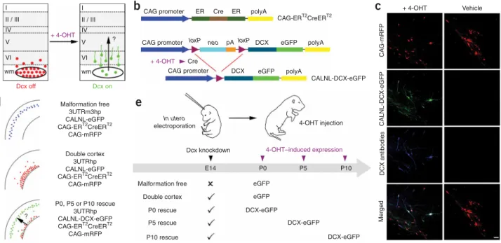
The purpose of the present study was to investigate whether neo-cortical lamination deficits and SBH malformations can be reduced by reexpression of Dcx after birth. Our approach was to initiate SBH formation and laminar displacement byin uteroRNAi ofDcxand then reexpress Dcx in mispositioned neurons (Fig. 1). To accomplish this, we adapted a previously developed in vivo conditional transgene expression system28to a conditional RNAi rescue approach. Because endogenousDcxexpression decreases with neuronal development, we could not achieve conditional reexpression by turning offDcxRNAi. Instead we created a system in which a version of Dcx that is insensitive toDcxRNAi was turned on in cells in which endogenous Dcxwas knocked down by RNAi. We constructed a conditional DCX-eGFP expression vector (CALNL-DCX-DCX-eGFP) that contains a stop codon flanked by twoloxPsites downstream from the CAG promoter which ubiquitously drives expression and upstream from the DCX-eGFP sequence (Fig. 1b). TheDcxsequence in this plasmid vector is missing the 3¢ untranslated region (UTR) of Dcx, and the vector encoding short hairpin RNA (shRNA) againstDcx(3UTRhp) that we previously developed26targets this region. Thus, unlike endogenous Dcx, DCX-eGFP expressed from CALNL-DCX-eGFP is not targeted byDcxRNAi. Another requirement of this strategy is that DCX-eGFP be expressed only after the addition of 4-hydroxytamoxifen (4-OHT). To test for such controlled reexpression in vivo, we trans-fected neocortical neuronal progenitors in utero at embryonic day 14 (E14) with the plasmid DNA vectors CALNL-DCX-eGFP, CAG-ERT2CreERT2 and 3UTRhp (see Methods). At birth, rat pups were given single intraperitoneal injections of either 4-OHT or vehicle
control. In postnatal day 15 (P15) neocortical sections from 4-OHT– injected rats, transfected cells were positive for eGFP signal and were immunopositive for Dcx (Fig. 1c). In addition, DCX-eGFP expression was detected as early as 1 d after 4-OHT injection and peaked to over 80% of transfected cells after 2 d (Supplementary Fig. 1online). In contrast, in littermate controls receiving the same in utero transfec-tions but injected with vehicle only, Dcx and eGFP expression were not detected (Fig. 1c). Thus, this system allows for temporally controlled reexpression of Dcx at times when expression of endogen-ous Dcx is decreased.
Reexpression of Dcx reduces SBH and restores laminar position To determine when to first reexpress Dcx in mispositioned neurons, we next established when SBH and lamination deficits were present after birth in theDcxRNAi model26.Dcxknockdown caused laminar displacement and prominent SBH by the day of birth (P0;Fig. 2a). Consequently, we conducted two types of experiments to induce reexpression of Dcx in mispositioned neurons on P0 and analyzed the rats at P20. In the first, we controlled for the addition of 4-OHT and for Cre recombinase activity and varied whether eGFP or DCX-eGFP was conditionally expressed from DCX-eGFP or CALNL-DCX-eGFP, respectively, in rats electroporated at E14 with shRNA vectors targeting Dcx (Fig. 1d,e). This experiment controlled for possible effects of Cre recombinase, as both groups received transfec-tion of the plasmid CAG-ERT2CreERT2 and both received 4-OHT injections. We found that, when DCX-eGFP was expressed at P0, SBH malformations were significantly reduced in size (Fig. 2b–e) relative to when eGFP was expressed. In addition, whereas transfected cells in the eGFP group accumulated in white matter and in deep layers, CAG promoter I
a
d
e
b
c
II / III IV + 4-OHT 4-OHT–induced expression 4-OHT injection + 4-OHT Vehicle Merged DCX antibodies CALNL-DCX-eGFP CA G-mRFP V VI wm I II / III IV V VI wm Dcx off Dcx on CAG promoter CAG promoter Malformation free 3UTRm3hp CALNL-eGFP CAG-ERT2CreERT2 CAG-mRFP Double cortex 3UTRhp CALNL-eGFP CAG-ERT2CreERT2CAG-mRFP Malformation free
Dcx knockdown E14 P0 P5 P10 In utero electroporation eGFP eGFP DCX-eGFP DCX-eGFP DCX-eGFP Double cortex P0 rescue P5 rescue P10 rescue P0, P5 or P10 rescue 3UTRhp CALNL-DCX-eGFP CAG-ERT2CreERT2 CAG-mRFP ? ? + 4-OHT Cre
loxP neo loxP DCX
DCX eGFP eGFP polyA polyA CALNL-DCX-eGFP pA ER polyA CAG-ERT2CreERT2 ER Cre
Figure 1 Temporal control of Dcx expression in mispositioned neurons by using Cre-dependent expression vectors and 4-OHT–activatable Cre recombinase. (a) A schematic diagram of the experimental approach used to test the hypothesis that reexpression of Dcx can reactivate migration and regress
malformation. (b) A 4-OHT–activatable Cre recombinase composed of two estrogen receptor (ER) binding domains is expressed under the control of the CAG promoter (CAG-ERT2CreERT2). The Cre-dependent Dcx expression vector contains a neomycin resistance gene (neo) with a stop codon flanked byloxPsites
(CALNL-DCX-eGFP). In the presence of 4-OHT, recombination occurs and DCX-eGFP is expressed. pA and polyA indicate the polyadenosine sequence in the plasmids. (c) Confocal images showing transfected neurons in neocortex of P15 rats that have received 4-OHT or vehicle injection at birth. Rats were electroporated at E14 with four plasmids: CAG-mRFP, CAG-ERT2CreERT2, CALNL-DCX-eGFP and 3UTRhp. In 4-OHT–treated rats (left), DCX-GFP is
expressed and Dcx is detectable with antibodies in transfected mispositioned neurons. In vehicle-treated rats (right), no signal is detected in the green channel or with Dcx antibodies. (d) Transfection conditions. (e) Schematic diagram of experiments. Scale bar, 50mm.
©
2009
Nature
America,
Inc.
All rights reser
transfected cells in the DCX-eGFP group were located predominantly in upper layers in a pattern similar to that found in rats that received a shRNA vector (3UTRm3hp) ineffective in decreasing Dcx expression (Fig. 2c).
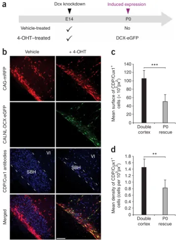
In a second version of the delayed rescue experiment, we controlled for transfection of all plasmids, but varied whether the rats in a litter received an injection of 4-OHT or vehicle solution (Fig. 3a). The results of this experiment were nearly identical to the first experiment. SBH size, assessed either by the density of neurons within the white matter that were positive for the upper layer neuron marker CAAT displacement protein, known as CDP/Cux1, or by the cross sectional area of the malformation defined by accumulation of CDP/Cux1+ neurons, was significantly reduced in the group that received 4-OHT compared to the vehicle control group (Fig. 3b–d). In both types of reexpression experiments, DCX-eGFP expression caused a redistribu-tion of transfected cells from aberrant posiredistribu-tions to upper neocortical layers without a significant change in total number of cells (Fig. 3). Together, these results indicate that cells normally fated for the upper layers of the neocortex but stalled at aberrant positions were induced by Dcx reexpression to migrate to their normally fated layer.
Rescued neurons are morphologically normal We next assessed whether the mispositioned neurons induced to migrate to upper layers by Dcx reexpression were morphologically similar to neurons that migrated normally to their fated upper layer positions. We compared several morphological properties
of apical dendrites in two conditions: a malformation-free condition, in which neurons were treated with an ineffectiveDcxRNAi and were instead induced to express eGFP starting at P0, and a P0 rescue group, in which neurons were initially arrested in SBH byDcxRNAi and then stimulated to migrate by induced expression of DCX-eGFP (Fig. 4a–
d). Labeled neurons in both groups were uniformly positive for the upper layer marker CDP/Cux1 (Fig. 4a–d). Additionally, there were no significant differences between the morphologies of labeled neu-rons in the two groups, as assessed by the number of dendritic processes (Fig. 4e), the average length of the processes (Fig. 4f) and the total process length (Fig. 4g). In addition to having normal dendritic development, the rescued neurons extended axons across the corpus callosum, and these axons projected to appropriate laminar territories in the contralateral hemisphere (Supplementary Fig. 2
online). We also found no significant difference in the number of interneurons in neocortex containing SBH, in rescued neocortex or in malformation-free contralateral hemispheres (Supplementary Figs. 3
and4online). These results indicate that rescued neurons stimulated to migrate away from aberrant positions settled in the appropriate layers, continued to express laminar-specific markers and showed appropriate dendritic and axonal differentiation patterns.
12
CAG-mRFP and 3UTRhp E14 : P0
a
b
c
d
e
3UTRhp 3UTRm3hpE14 : P5 E14 : P10 E14 : P15
CALNL-DCX-eGFP Merged (+ Blue Nissl)
11 NS *** *** *** *** *** *** *** * NS NS NS Relativ e position of tr ansf ected cells in the g re y matter (bin n u mber) 10 0 10 20 30 40 Percentage of cells Double cortex 0 50 100 Doub le cor tex P0 rescue 150 200 * Mean surf ace of heterotopia ( × 10 3 px 2) P0 rescue 50 60 9 8 7 6 5 4 3 2 1 Malformation free P0 rescue Double cortex
Figure 2 Restoration of neocortical lamination and regression of SBH after reexpression of Dcx at P0. (a) Representative neocortical sections showing laminar position of transfected cells in rats electroporated at E14 with either ineffective, control shRNA (3UTRm3hp; top row) or effective DCX-targeting shRNA vectors (3UTRhp; bottom row) together with CAG-mRFP and processed postnatally from P0 to P15. (b) Representative neocortical sections showing restoration of neocortical lamination at P20 after reexpression of Dcx in mispositioned neurons at P0. Four plasmids were electroporated at E14: CAG-mRFP, CAG-ERT2CreERT2, CALNL-DCX-eGFP and
3UTRhp, and 4-OHT was administered at birth. (c) Quantification of transfected cell distribution within the neocortical gray matter after induction of eGFP or DCX-eGFP expression in rats electroporated at E14 with either effective (3UTRhp) or noneffective (3UTRm3hp)Dcx -targeting shRNA vector together with CAG-mRFP, CAG-ERT2CreERT2and either CALNL-DCX-eGFP
or CALNL-eGFP (10–12 sections from two or three rats per condition). NS, not significant. (d) Size and position of SBH at three rostro-caudal levels after induction of eGFP (top) or DCX-eGFP (bottom) expression in rats electroporated at E14 with CAG-mRFP, CAG-ERT2CreERT2, 3UTRhp and either
CALNL-eGFP (top) or CALNL-DCX-CALNL-eGFP (bottom). (e) Quantification of SBH surface after induction of eGFP or DCX-eGFP in the same experimental conditions (eight or nine sections from two or three rats per condition; area units in square pixels, px2¼1mm2). ***Po0.001, *Po0.05. Scale bars, 500mm. © 2009 Nature America, Inc.
All rights reser
Heterotopia reduction becomes less effective with age
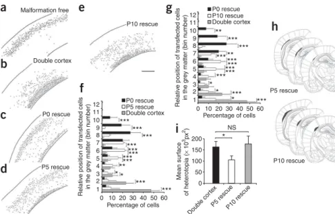
To identify the latest developmental time point at which induced Dcx reexpression could still rescue neocortical lamination and cause SBH to regress, we administered 4-OHT at P5 or P10 and compared the results to those in pups that received 4-OHT at P0. The results of these three rescue treatments were also compared to those in rats that received only induction of eGFP at P0 and thus developed SBH. The total number of cells, the positions of the transfected cells and the size of SBH were analyzed at P20 (Fig. 5). There was no significant difference in the total number of transfected cells per section between any of the conditions, indicating that there was no cell death induced by either eGFP or DCX-eGFP expression at P0, P5 or P10 (Fig. 5a–e). In contrast, in both the P5 and the P10 rescue groups there was a significant shift of neurons toward upper layer positions relative to
neurons in rats that received no rescue (Fig. 5f,g). Although neurons in the P5 and P10 rescue groups migrated further into upper layers than neurons in the unrescued group, neurons rescued at P5 or P10 did not migrate as far into upper layers as did neurons that were rescued at P0 (Fig. 5f,g). However, the P5 rescue group, similar to the P0 rescue group, had significantly smaller SBH malformations than did the P10 rescue group or the group that received no rescue (Fig. 5h,i). These results indicate that reexpression of Dcx at P5 or P10 can induce partial recovery of neuronal position, that reexpression at P5 can cause SBH to regress and that by P10 reexpression is no longer effective at reducing the size of SBH malformations.
Reduction of SBH reduces seizure susceptibility and severity SBH malformations in humans are a well known risk factor for epilepsy11,12. Therefore, we investigated whether the susceptibility to pentylenetetrazol-induced seizures was changed in rats with regressed SBH relative to unrescued rats. A series of subconvulsive doses of pentylenetetrazol (25 mg kg–1) were administrated intraperitoneally every 10 min to rats in three different groups (described inFig. 1d,e): 140 Vehicle Vehicle-treated
a
b
c
d
E14Dcx knockdown Induced expression
P0 No DCX-eGFP 4-OHT–treated + 4-OHT Mean surf ace of CDP/Cux1 + cells ( × 10 3px 2)
Mean density of CDP/Cux1
+
cells (cells per 10
3px 2) 120 100 80 60 40 1.8 ** *** VI VI SBH SBH 1.6 1.4 1.2 1.0 Merged CDP/Cux1 antibodies CALNL-DCX-eGFP C A G -mRFP 0.8 0.6 0.4 0.2 Double cortex P0 rescue 0 Double cortex P0 rescue 20 0
Figure 3 Migration of fated upper layer neurons from SBH. (a) A schematic diagram of experiments. Four plasmids were electroporated at E14: CAG-mRFP, CAG-ERT2CreERT2, CALNL-DCX-eGFP and 3UTRhp. 4-OHT or
its vehicle solution was administered at birth. DCX-eGFP expression is induced in the 4-OHT–treated group only. (b) Immunohistochemistry of P20 neocortical sections showing E14 transfected and nontransfected cells immunopositive for the upper layers neurons marker CDP/Cux1 within SBH at P20 in the absence of Dcx reexpression (left) or after Dcx reexpression (right). Some transfected cells extend out of the SBH into neocortical layer VI. (c,d) Quantifications of surface area occupied by SBH (c) and density (d) of CDP/Cux1+cells within SBH in the same experimental conditions (six
sections from two rats per condition). ***Po0.001, **Po0.01. Scale bar, 150mm. 7 1,200
a
b
c
d
e
f
g
II / III II / III IV IV Malformation free P0 rescue 1,000 2,500 P0 rescue Malformation free 2,000 1,500 1,000 500 0 800 600 400 200 0 6 5 Mean n u mberof processes Mean length of processes Total length
4 3 2 1 0 Secondar y Secondar y Primar y Tertiar y Tertiar y Quater nar y Quater nar y Figure 4 Morphology of rescued neurons. (a,b) Immunohistochemistry
for the upper layer neuron marker CDP/Cux1 on P20 frontal neocortical sections. Rats were electroporated at E14 with either noneffective (3UTRm3hp;a) or effective (3UTRhp;b)Dcx-targeting shRNA vectors together with CAG-mRFP, CAG-ERT2CreERT2and either CALNL-eGFP
(a) or CALNL-DCX-eGFP (b) and injected with 4-OHT at birth. Both initially correctly positioned transfected neurons (a) and initially mispositioned transfected neurons induced to migrate to appropriated positions after Dcx reexpression (b) (both green and red) are located within the CDP/Cux1+
band of upper layer neurons (in blue). Transfected cells were located within neocortical layers II/III and IV. (c,d) Reconstructed cortical neurons incand
dshowing dendritic arborization patterns from the same experimental conditions as inaandb, respectively. (e–g) Quantification of dendritic arborization: mean number of apical processes per neuron (e), their mean length (f) and total length (g) (36 or 37 reconstructed neurons from three or four rats per condition). Scale bar, 200mm.
©
2009
Nature
America,
Inc.
All rights reser
one that had received 4-OHT to induce DCX-eGFP expression at P0 (P0 rescue), one that received 4-OHT at P0 to induce eGFP expression (double cortex, no rescue) and a third group that received ineffective shRNAs and so was free of malformations (malformation free). Injections of pentylenetetrazol were continued until each rat had a generalized seizure. Seizure susceptibility and severity was quantified with the canonical Racine’s scale of seizure severity, including dose and latency to onset of the first minimal motor seizure and the first generalized tonic-clonic seizure (Fig. 6). Generalized seizures in the double cortex group occurred at significantly lower doses of pentyle-netetrazol (Fig. 6a) and with significantly shorter latencies (Fig. 6b) than they did in either malformation-free or P0 rescue rats. Notably, malformation-free and P0 rescue groups showed similar seizure thresholds by all measures (Fig. 6a–c). These results suggest that Dcx reexpression at P0 restored levels of seizure risk back to levels seen in rats that had no neocortical malformation.
DISCUSSION
To our knowledge, this is the first study to demonstrate that a molecular intervention can reduce the size and functional effects of
a preexisting disruption in neuronal migration. In general, our study raises the possibility that, in some contexts, neuronal migration is a form of neuronal plasticity that may be engaged to induce neural repair. Previous examples of continued neuronal migration in the brain include neuronal migration of endogenous neurons to the olfactory bulb and dentate gyrus throughout life29as well as migration of transplanted neuronal progenitors into neocortex and other regions of the central nervous sytem30. Our study extends this phenomenon of continued migration to include induced migration of mispositioned neurons in the neocortex. Our study also suggests that reversing a migration disruption in development can reduce aberrant excitability levels in more mature brain.
In this study, we show that there are considerable developmental constraints on stimulating migration in the postnatal neocortex induced by reexpression of Dcx. Whereas Dcx reexpression at P0 induced a migration that led to marked regression of heterotopia and restored neocortical lamination, reexpression at P5 led to partial restoration of neuronal position with heterotopia regression, and reexpression at P10 led to partial recovery of position without significant heterotopia reduction. These ages in rats correspond in
12 P0 rescue P0 rescue P10 rescue P5 rescue P10 rescue P5 rescue P5 rescue Double cortex Double cortex Malformation free
a
b
c
d
e
f
g
h
i
11 10 200 NS 150 100 Doub le cor tex P5 rescueP10 rescue 50 0 9 8 7 6 5 4 3 2 1 12 P0 rescue Double cortex P10 rescue 11 10 9 8 7 6 5 4 3 2 1 0 10 20 30 40 Percentage of cells Relativ e position of tr ansf ected cells in the g re y matter (bin n u mber) Mean surf ace of heterotopia ( × 10 3px 2) Relativ e position of tr ansf ected cells in the g re y matter (bin n u mber) 50 60 0 10 20 30 40 Percentage of cells 50 60 *** *** *** *** *** *** *** *** ****** *** *** *** *** ** ** * * * *** *** *** *** ** *** *** * * *Figure 5 Crucial developmental period for regressing malformation. (a–e) Representative reconstructed neocortical sections showing the laminar position of neurons at P20 after induction of eGFP (a,b) or DCX-eGFP expression at P0 (a–c), P5 (d) and P10 (e). Black dots represent positions of transfected cells. Four plasmids were electroporated at E14: CAG-mRFP (a–e), CAG-ERT2CreERT2(a–e), either
CALNL-eGFP (a,b) or CALNL-DCX-eGFP (c–e) and either 3UTRhp (c–e) or 3UTRm3hp (a). 4-OHT was administered at P0 (a–c), P5 (d) or P10 (e). (f,g) Quantification of transfected cell distribution within the neocortical gray matter after induction of eGFP or DCX-eGFP expression at P5 (f) and P10 (g), compared to Dcx reexpression at P0 (8–12 sections from two or three rats per condi-tion). (h) Size and position of SBH at three rostro-caudal levels after induction of Dcx expres-sion at P5 (top) and P10 (bottom). (i) Quanti-fication of SBH surface after induction of Dcx expression at P5 and P10 (eight sections from two or three rats per condition). ***Po0.001, **Po0.01, *Po0.05. Scale bar, 500mm. 175
a
d
b
c
NS NS NS *** *** * ** 60 50 40 30 20 10 0 40 30 20 10 0 150 125 Cum ulativ e dose of PTZinducing tonic-clonic seizures
(mg kg –1 ) P0 rescue Doub le cor te x
Mean latency to induce tonic-clonic seizures
(min)
Inter
v
al betw
een minimal
and tonic-clonic seizures
(min) 100 75 50 25 Malformation free Double cortex P0 rescue Malformation free Double cortex P0 rescue * * Malformation free Double cortex P0 rescue 0 31′24 31′27 31′30 31′33 31′36 33′22
Figure 6 Decreased seizure susceptibility to pentylenetetrazol-induced seizures. Quantification of doses of pentylenetetrazol (PTZ;a), latency to induce generalized tonic-clonic seizures (b) and interval between minimal and generalized seizures (c) in three groups of rats: malformation-free rats (ineffective shRNAs and eGFP expression induced at P0), double cortex rats with SBH and lamination deficits (effective shRNAs and eGFP expression induced at P0) and P0 rescue rats with restored lamination and regressed SBH (effective shRNAs and DCX-eGFP expression induced at P0). (d) Example of a double cortex rat experiencing rearings, fallings and convulsions (top) and a P0 rescue rat at the same dose of PTZ and same time after injection (bottom). The P0 rescue rat experienced seizures 10 min later, after an additional injection of PTZ was given. ***Po0.001, **Po0.01, *Po0.05. © 2009 Nature America, Inc.
All rights reser
humans roughly to preterm gestational weeks 25–35 (for P0) and to full-term infants (for P10). We do not currently know the reason for the absence of regression at P10 or the reduced migration after P5 intervention. It is likely, however, that developmental changes in neocortical tissue and within differentiating neurons limit continued neuronal migration into the postnatal period. Migration of pyramidal neurons during normal neocortical development is guided by radial glial processes31, and it is likely that developmental loss of radial glia, which normally occurs by the middle of the second postnatal week in the rat, reduces the potential for continued migration32,33. In addition, the developmental window for migration out of SBH may close because of neuronal maturation. Neurons within SBH form synapses and send axonal projections to subcortical targets26,27, and such neuronal differentiation may preclude induced migration by reexpres-sion of Dcx. However, it is noteworthy that even as late as at P10 our results show that there is still a capacity for plasticity and that mispositioned neurons can be shifted from deeper to more superficial positions by expression of Dcx. In the future it will be useful to determine which specific cellular changes are responsible for developmental restriction of induced remigration and whether these can be manipulated to allow for heterotopia regression in even more mature rats.
Virtually all neuronal migration disorders, including lissencephaly, periventricular nodular heterotopias and subcortical band heterotopia, are risk factors for seizures34–36. In addition, focal cortical dysplasias are estimated to be associated with 30% of intractable temporal lobe epilepsies37,38. The specific mechanisms that cause dysplastic cortices to cause epileptiform activity remain unclear. For example, the point in development at which the presence of a neocortical malformation first disposes cortical tissue to overexcitability has remained unknown1,35,39–41. Our results suggest that an underlying cause of seizure susceptibility to reduced pentylenetetrazol dose is directly related to aberrant neuronal positioning and to the malformation. We cannot yet distinguish from our results at what point in develop-ment migration impairdevelop-ment causes increased excitability, but regres-sion initiated after birth is sufficient to restore pentylenetetrazol thresholds to control levels. Future experiments can now be directed at determining the time course of changes in synaptic and cellular physiology that correlate with reduced seizure susceptibility.
The specific experimental intervention we used to restore migration patterns, electroporation of an inducible construct, is clearly not applicable to humans. There are at least two possible directions indicated by our findings for interventions that may one day be therapeutically viable. First,Dcxexpression may be induced by viral transduction of cells within SBH malformations. Our results currently suggest that such gene therapy would probably need to be applied perinatally but could be restricted to mispositioned neurons within SBH. An alternative to gene therapy could be to enhance the function of other members of the DCX superfamily that may have redundant function with DCX. For example, in mouse knockouts of Dcx there are no cortical dysplasias42, and only when Dcx and the related geneDclkare both disrupted do migration disorders similar to human lissencephaly appear43. Thus,Dclkin the mouse seems to be capable of compensating for Dcx function. There are now multiple defined members of the DCXsuperfamily with similar cellular and biochemical functions44, and perhaps pharmacological enhancement of the function of one or several of these may facilitate regression of SBH in humans. Finally, the present study serves as a proof of concept that mismigrated cells can be manipulated to restore a normal morphological pattern and level of neuronal excitability in the cerebral cortex.
METHODS
In uteroelectroporation and Dcx RNA interference.We performedin utero
electroporation as described before26. We used shRNAs targeting the 3¢UTR of
Dcx(3UTRhp) or an ineffective shRNA with three point mutations creating
mismatches (3UTRm3hp). We previously showed26that the 3UTRhp construct
efficiently knocks down Dcx expression in vivo, whereas the 3UTRm3hp
construct is ineffective in causingDcxknockdown. In addition, theDcx-specific
RNAi–induced phenotype is rescued by Dcx expression, confirming the
specificity of shRNAs27. We electroporated plasmids encoding mRFP
(CAG-mRFP, 0.5mgml–1) to fluorescently label transfected cells along with plasmids
expressing shRNAs against Dcx (3UTRhp or 3UTRm3hp, 0.5 mg ml–1), a
4-OHT–activatable form of Cre recombinase (CAG-ERT2CreERT2, 1.0mgml–1)
and Cre-dependent inducible expression vectors (CALNL-eGFP, 1.5mgml–1; a
gift from T. Matsuda and C. Cepko) and the plasmid CALNL-DCX-eGFP
(1.5mgml–1), which was made for this study. We constructed a
pCAGGS-DCX-eGFP plasmid on the basis of the previously described pCAGGS-DCX plasmid27
by fusing eGFP to the carboxy terminus. To generate CALNL-DCX-eGFP, we subcloned the DCX-eGFP fusion fragment from pCAGGS-DCX-eGFP into theEcoRI andNotI sites of the CALNL-eGFP plasmid. All rat protocols were approved by the University of Connecticut Institutional Animal Care and Use Committee.
4-hydroxytamoxifen administration. We dissolved 4-OHT (Sigma) in 95%
ethanol at a concentration of 20 mg ml–1and diluted it with nine volumes of
corn oil. We administered diluted 4-OHT (2 mg per 100 g body weight) to the rats through intraperitoneal injections. We injected vehicle-treated rats with the same solution without 4-OHT.
Histological procedures and microscopy. We transcardially perfused rats
under deep anesthesia with 4% paraformaldehyde in PBS. We removed their brains and post-fixed them for 48 h in the same fixative solution before frontal sectioning with a vibratome (Leica). We processed brain sections for immuno-histochemistry as floating sections. We used the following primary antibodies: goat antibody to doublecortin (1 in 100, Santa Cruz Biotechnology), rabbit antibody to CDP/Cux1 (1 in 200, Santa Cruz Biotechnology), mouse and rabbit antibodies to GFP (1 in 2,000 and 1 in 3,000, respectively, Molecular Probes), rabbit antibody to RFP (1 in 3,000, Chemicon). We used the appropriate secondary antibodies (1 in 200) conjugated to Alexa 488, Alexa 568, Alexa 633 (Chemicon) or Cy5 (Jackson ImmunoResearch) and conter-stained sections with the fluorescent Nissl staining NeuroTrace 435/455 (1 in 200, Molecular Probes).
We took photomicrographs with a Nikon Eclipse E400 microscope equipped with a digital Spot Camera (Diagnostic Instruments) or with a Leica TCS SP2 confocal microscope.
Seizure induction with pentylenetetrazol. We dissolved pentylenetetrazol
(Sigma) in 0.9% saline and administered it intraperitoneally to rats at P30 at a concentration of 25 mg per kg of body weight. There was no statistically significant difference in weight or sex ratio between groups of rats. Rats were placed in Plexiglass cages, observed by an experimenter naive to the status of
the rat and their behavior was scored with the Racine’s scale45. We administered
pentylenetetrazol every 10 min until generalized seizures occurred.
Quantifications and statistical analyses.We performed quantifications with
the image analysis software ImageJ 1.39e on coronal sections (60mm) located
from bregma –3.00 to bregma –5.00 according to the Rat Brain Atlas46. We
quantified the relative positions of transfected cells in the cortex by counting
mRFP+cells in 12 areas of interests normalized in individual sections to fit
within the whole thickness of the cortical wall, from deep to superficial layers and excluding the white matter. We quantified the size of SBH malformations in square pixels with ImageJ measuring tools. We made morphological analyses of dendritic arborization after neuronal reconstructions with the ImageJ plug-in NeuronJ 1.2 (Erik Meijerplug-ing).
We performed statistical tests with InStat 3.0 (GraphPad). We checked the normality of the data distribution with the Kolmogorov and Smirnov method.
We used two-sample Student’st-test to compare means of two independent
groups. When three groups of data were compared, we used one-way analysis of variance with the Tukey-Kramer post-test for multiple comparisons. We
©
2009
Nature
America,
Inc.
All rights reser
considered values as significant when Po0.05. All data are presented as means ± s.e.m.
Note: Supplementary information is available on the Nature Medicine website.
ACKNOWLEDGMENTS
CALNL-eGFP and CAG-ERT2CreERT2were gifts from T. Matsuda and C. Cepko,
Harvard Medical School. ImageJ software was from W. Rasband, US National Institutes of Health. NeuronJ 1.2 was from E. Meijering (Erasmus University Medical Center Rotterdam). This work was supported by the US National Institutes of Health (MH056524 and NS062416 to J.J.L.) and the Jerome Lejeune foundation (research fellowship to J.-B.M.).
AUTHOR CONTRIBUTIONS
J.-B.M. and J.J.L. contributed to all aspects of the project; J.-B.M. conducted histological procedures, seizure induction experiments, quantifications and data analysis; Y.W. performed intrauterine surgeries and interneuron analysis; Y.C. performed confocal microscopy, interneuron analysis and contributed to seizure induction experiments and M.P. constructed the plasmid vectors and contributed to confocal microscopy.
Published online at http://www.nature.com/naturemedicine/
Reprints and permissions information is available online at http://npg.nature.com/ reprintsandpermissions/
1. Jacobs, K.M., Kharazia, V.N. & Prince, D.A. Mechanisms underlying epileptogenesis in cortical malformations.Epilepsy Res.36, 165–188 (1999).
2. Chevassus-au-Louis, N., Baraban, S.C., Gaı¨arsa, J.L. & Ben-Ari, Y. Cortical malforma-tions and epilepsy: new insights from animal models.Epilepsia40, 811–821 (1999). 3. Schwartzkroin, P.A. & Walsh, C.A. Cortical malformations and epilepsy.Ment. Retard.
Dev. Disabil. Res. Rev.6, 268–280 (2000).
4. Kuzniecky, R.I. & Barkovich, A.J. Malformations of cortical development and epilepsy. Brain Dev.23, 2–11 (2001).
5. Sisodiya, S.M. Malformations of cortical development: burdens and insights from important causes of human epilepsy.Lancet Neurol.3, 29–38 (2004).
6. Sisodiya, S.M. Surgery for malformations of cortical development causing epilepsy.
Brain123, 1075–1091 (2000).
7. Guerrini, R. Genetic malformations of the cerebral cortex and epilepsy.Epilepsia
46Suppl 1, 32–37 (2005).
8. Barkovich, A.J., Jackson, D.E.J. & Boyer, R.S. Band heterotopias: a newly recognized neuronal migration anomaly.Radiology171, 455–458 (1989).
9. Dobyns, W.B. et al.X-linked malformations of neuronal migration.Neurology 47, 331–339 (1996).
10. Bernasconi, A.et al. Surgical resection for intractable epilepsy in double cortex syndrome yields inadequate results.Epilepsia42, 1124–1129 (2001).
11. Barkovich, A.J. et al. Band heterotopia: correlation of outcome with magnetic resonance imaging parameters.Ann. Neurol.36, 609–617 (1994).
12. Guerrini, R. & Carrozzo, R. Epilepsy and genetic malformations of the cerebral cortex. Am. J. Med. Genet.106, 160–173 (2001).
13. Pinard, J.M.et al.Subcortical laminar heterotopia and lissencephaly in two families: a single X linked dominant gene. J. Neurol. Neurosurg. Psychiatry 57, 914–920 (1994).
14. des Portes, V.et al.doublecortin is the major gene causing X-linked subcortical laminar heterotopia (SCLH).Hum. Mol. Genet.7, 1063–1070 (1998).
15. Gleeson, J.G.et al.Doublecortin, a brain-specific gene mutated in human X-linked lissencephaly and double cortex syndrome, encodes a putative signaling protein.Cell
92, 63–72 (1998).
16. Matsumoto, N.et al.Mutation analysis of theDCXgene and genotype/phenotype correlation in subcortical band heterotopia.Eur. J. Hum. Genet.9, 5–12 (2001). 17. D’Agostino, M.D.et al.Subcortical band heterotopia (SBH) in males: clinical, imaging
and genetic findings in comparison with females.Brain125, 2507–2522 (2002). 18. Lee, K.S.et al.A genetic animal model of human neocortical heterotopia associated
with seizures.J. Neurosci.17, 6236–6242 (1997).
19. Chevassus-Au-Louis, N., Rafiki, A., Jorquera, I., Ben-Ari, Y. & Represa, A. Neocortex in the hippocampus: an anatomical and functional study of CA1 heterotopias after
prenatal treatment with methylazoxymethanol in rats. J. Comp. Neurol. 394, 520–536 (1998).
20. Baraban, S.C. & Schwartzkroin, P.A. Flurothyl seizure susceptibility in rats following prenatal methylazoxymethanol treatment. Epilepsy Res. 23, 189–194 (1996).
21. Baraban, S.C., McCarthy, E.B. & Schwartzkroin, P.A. Evidence for increased seizure susceptibility in rats exposed to cocaine in utero.Brain Res. Dev. Brain Res.102, 189–196 (1997).
22. Roper, S.N., Gilmore, R.L. & Houser, C.R. Experimentally induced disorders of neuronal migration produce an increased propensity for electrographic seizures in rats.Epilepsy Res.21, 205–219 (1995).
23. Jacobs, K.M., Gutnick, M.J. & Prince, D.A. Hyperexcitability in a model of cortical maldevelopment.Cereb. Cortex6, 514–523 (1996).
24. Luhmann, H.J. & Raabe, K. Characterization of neuronal migration disorders in neocortical structures: I. Expression of epileptiform activity in an animal model. Epilepsy Res.26, 67–74 (1996).
25. Nosten-Bertrand, M.et al.Epilepsy in Dcx knockout mice associated with discrete lamination defects and enhanced excitability in the hippocampus.PLoS ONE3, e2473 (2008).
26. Bai, J.et al.RNAi reveals doublecortin is required for radial migration in rat neocortex. Nat. Neurosci.6, 1277–1283 (2003).
27. Ramos, R.L., Bai, J. & LoTurco, J.J. Heterotopia formation in rat but not mouse neocortex after RNA interference knockdown of DCX.Cereb. Cortex16, 1323–1331 (2006).
28. Matsuda, T. & Cepko, C.L. Controlled expression of transgenes introduced by in vivo electroporation.Proc. Natl. Acad. Sci. USA104, 1027–1032 (2007).
29. Lim, D.A., Huang, Y. & Alvarez-Buylla, A. The adult neural stem cell niche: lessons for future neural cell replacement strategies.Neurosurg. Clin. N. Am. 18, 81–92 ix (2007).
30. Imitola, J.et al.Directed migration of neural stem cells to sites of CNS injury by the stromal cell-derived factor 1a/CXC chemokine receptor 4 pathway.Proc. Natl. Acad.
Sci. USA101, 18117–18122 (2004).
31. Rakic, P. Mode of cell migration to the superficial layers of fetal monkey neocortex. J. Comp. Neurol.145, 61–83 (1972).
32. Stichel, C.C., Muller, C.M. & Zilles, K. Distribution of glial fibrillary acidic protein and vimentin immunoreactivity during rat visual cortex development.J. Neurocytol.20, 97–108 (1991).
33. Kalman, M. & Ajtai, B.M. A comparison of intermediate filament markers for presumptive astroglia in the developing rat neocortex: immunostaining against nestin reveals more detail, than GFAP or vimentin.Int. J. Dev. Neurosci.19, 101–108 (2001).
34. Guerrini, R., Canapicchi, R. & Dobyns, W.B. Epilepsy and malformations of the cerebral cortex.Neurologia14Suppl 3, 32–47 (1999).
35. Baraban, S.C. Epileptogenesis in the dysplastic brain: a revival of familiar themes. Epilepsy Curr.1, 6–11 (2001).
36. Sheen, V.L. & Walsh, C.A. Developmental genetic malformations of the cerebral cortex. Curr. Neurol. Neurosci. Rep.3, 433–441 (2003).
37. Crino, P.B. Malformations of cortical development: molecular pathogenesis and experimental strategies.Adv. Exp. Med. Biol.548, 175–191 (2004).
38. Cepeda, C.et al.Pediatric cortical dysplasia: correlations between neuroimaging, electrophysiology and location of cytomegalic neurons and balloon cells and glutamate/ GABA synaptic circuits.Dev. Neurosci.27, 59–76 (2005).
39. Chae, T.et al.Mice lacking p35, a neuronal specific activator of Cdk5, display cortical lamination defects, seizures and adult lethality.Neuron18, 29–42 (1997). 40. Hablitz, J.J. & DeFazio, T. Excitability changes in freeze-induced neocortical
micro-gyria.Epilepsy Res.32, 75–82 (1998).
41. Andres, M.et al.Human cortical dysplasia and epilepsy: an ontogenetic hypothesis based on volumetric MRI and NeuN neuronal density and size measurements.Cereb. Cortex15, 194–210 (2005).
42. Corbo, J.C.et al.Doublecortin is required in mice for lamination of the hippocampus but not the neocortex.J. Neurosci.22, 7548–7557 (2002).
43. Koizumi, H., Tanaka, T. & Gleeson, J.G. Doublecortin-like kinase functions with doublecortin to mediate fiber tract decussation and neuronal migration.Neuron49, 55–66 (2006).
44. Coquelle, F.M.et al.Common and divergent roles for members of the mouse DCX superfamily.Cell Cycle5, 976–983 (2006).
45. Racine, R.J. Modification of seizure activity by electrical stimulation. I. After-discharge threshold.Electroencephalogr. Clin. Neurophysiol.32, 269–279 (1972). 46. Paxinos, G. & Watson, C.The Rat Brain in Stereotaxic Coordinates(Elsevier Academic
Press, Oxford, 2005). © 2009 Nature America, Inc.
All rights reser