Frontiers in Dentistry
Shear Bond Strength of Molar Tubes to Enamel
Using an Orthodontic Resin-Modified Glass
Ionomer Cement Modified with Amorphous
Calcium Phosphate
Behrad Tanbakuchi
1,2, Tabassom Hooshmand
3,4, Mohammad Javad
Kharazifard
1, Kiana Shekofteh
3, Arian Hesam Arefi
5*1. Dental Research Center, Dentistry Research Institute, Tehran University of Medical Sciences, Tehran, Iran 2. Department of Orthodontics, School of Dentistry, Tehran University of Medical Sciences, Tehran, Iran 3. Department of Dental Biomaterials, School of Dentistry, Tehran University of Medical Sciences, Tehran, Iran 4. Research Center for Science and Technology in Medicine, Tehran University of Medical Sciences, Tehran, Iran 5. Dental Research Center, Zahedan University of Medical Sciences, Zahedan, Iran
Article Info A B S T R A C T
Article type:
Original Article Objectives: to the enamel surface of molar teeth using a resin-modified glass ionomer (RMGI) This study aimed to assess the shear bond strength (SBS) of molar tubes cement modified with amorphous calcium phosphate (ACP).
Materials and Methods: In this in-vitro study, 60 extracted human third molars were randomly divided into four groups for bonding of molar tubes to the enamel surface. Fuji Ortho LC and Fuji Ortho LC modified with ACP (1.55 wt%) were used in groups 1 and 2, respectively. In group 3, the enamel surface was sandblasted, and bonding was then performed using Fuji Ortho LC glass ionomer modified with ACP. In group 4, molar tubes were conventionally bonded using Transbond XT composite. The SBS was measured using a universal testing machine.
Results: The mean SBS of groups 1 to 4 was 10.22, 6.88, 9.4, and 13.68 MPa, respectively. Only the SBS of group 1 was not significantly different from that of groups 3 and 4 (P>0.05). Comparison of adhesive remnant index (ARI) scores of the groups revealed significant differences only between groups 1 and 4 (P<0.001) and between groups 1 and 2 (P=0.002).
Conclusion: The results revealed that the addition of ACP to Fuji Ortho LC significantly decreased the SBS of molar tubes bonded to enamel compared to the conventional resin bonding system. Sandblasting of the enamel surface significantly increased the bond strength. Fuji Ortho LC modified with ACP is recommended for bonding of molar tubes to posterior teeth considering its cariostatic property.
Keywords: Shear Strength; Fuji Ortho LC; Glass Ionomer Cements; Amorphous Calcium Phosphate
Article History:
Received: 7 February 2019 Accepted: 11 August 2019 Published: 15 October 2019
* Corresponding author :
Dental Research Center, Zahedan University of Medical Sciences, Zahedan, Iran
Email: hesama891@gmail.com
370 Front Dent, Vol. 16, No. 5, Sep-Oct 2019 INTRODUCTION
Increased risk of occurrence of enamel white spot lesions during orthodontic treatment is due to unhealthy nutritional habits, high consumption of sugary substances, high levels of oral cariogenic bacteria, decreased buffering capacity of the saliva, long-term course of orthodontic treatment, and difficult oral hygiene maintenance due to the presence of orthodontic appliances [1,2]. The prevalence of enamel caries during or after orthodontic treatment varies from 46% to 97% [1,2]. Also, evidence shows a high prevalence of caries in maxillary and mandibular first molars after the termination of orthodontic treatment [3-5].
Several materials, such as fluoride, xylitol, casein phosphopeptide-amorphous calcium phosphate (CPP-ACP), and bioactive glasses, and several products, such as toothpastes and varnishes, are used to enhance enamel
remineralization. Amorphous calcium
phosphate (ACP) in combination with casein phosphopeptide (CPP) has shown positive clinical efficacy for the prevention of enamel white spot lesions [6]. In the process of enamel remineralization, ACP is used as a mediator for the formation of hydroxyapatite [7]. ACP is the first deposited phase in the supersaturated solution of calcium phosphate [8]. In the acidic oral environment, we can benefit from the calcium and orthophosphate ion release potential of ACP by the application of bio-composites containing ACP; these ions play a role in remineralization [9].
To date, ACP has been used in combination with different materials, such as composite
resins, glass ionomer cements, and
orthodontic adhesives [10-12]. The addition of 3 wt% of CPP-ACP to glass ionomer increases the release of calcium and phosphate ions with no significant change in fluoride release and no adverse effect on surface hardness or material mass [13,14].
Resin-modified glass ionomer (RMGI)
cements were introduced to benefit from the optimal physical properties of composite resins in combination with the cariostatic properties of glass ionomers. RMGI cements are synthesized by the addition of 4.5% to 6%
resin to the glass ionomer matrix [15]. They show favorable cariostatic properties [16,17]. However, some studies have reported a higher rate of bond failure for orthodontic brackets bonded to the enamel using RMGI compared to composite resins [18,19]. Nonetheless, both RMGI and composite resin have shown an acceptably low rate of bracket bond failure in the clinical setting.
Several methods have been employed to enhance the bond strength of brackets bonded with RMGI cement to the enamel, such as acid-etching of the enamel surface, laser irradiation, light radiation with different intensities for curing of RMGI, and
sandblasting [20-24]. Sandblasting in
combination with acid-etching increases the shear bond strength (SBS) of brackets bonded to the enamel using orthodontic adhesives [25,26]. On the other hand, sandblasting of the enamel surface has no destructive effect on the
enamel [27]. Sandblasting creates
Fig. 1. X-ray diffraction for confirming the amorphous structure of calcium phosphate
On the other hand, the risk of the formation of an unacceptable etching pattern is higher in posterior teeth [31]. Moreover, heavier masticatory forces in the molar region and non-uniform thickness of resin between the enamel and bracket base in posterior teeth may contribute to a higher rate of bracket bond failure in such teeth [32]. Difficult isolation of posterior teeth and the presence of higher percentages of aprismatic enamel in molars may also play a role in this respect [33]. Evidence shows that a bond strength between 6 to 8 MPa is required for adequate resistance to masticatory forces [34,35].
To the best of the authors’ knowledge, no previous study has assessed the SBS of molar tubes bonded to the enamel using Fuji Ortho LC modified with ACP. Thus, this study aimed to assess the SBS of molar tubes bonded to the enamel surface using orthodontic RMGI modified with ACP. The efficacy of sandblasting to enhance the SBS was also evaluated.
MATERIALS AND METHODS
This experimental in-vitro study has been approved by the Ethics Committee on Research of the Dentistry Research Institute, Tehran University of Medical Sciences,
Tehran, Iran
(IR.TUMS.DENTISTRY.REC.1397.112).
Sixty extracted human third molars were immersed in 1% thymol solution (chloramine-T) for one week after extraction for disinfection. They were then stored in
distilled water at 4°C. The surface of all teeth was cleaned using a rubber cup, non-fluoridated pumice paste, and water for 10 seconds to eliminate debris from the tooth surface. After five times of use, the rubber cup was replaced to ensure its proper function. The teeth were then mounted on a wax sheet, and their buccal surfaces were inspected for
enamel defects under an optical
stereomicroscope (SMZ800; Nikon, Japan) at x10 magnification. This was done to ensure the absence of enamel cracks and structural defects. The teeth with enamel defects were replaced with sound teeth.
Synthesis of ACP:
The ACP was synthesized by the deposition and freeze-drying technique. For this purpose, 6.298 g of calcium nitrate [Ca(NO3)2.4H2O] and
3.352 g of magnesium nitrate
[Mg(NO3)2.6H2O] were dissolved in 51 ml of
deionized water containing 4 ml of ammonia such that the molar ratio of Mg/Ca was 0.3. Next, 5.44 g of diammonium hydrogen
phosphate [(NH4)2HPO4] was dissolved in 126
372 Front Dent, Vol. 16, No. 5, Sep-Oct 2019
ACP powder was then added to RMGI powder in 1.55 wt% [36].
The teeth were randomly divided into four groups of 15. The tubes were bonded to molar teeth in each group as follows:
Group 1. The etching was performed with 37% phosphoric acid gel (Ultra-Etch; Ultradent, South Jordan, UT, USA) for 20 seconds. The teeth were then rinsed for 20 seconds and dried to obtain a frosty appearance. Bonding was performed using Fuji Ortho LC (GC Corp., Tokyo, Japan) with a standard powder to liquid ratio of 3 g to 1 g and 20 seconds of curing time for each surface (a total of 80 seconds).
Group 2. The enamel surface was prepared as in group 1. Bonding was performed using Fuji Ortho LC (GC Corp., Tokyo, Japan) modified with ACP with a standard powder to liquid ratio of 3 g to 1 g and 20 seconds of curing of each surface (a total of 80 seconds) using a light-curing unit (Woodpecker; Guilin, China). Group 3. The buccal surface of the teeth was sandblasted with 50-µ aluminum oxide particles for 10 seconds (MicroEtcher ERC; Danville Engineering Co., Danville, CA, USA). Acid-etching was performed as in groups 1 and 2, and then, bonding was performed using Fuji Ortho LC (GC Corp., Tokyo, Japan) modified with ACP with a standard powder to liquid ratio of 3 g to 1 g. Curing was performed for 20 seconds on each surface (a total of 80 seconds).
Group 4. The etching was performed using 37% phosphoric acid for 20 seconds. The teeth were then rinsed for 20 seconds and dried to obtain a frosty appearance. Next, Transbond XT Primer (3M Unitek, Monrovia, CA, USA) was applied on the tooth surface using a small microbrush and cured for 10 seconds. Transbond XT (3M Unitek, Monrovia, CA, USA) composite was used for bonding with a curing time of 40 seconds (10 seconds from each side).
In all four groups, the adhesive was applied to the back of stainless steel maxillary and mandibular first molar tubes (Ortho-Cast, Dentaurum, Ispringen, Germany). The tubes were then placed at the center of the clinical crown and along the buccal groove of each
tooth. A 0.025×0.019-inch stainless steel wire was passed through the tube. Assuming that the wire was parallel to the horizon, we tried to align the buccal surface of the teeth perpendicular to the horizon, and the tubes were bonded perpendicular to the surface. The bonding of all samples was performed by one operator. After positioning, the tube was compressed to the tooth surface in all four groups to decrease the thickness of the bonding agent. Next, excess bonding material was removed from around the tubes using an explorer. After completion of the bonding procedure, the teeth were immersed in distilled water at room temperature for 24 hours. Next, each tooth was mounted in a
rectangular acrylic block measuring
10×10×10 mm3. The teeth then underwent
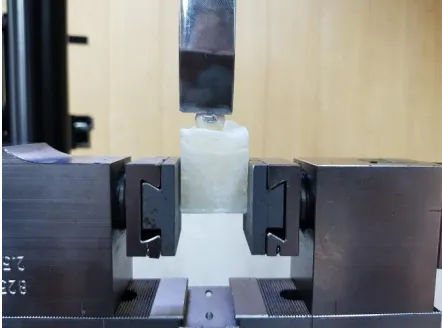
thermocycling (Vafaei Industrial Factory, Tehran, Iran) between 5-55°C for 3000 cycles to simulate oral clinical conditions. The SBS was measured using a universal testing machine (Zwick/Roell, Ulm, Germany) at a crosshead speed of 1 mm/minute. The blade applied the load perpendicular to the superior bonding interface of the tube and the tooth (Fig. 2).
Fig. 2. Universal testing machine blade applied load perpendicular to the superior bonding interface of the tube and tooth.
The load at failure was recorded in Newton (N) and divided by the surface area of the tube
base in square-millimeters (mm2) to calculate
the SBS in megapascal (MPa).
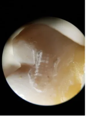
Fig. 3. Stereomicroscope view for evaluation of ARI tooth was evaluated under a stereo-microscope (SMZ800, Nikon, Japan) at x10 magnification to determine the adhesive remnant index (ARI) score (Fig. 3) according to the criteria proposed by Årtun and Bergland [37], as follows:
Score 0: No adhesive remained on the tooth surface.
Score 1: Less than 50% of the adhesive remained on the tooth surface.
Score 2: More than 50% of the adhesive remained on the tooth surface.
Score 3: All the adhesive remained on the tooth surface.
The data were analyzed using SPSS version 22 (SPSS Inc., Chicago, IL, USA). One-way analysis of variance (ANOVA) was applied to compare the SBS between the test and control groups. The Kruskal-Wallis test was applied to compare the ARI scores of the groups. P<0.05 was considered statistically significant. Data were analyze using SPPP25(IBM,Chicago,Ille).
RESULTS
As shown in Table 1, the maximum SBS was noted in group 4 (positive control) with a mean value of 13.68 MPa followed by group 1 (10.22 MPa), group 3 (9.4 MPa), and group 2 (6.88 MPa).
Table 1. Mean shear bond strength (MPa) and standard deviation (SD) of the groups
Group Min Max Mean SD
1 4.11 16.30 10.23 3.65
2 3.07 10.55 6.88 2.19
3 5.29 13.16 9.40 1.97
4 4.99 20.13 13.68 4.73
As shown in Table 2, no significant difference was noted in the SBS of group 1 with that of groups 3 and 4 (P>0.05). Significant differences were noted in the SBS of other groups (P<0.001).
Table 2. Pairwise comparisons of the groups in terms of the shear bond strength (SBS)
Group Mean Difference (I-J) SE P-value Lower Bound 95% Confidence interval Upper Bound
1
2 3.35* 1.10 0.034 0.18 6.51
3 0.833 1.074 0.97 -2.27 3.92
4 -3.46 1.54 0.19 -7.84 0.93
2
1 -3.35* 1.10 0.03 -6.51 -0.18
3 -2.52* 0.76 0.02 -4.67 -0.37
4 -6.80* 1.35 <0.001 -10.73 -2.87
3
1 -0.83 1.07 0.97 -3.92 2.27
2 2.519* 0.76 0.02 0.37 4.67
4 -4.28* 1.32 0.03 -8.17 -0.39
4
1 3.46 1.54 0.19 -0.93 7.84
2 6.80* 1.35 <0.001 2.87 10.73
3 4.28* 1.32 0.03 0.39 8.17
374 Front Dent, Vol. 16, No. 5, Sep-Oct 2019
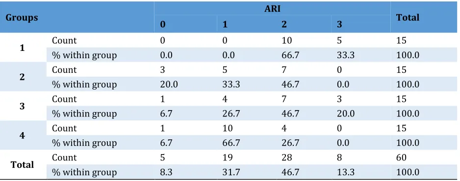
Table 3. Frequency distribution of the adhesive remnant index (ARI) scores of the groups
Groups ARI Total
0 1 2 3
1 Count 0 0 10 5 15
% within group 0.0 0.0 66.7 33.3 100.0
2 Count 3 5 7 0 15
% within group 20.0 33.3 46.7 0.0 100.0
3 Count 1 4 7 3 15
% within group 6.7 26.7 46.7 20.0 100.0
4 Count 1 10 4 0 15
% within group 6.7 66.7 26.7 0.0 100.0
Total Count 5 19 28 8 60
% within group 8.3 31.7 46.7 13.3 100.0
Regarding the ARI scores, the highest frequency of score zero was noted in group 2. Table 3 presents the frequency of the ARI scores in different groups.
Significant differences were noted in the ARI scores between groups 4 and 1 (P<0.001) and between groups 2 and 1 (P=0.002). Other groups were not significantly different in terms of the ARI scores (P>0.01).
DISCUSSION
Although the results of the current study revealed that the SBS after bonding the molar tubes using Fuji Ortho LC glass ionomer was within the clinically acceptable range, it was still lower than that related to Transbond XT. Thus, this study recommends the cautious use of Fuji Ortho LC glass ionomer for the bonding of molar tubes to the enamel surface, mainly in caries prone patients.
The addition of ACP to enhance the remineralizing potential of Fuji Ortho LC glass ionomer decreases the SBS (significantly lower than that of the control group). However, enamel surface sandblasting effectively enhances the SBS of molar tubes bonded to the enamel with Fuji Ortho LC modified with ACP.
This study revealed a significant difference in the ARI scores between groups 1 and 4 and between groups 1 and 2. The highest frequency of ARI score zero was noted in
group 2, which was in agreement with the decreased bond strength in this group. The significant difference between groups 1 and 2 was probably due to the weaker bond to the enamel in group 2.
The difference between groups 1 and 4 can be due to the difference in the bond strength of the two groups. Since the ARI is a qualitative variable, its pairwise comparisons are not as reliable as that for the SBS. Thus, the ARI scores are not always in agreement with the SBS.
with Fuji Ortho LC glass ionomer modified with ACP had borderline SBS for use in the clinical setting. However, a comparison of their study and ours is difficult since we used buccal tubes and Fuji Ortho LC glass ionomer instead of orthodontic bands and a conventional glass ionomer. Dandachli [38] reported a 15.7% rate of clinical bond failure when using RMGI cement for bonding of ceramic brackets to the enamel, which was higher than the 7% failure rate in the group bonded with Transbond XT. This finding was also confirmed in our study. Similar to the samples in group 2 of our study, the most common location of debonding of the tubes in the cited study was at the enamel-adhesive interface, which is attributed to the weak bond strength of the glass ionomer cement to the enamel [38].
Yassaei et al [19] reported that brackets bonded with Fuji Ortho LC glass ionomer showed significantly lower SBS compared to the composite resin group. In the present study, the difference between Fuji Ortho LC glass ionomer and the control group was not significant. This difference may be attributed to the use of an acid etchant instead of a conditioner in the present study, which enhances the bond strength of Fuji Ortho LC to the enamel. A systematic review regarding the SBS of metal brackets bonded with RMGI cement and composite revealed no significant difference between the two groups in terms of the frequency of bond failure at 12 months [39]. Their results, similar to our findings, confirmed the acceptable SBS of RMGI. Godoy-Bezerra et al [22] reported higher SBS in a wet environment with the use of an acid etchant and RMGI cement compared to the use of Transbond XT. Considering the higher risk of faulty isolation in the posterior region of the oral cavity, another advantage of Fuji Ortho LC glass ionomer is its favorable resistance to moisture contamination. Summers et al [40] showed that although the bonding of orthodontic attachments with RMGI in vitro yielded a lower bond strength compared to the use of composite resins, it was strong enough for use in the clinical setting. This finding was in line with our results regarding the
acceptable bond strength of molar tubes bonded with Fuji Ortho LC glass ionomer. Elnafar et al [41] performed sandblasting without applying a conditioner to enhance the SBS of RMGI. They concluded that enamel surface preparation with sandblasting alone yielded a significantly lower SBS compared to acid-etching when bonding metal brackets. However, this value was still within the clinically acceptable range [41]. In the present study, acid-etching was performed in all groups using Fuji Ortho LC glass ionomer. In our study, similar to that of Elnafar et al [41], Fuji Ortho LC glass ionomer yielded a clinically acceptable SBS. In our study, sandblasting along with acid-etching increased the SBS to the clinically acceptable range probably due to the elimination of aprismatic enamel of molar teeth and the formation of mechanical retention. Sharma et al [29] indicated that sandblasting of the bracket base and enamel significantly decreases the failure rate of brackets bonded with RMGI. This finding was also confirmed in our study. However, acid-etching should be performed along with sandblasting to increase microscopic and macroscopic porosities and to enhance the mechanical retention of Fuji Ortho LC glass ionomer. Evidence shows that sandblasting alone is not a suitable replacement for acid-etching [25]. In the present study, simultaneous sandblasting and acid-etching enhanced the bond strength of Fuji Ortho LC glass ionomer modified with ACP to the enamel. Uysal et al [42] demonstrated the high remineralization potential of RMGI containing ACP by laser fluorescence (DIAGNOdent). Considering the favorable remineralizing properties of ACP-containing RMGI and high risk of dental caries in posterior teeth during orthodontic treatment, the application of this material for bonding of molar tubes seems logical [3,13]. The only problem is the low SBS of this cement, which can be increased to the clinically acceptable level by sandblasting and acid-etching.
376 Front Dent, Vol. 16, No. 5, Sep-Oct 2019
calcium, phosphate, and fluoride ions in neutral and acidic pH [43]. In the present study, ACP was added to Fuji Ortho LC glass ionomer powder in 1.55 wt%. The addition of ACP to Fuji Ortho LC glass ionomer probably increases the release of calcium, phosphate, and fluoride ions and decreases the risk of caries and demineralization of posterior teeth. Uysal et al [44] measured the SBS of brackets bonded with conventional and ACP-modified composites. Similar to our study, the addition of ACP to the bonding agent decreased the SBS [44]. In their study, in contrast to ours, sandblasting was not performed to increase the SBS of the composite containing ACP to the enamel. Millett et al [28] bonded molar tubes and reported that RMGI cement yielded a higher SBS than Transbond XT; this finding was different from the results of the current study. This difference may be attributed to the use of molar tubes with different cross-sections, different storage times of the samples before SBS testing, and different methods of enamel surface preparation in the two studies. Millett et al [28] maintained the enamel surface moist after etching and before the application of Fuji Ortho LC glass ionomer, which was in contrast to our study.
We should mention that the lack of evaluation of the amount and duration of ion release and cariostatic properties of the tested bonding material is the major limitation of this study, and our assumptions are based on previous similar studies. Considering the possible cariostatic properties of Fuji Ortho LC glass ionomer containing ACP, clinical studies are required to assess the remineralizing potential and durability of bonding of molar tubes bonded to the enamel using this cement.
CONCLUSION
Within the limitations of this study, the results showed that:
1. Although the bonding of molar tubes
with Fuji Ortho LC glass ionomer (as opposed to Transbond XT) decreased SBS, the resultant SBS was still within a clinically acceptable range.
2. Addition of ACP to Fuji Ortho LC glass
ionomer to enhance its remineralizing potential significantly decreased the SBS of
molar tubes to the enamel surface of molar teeth compared to the control group.
3. Sandblasting of the enamel surface
before the application of an acid etchant increases the SBS of tubes bonded with Fuji Ortho LC glass ionomer modified with ACP. The resultant SBS is within the clinically acceptable range. Considering the possibly higher remineralizing potential of glass ionomer modified with ACP, sandblasting of the enamel surface of molar teeth is recommended as a surface treatment to enhance the bond strength of molar tubes bonded to the enamel using Fuji Ortho LC glass ionomer modified with ACP.
ACKNOWLEDGMENTS
This study was funded and supported by Dental Research Center, Dentistry Research Institute, Tehran University of Medical Sciences, Grant No: 97-01-70-37695.
CONFLICT OF INTEREST STATEMENT
None declared.
REFERENCES
1 . Tufekci E, Dixon JS, Gunsolley JC,
Lindauer SJ. Prevalence of white spot lesions during orthodontic treatment with fixed
appliances. Angle Orthod. 2011
Mar;81(2):206-10.
2. Boersma JG, Van der Veen MH,
Lagerweij MD, Bokhout B, Prahl-Andersen B. Caries prevalence measured with QLF after treatment with fixed orthodontic appliances: influencing factors. Caries Res. 2005 Jan-Feb;39(1):41-7.
3. Gorelick L, Geiger AM, Gwinnett AJ.
Incidence of white spot formation after bonding and banding. Am J Orthod. 1982 Feb;81(2):93-8.
4. Khalaf K. Factors Affecting the
Formation, Severity and Location of White
Spot Lesions during Orthodontic Treatment with Fixed Appliances. J Oral Maxillofac Res. 2014 Apr 1;5(1):e4.
5. Lucchese A, Gherlone E. Prevalence of
white-spot lesions before and during
orthodontic treatment with fixed appliances.
6. Yengopal V, Mickenautsch S. Caries preventive effect of casein phosphopeptide-amorphous calcium phosphate (CPP-ACP): a
meta-analysis. Acta Odontol Scand.
2009;67(6):321-32.
7. Reynolds EC. Remineralization of
enamel subsurface lesions by casein phosphopeptide-stabilized calcium phosphate solutions. J Dent Res. 1997 Sep;76(9):1587-95.
8. Zhao J, Liu Y, Sun WB, Zhang H.
Amorphous calcium phosphate and its application in dentistry. Chem Cent J. 2011 Jul 8;5:40.
9 . Dorozhkin SV. Amorphous calcium
(ortho)phosphates. Acta Biomater. 2010 Dec;6(12):4457-75.
10. Skrtic D, Antonucci JM, Eanes ED.
Amorphous calcium phosphate-based
bioactive polymeric composites for
mineralized tissue regeneration. J Res Natl Inst Stand Technol. 2003 Jun 1;108(3):167-82.
11. Uysal T, Yilmaz E, Ramoglu SI.
Amorphous calcium phosphate-containing orthodontic cement for band fixation: an in vitro study. World J Orthod. 2010 Summer;11(2):129-34.
12 . Melo MA, Cheng L, Zhang K, Weir MD,
Rodrigues LK, Xu HH. Novel dental adhesives containing nanoparticles of silver and amorphous calcium phosphate. Dent Mater. 2013 Feb;29(2):199-210.
13 . Zalizniak I, Palamara JE, Wong RH,
Cochrane NJ, Burrow MF, Reynolds EC. Ion release and physical properties of CPP-ACP modified GIC in acid solutions. J Dent. 2013 May;41(5):449-54.
14. Al Zraikat H, Palamara JE, Messer HH,
Burrow MF, Reynolds EC. The incorporation of casein phosphopeptide-amorphous calcium phosphate into a glass ionomer cement. Dent Mater. 2011 Mar;27(3):235-43.
15 . Nagaraja Upadhya P, Kishore G. Glass
ionomer cement - The different generations.
Trends Biomater Artif Organs.
2005;18(2):158-65.
16 . Friedl KH, Schmalz G, Miller KA, Shams
M. Resin‐modified glass ionomer cements:
fluoride release and influence on
Streptococcus mutans growth. Eur J Oral Sci. 1997 Feb;105(1):81-5.
17. Chung CK, Millett DT, Creanor SL,
Gilmour WH, Foye RH. Fluoride release and cariostatic ability of a compomer and a resin-modified glass ionomer cement used for orthodontic bonding. J Dent. 1998 Jul-Aug;26(5-6):533-8.
18. Gaworski M, Weinstein M, Borislow
AJ ,Braitman LE. Decalcification and bond failure: A comparison of a glass ionomer and a composite resin bonding system in vivo. Am J
Orthod Dentofacial Orthop. 1999
Nov;116(5):518-21.
19. Yassaei S, Davari A, Goldani
Moghadam M, Kamaei A. Comparison of shear bond strength of RMGI and composite resin for orthodontic bracket bonding. J Dent (Tehran). 2014 May;11(3):282-89.
20 . Bishara SE, Olsen ME, Damon P,
Jakobsen JR. Evaluation of a new light-cured orthodontic bonding adhesive. Am J Orthod Dentofacial Orthop. 1998 Jul;114(1):80-7.
21 . Jafari A, Shahabi S, Chiniforush N,
Shariat A. Comparison of the shear bond strength of resin modified glass ionomer to enamel in bur-prepared or lased teeth
(Er:YAG). J Dent (Tehran). 2013
Mar;10(2):119-23.
22 . Godoy-Bezerra J, Vieira S, Oliveira JH,
Lara F. Shear bond strength of resin-modified glass ionomer cement with saliva present and different enamel pretreatments. Angle Orthod. 2006 May;76(3):470-4.
23 . van Waveren Hogervorst WL, Feilzer
AJ, Prahl-Andersen B. The air-abrasion technique versus the conventional acid-etching technique: A quantification of surface enamel loss and a comparison of shear bond strength. Am J Orthod Dentofacial Orthop. 2000 Jan;117(1):20-6.
24 . Ulusoy Ç, Irmak Ö, Bağiş YH, Ulusoy Öİ.
Temperature rise and shear bond strength of bondable buccal tubes bonded by various light sources. Eur J Orthod. 2008 Aug;30(4):413-7.
25 . Canay S, Kocadereli I, Ak"ca E. The
effect of enamel air abrasion on the retention of bonded metallic orthodontic brackets. Am J
Orthod Dentofacial Orthop. 2000
Jan;117(1):15-9.
26 . Newman GV, Newman RA, Sun BI, Ha
378 Front Dent, Vol. 16, No. 5, Sep-Oct 2019
effect on the bond strength of metal brackets. Am J Orthod Dentofacial Orthop. 1995 Sep;108(3):237-41.
27 . Kim SS, Park WK, Son WS, Ahn HS, Ro
JH, Kim YD. Enamel surface evaluation after removal of orthodontic composite remnants by intraoral sandblasting: a 3-dimensional surface profilometry study. Am J Orthod Dentofacial Orthop. 2007 Jul;132(1):71-6.
28 . Millett DT, Letters S, Roger E,
Cummings A, Love J. Bonded molar tubes--an in vitro evaluation. Angle Orthod. 2001 Oct;71(5):380-5.
29 . Sharma S, Tandon P, Nagar A, Singh
GP, Singh A, Chugh VK. A comparison of shear bond strength of orthodontic brackets bonded with four different orthodontic adhesives. J Orthod Sci. 2014 Apr;3(2):29-33.
30 . Johnston CD, Burden DJ, Hussey DL,
Mitchell CA. Bonding to molars--the effect of etch time (an in vitro study). Eur J Orthod. 1998 Apr;20(2):195-9.
31 . Hobson RS, Rugg-Gunn AJ, Booth TA.
Acid-etch patterns on the buccal surface of human permanent teeth. Arch Oral Biol. 2002 May;47(5):407-12.
32 . Knoll M, Gwinnett AJ, Wolff MS. Shear
strength of brackets bonded to anterior and posterior teeth. Am J Orthod. 1986 Jun;89(6):476-9.
33 . Sunna S, Rock WP. Effect of
sandblasting on the retention of orthodontic brackets: a controlled clinical trial. J Orthod. 2008 Mar;35(1):43-8.
34 . Powers JM, Kim HB, Turner DS.
Orthodontic adhesives and bond strength testing. Semin Orthod. 1997 Sep;3(3):147-56.
35 . Trimpeneers LM, Verbeeck RMH,
Dermaut LR, Moors MG. Comparative shear bond strength of some orthodontic bonding resins to enamel. Eur J Orthod. 1996 Feb;18(1):89-95.
36 . Heravi F, Omidkhoda M, Koohestanian
N, Hooshmand T, Bagheri H, Ghaffari N. Retentive Strength of Orthodontic Bands
Cemented with Amorphous Calcium
Phosphate-Modified Glass Ionomer Cement: An In-Vitro Study. J Dent (Tehran). 2017 Jan;14(1):13-20.
37 . Årtun J, Brobakken BO. Prevalence of
carious white spots after orthodontic treatment with multibonded appliances. Eur J Orthod. 1986 Nov;8(4):229-34.
38 . Dandachli MG. Bond failure rate of
MBT brackets bonded with either self-etching primer or resin modified glass ionomer vs conventional method - an in vivo study. Dent Med Probl. 2015 Jan;52(4):440-6.
39 . Mickenautsch S, Yengopal V, Banerjee
A. Retention of orthodontic brackets bonded with resin-modified GIC versus composite resin adhesives--a quantitative systematic review of clinical trials. Clin Oral Investig. 2012 Feb;16(1):1-14.
40 . Summers A, Kao E, Gilmore J, Gunel E,
Ngan P. Comparison of bond strength between a conventional resin adhesive and a resin-modified glass ionomer adhesive: an in vitro and in vivo study. Am J Orthod Dentofacial Orthop. 2004 Aug;126(2):200-6; quiz 254-5.
41 . Elnafar AAS, Alam MK, Hassan R,
Purmal K. Enamel surface preparations and shear bond strength of orthodontic brackets: a review. Int Med J. 2015 Jun;22(3):194-8.
42. Uysal T, Amasyali M, Koyuturk AE,
Sagdic D. Efficiency of amorphous calcium phosphate-containing orthodontic composite and resin modified glass ionomer on
demineralization evaluated by a new laser
fluorescence device. Eur J Dent. 2009 Apr;3(2):127-34.
43 . Piekarz C, Ranjitkar S, Hunt D,
McIntyre J. An in vitro assessment of the role of Tooth Mousse in preventing wine erosion. Aust Dent J. 2008 Mar;53(1):22-5.
44. Uysal T, Ustdal A, Nur M, Catalbas B.