International Journal of Medical Science and Current Research (IJMSCR)
Available online at: www.ijmscr.com Volume2, Issue 2,Page No: 361-366 March-April 2019
361
Medicine ID-101739732
Spectrum of Fine Needle Aspiration Cytology of Neck Swellings
Dr. Ekta Rani 1 MD, Dr. Vijay Suri2, MD, Dr Yadwinder Kaur Virk3 MBBS, Dr. G L Sharma4 MD, Dr. Darshan Goyal5 MD, Dr. Krishna Jindal6 MD
1Assistant Professor, 2,4,5,6 Professors, 3Junior Resident 1, 2, 3,6
Department of Pathology, 4Department of Radiology, 5Department of ENT Adesh Institute Of Medical Sciences And Research, Bathinda,Punjab, India
*Corresponding Author:
Dr. Ekta Rani
Department of pathology
Adesh Institute Of Medical Sciences And Research, Bathinda,Punjab, India
Type of Publication: Original Research Paper Conflicts of Interest: Nil
ABSTRACT Objective:
To study the spectrum of FNAC of neck swellings
Material and Methods:
The is a prospective study of one year carried out in the Department of Pathology, Adesh Institute from 1st January 2017 to 31st December 2017. A total of 70 patients with neck swelling constituted the material for study. The FNAC was performed of all the swellings.
Results: Total numbers of cases were 70. Out of which 44 cases were of thyroid, 10 cases of lipomatous lesion, 07(10%) cases of lymph node, 06(8.5%) cases of cystic lesions and 03 (4.2%) cases of salivary gland. Thyroid swellings included maximum number of cases and there were (22) cases of colloid goiter followed by multinodular goiter (18) cases and 2 cases each of subacute thyroiditis and papillary carcinoma of thyroid, 10 cases of lipomatous lesion, 07 cases of lymph node included 02 cases of tuberculous lymphadenitis and 01 case each of reactive lymph node, hodgkin’s lymphoma, granulomatous inflammatory pathology, adenomatous metastatic deposits and metastatic deposits of squamous cell carcinoma. 3 cases of salivary gland each of chronic sialadenitis, warthin’s tumor and pleomorphic adenoma. Cystic lesions included epidermal inclusion cyst, sebaceous cyst, dermoid cyst.
Male patients were 41.43% and 58.57% were female patients and male to female ratio was 1:1.41. Maximum numbers of the patients were in age group of 41 to 50 years (24.29%).
Majority of the patients, 65(92.86%) had benign lesions while malignant lesions were diagnosed in 5(7.14%) patients.
Conclusion:
FNAC is not a substitute for conventional surgical histopathology but is regarded as an extremely valuable complement in diagnosis. Hence FNAC should be treated as a first-line diagnostic test for neck swellings.
Keywords: NIL
INTRODUCTION
The neck is the part of the body, which separates head from the torso. It contains blood vessels and nerves that supply structures of the body. These includes a part of the esophagus, the larynx, trachea, and thyroid gland, major blood vessels including the carotid arteries and jugular vein and the top part of the spinal cord. Anatomically, it is further divided into various triangles. Demarcation of neck triangles help in localizing and defining the site of swellings.
e
362
e
362
e362
e
362
e362
e
362
e362
e
362
e362
e
362
e
362
e
362
e
362
e362
e
362
e362
e
362
e362
e
362
e362
e
362
key in providing treatment in General Practice, or is an indication for referral for further investigations, including radioimaging and for planning surgery. Neck swellings in adults (over 40 years) are likely to be considered malignant and can be excluded by examination of the mucosal surfaces of the head and neck region, and by Fine needle aspiration cytology.1
Fine Needle Aspiration Cytology (FNAC) is a simple, inexpensive and quick method used for sampling superficial masses found in the neck and is usually performed in the outpatient clinic. Minimal trauma is caused to the patient and it carries no risk of complications. Masses located in the neck, including salivary gland and thyroid gland lesions can be readily diagnosed using this technique.2
Now Fine-needle aspiration (FNA) is frequently used to diagnose neck swellings discovered by either palpation or imaging studies. Despite a long history of clinical application of FNA in the diagnosis of neck swellings and attempts by several professional organizations to clarify appropriate utilization, terminology, diagnostic criteria, post-FNA follow-up, and therapeutic options, no universally accepted guidelines or recommendations exist. Clinical practice guidelines or recommendations have been developed by many, including the Papanicolaou Society, the American Thyroid Association, the American Association of Clinical Endocrinologists, and the Italian Association for Medical Endocrinology.3
CLASSIFICATION OF NECK SWELLINGS
I. Congenital and Developmental
1. Thyroglossal cyst
2. Branchial cyst
3. Sebaceous cyst
4. Dermoid cyst
II. Inflammatory
1. Reactive lymphadenitis
2. Tubercular lymphadenitis
3. Infected epidermal inclusion cyst
III. Neoplastic
a) Benign
1. Lipoma
2. Fibroma
3. Hemangioma
4. Epidermal inclusion cyst/sebaceous cyst
b) Malignant
1. Primary tumors of neck region (salivary gland, skin adnexal tumors, thyroid, parathyroid gland, lymphomas)
2. Secondary tumors 4
MATERIAL AND METHOD
This is a prospective study carried out in the Department of Pathology, Adesh Institute. The present study was conducted from 1st January 2017 to 31st December 2017. A total of 70 patients with neck swelling constituted the material for study. The aspirations were performed without USG guidance in this study. The success of FNA depends on getting the sample which is adequately representative of the underlying pathology. The standard technique was followed while performing the procedure.
The slides were carefully labelled, few slides were air dried and stained with MGG stain, other slides were wet fixed by using ethanol and were stained with Papanicolaou method. Diagnosis of cytological smears was made according to standard criteria defined by various authors.
RESULTS
This one year prospective study was conducted from January 2017 to December 2017. A total of 70 eligible patients with neck swellings referred to the Adesh Institute of Medical Sciences (AIMSR), Bathinda during the study period were studied.
The data was analyzed and the final results and observations were tabulated and interpreted as below.
Table 1: Distribution of patients according to the gender
Gender Distribution (n=70)
Number Percentage
Male 29 41.43
Pag
e
363
Pag
e
363
Pag
e
363
Pag
e
363
Pag
e
363
Pag
e
363
Pag
e
363
Pag
e
363
Pag
e
363
Pag
e
363
Pag
e
363
Pag
e
363
Pag
e
363
Pag
e
363
Pag
e
363
Pag
e
363
Pag
e
363
Pag
e
363
Pag
e
363
Pag
e
363
Pag
e
363
Total 70 100.00
Table 1: Shows 58.57% of the patients were females and 41.43% were males. The male to female ratio was 1:1.41.
Table 2: Distribution of patients according to the age
Age group (Years) Distribution (n=70)
Number Percentage
<18 4 5.71
18 to 30 18 25.71
31 to 40 13 18.57
41 to 50 15 21.43
51 to 60 17 24.29
61 to 70 2 2.86
71 to 80 1 1.43
Total 70 100.00
Table 2 shows most of the patients were aged between 41 to 50 years (24.29%). However, 5.71% of the patients were aged < 18 years and 4.28% of the patients were aged > 60 years. The mean age was 40.23±15.13 years. The median age was 40.50 years and ranged between to 72 years.
Table 3: Distribution of patients according to the site of swelling
Site of swelling Distribution (n=70)
Number Percentage
Thyroid 44 62.8
Nape of neck 10 14.2
Lymph node 07 10
Swellings
(miscellaneous) 09 12.8
Total 70 100.00
Table 3: In the present study the common site swelling was thyroid swelling (62.8%) followed by swellings on nape of neck (14.2%), lymph nodes (10%) and miscellaneous swellings (12.8%).
Table 4: Distribution of patients according to the Clinical presentation
Clinical Presentation
Distribution (n=70)
Number Percentage
Painful 13 18.57
Painless 57 81.43
Total 70 100.00
Table 4 shows 18.57% of the patients reported pain associated with the swellings.
Table 7: Distribution of patients according to the FNAC diagnosis
Lesions Diagnosis Distribution
No. %
Type of lesion (n=70)
Benign 65 92.86
Malignant 5 7.14
Total 70 100.00
Benign Lesions (n=65)
Colloid goitre 22 33.85
Nodular goiter 18 27.70
Lipomatous lesion 10 15.38
Infected epidermal cyst 5 7.69
Chronic sialdenitis/Pleomorphic Adenoma/
Warthin’s tumour 3 4.62
e
364
e
364
e364
e
364
e364
e
364
e364
e
364
e364
e
364
e
364
e
364
e
364
e364
e
364
e364
e
364
e364
e
364
e364
e
364
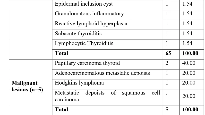
Epidermal inclusion cyst 1 1.54
Granulomatous inflammatory 1 1.54
Reactive lymphoid hyperplasia 1 1.54
Subacute thyroiditis 1 1.54
Lymphocytic Thyroiditis 1 1.54
Total 65 100.00
Malignant lesions (n=5)
Papillary carcinoma thyroid 2 40.00
Adenocarcinomatous metastatic depoists 1 20.00
Hodgkins lymphoma 1 20.00
Metastatic depoists of squamous cell
carcinoma 1 20.00
Total 5 100.00
Table 7 shows: Based on FNAC, majority of the patients (92.86%) had benign lesions while malignant lesions were diagnosed in 7.14% of the patients
DISCUSSION
Neck swellings are common and carry a low, but noticeable risk of malignancy. The most important challenge is differentiating benign from malignant swellings, and precise diagnosis and management of malignant swellings in the early stages.5
Conventional USG has been widely used to determine which lesion should be biopsied. There are several suspicious USG features that predict malignancy, such as hypoechogenicity, marked hypoechogenicity, a microlobulated or spiculated margin, micro- or macro-calcifications.6-8 Although conventional USG can provide meaningful information in neck swelling diagnosis, there has been considerable variation in diagnostic performances.9
This one year prospective diagnostic accuracy study was conducted from January 2017 to December 2017. 612 patients attended ear nose throat (ENT), Surgery and Paediatric OPD for neck swellings. After applying inclusion and exclusion criteria, a total of 70 patients were included in the present study during the study period and were studied. All the patients underwent FNAC .
Epidemiological data suggests that, neck swellings are less frequent in men than in women,10 accordingly in the present study females outnumbered males as 58.57% of the patients were
females and 41.43% were males with male to female ratio of 1:1.41. Similar findings were reported in a study from Pokhara Nepal where 92% were females.11 Recently Kumar A et al.12 (2017) reported male to female ratio as high as 1: 6.02. Another study by Handa U et al.13 (2008) also reported higher male to female ratio that is, 1:6.35. The female predominance suggests that hormonal factors may be involved and the literature also says that there can be biological changes occurring during pregnancy which may increase the risk.14
Pag e
365
Pag e365
Pag e365
Pag e365
Pag e365
Pag e365
Pag e365
Pag e365
Pag e365
Pag e365
Pag e365
Pag e365
Pag e365
Pag e365
Pag e365
Pag e365
Pag e365
Pag e36
5
Pag e365
Pag e365
Pag e365
et al.17 (2013) reported mean age as 40.57 years
which was sharply corroborates with the present study.
Out of 65 benign lesions, diagnosed on FNAC, colloid goiter was commonest contributing to 33.85% (22 cases) followed by nodular goiter constituting 27.7% (18 cases), 15.38% (10 cases) were of lipomatous lesion, 7.69% (5 cases) were of infected epidermal cyst. Salivary gland lesions included chronic sialdenitis, warthin’s tumor and pleomorphic adenoma constituting 4.69% (3 cases) of total cases. 4.69% (3 cases) were tubercular lymphadenitis. 1.54% that is 1 case each were contributed by epidermal inclusion cyst, granulomatous inflammatory pathology, reactive lymphoid hyperplasia, subacute and lymphocytic thyroiditis.
Among 5 malignant cases, on FNAC 2 cases were of papillary carcinoma thyroid followed by 1 case each of Adenocarcinomatous metastatic deposits,
Hodgkin’s lymphoma and metastatic deposits of squamous cell carcinoma.
CONCLUSION
FNAC of neck swellings has become one of the most useful, safe, accurate, relatively simple, inexpensive, less time-consuming OPD procedures, virtually painless, highly patient-compliance with highly accurate dependable tool in the diagnosis of neck pathology. Though, it is not a substitute for conventional surgical histopathology but is regarded as an extremely valuable complement in diagnosis. Hence FNAC should be treated as a first-line diagnostic test for neck swellings. By performing USG, followed by FNAC improved the accuracy and eliminating false positive cases for malignancy. FNAC can also yield material for ancillary diagnostic
techniques like flowcytometry,
immunohistochemistry, PCR in the workup of neck swellings.
REFERENCES
1. Neck Swellings BMJ 2014; 348:bmj.g1078.
Available from: URL:
https://www.bmj.com/content/348/bmj.g1078 Access Date: 18.10.2018
2. Rathod GB, Rai P, Rai S. A prospective study of ultrasonographic and FNAC correlation of thyroid pathology. IAIM 2015; 2(11):46-51. 3. Misiakos EP, Margari N, Meristoudis C,
Machairas N, Schizas D, Petropoulos K, et al. Cytopathologic diagnosis of fine needle aspiration biopsies of thyroid nodules. World J Clin Cases. 2016;4(2):38-48.
4. Koss MG, Melamed MR. Koss’s Diagnostic cytology and its histopathologic bases. 5th ed., New York: Williams and Wilkins; 2006. 5. Aliasgarzadeh A, Tarzemani MK, Raeisi M,
Mobasseri M, Mozayyan M, Ghojazadeh M. Diagnostic value of Elastography in thyroid nodules. J Anal Res Clin Med 2014;2(2):71-6. 6. Kwak JY, Kim EK. Ultrasound elastography for thyroid nodules: recent advances. Ultrasonography 2014;33(2):75-82.
7. Kim EK, Park CS, Chung WY, Oh KK, Kim DI, Lee JT, et al. New sonographic criteria for recommending fine-needle aspiration biopsy of nonpalpable solid nodules of the thyroid. AJR Am J Roentgenol 2002;178:687-91.
8. Moon WJ, Baek JH, Jung SL, Kim DW, Kim EK, Kim JY, et al. Ultrasonography and the ultrasound-based management of thyroid nodules: consensus statement and recommendations. Korean J Radiol 2011;12:1-14.
9. Gharib H, Papini E, Paschke R, Duick DS, Valcavi R, Hegedus L, et al. American Association of Clinical Endocrinologists, Associazione Medici Endocrinologi, and European Thyroid Association Medical Guidelines for Clinical Practice for the Diagnosis and Management of Thyroid Nodules. Endocr Pract 2010;16 Suppl 1:1-43. 10.Dankle SK, Griffing GT. Thyroid nodule.
Available from: URL:
https://emedicine.medscape.com/article/1274 91-overview#showall Access Date: 18.09.2018.
11.Bhatta S, Makaju R, Mohammad A. Role of fine needle aspiration cytology in the diagnosis of thyroid lesions. J Pathol Nepal 2012;2:186-8.
e
366
e
366
e366
e
366
e366
e
366
e366
e
366
e366
e
366
e
366
e
366
e
366
e366
e
366
e366
e
366
e366
e
366
e366
e
366
13.Gupta M, Gupta S, Gupta VB. Correlation of Fine needle Aspiration Cytology with Histopathology in the diagnosis of solitary thyroid nodule. J Thyroid Res 2010(2010); Article ID 379051.
14.Nagarkar R, Roy S, Akheel M, Palwe V, Kulkarni N, Pandit P. Incidence of Thyroid Disorders in India: An Institutional Retrospective Analysis. Int J Dent Med Spec 2015;2(2):19-23.
15.Fadda G, Livolsi VA. Histology and aspiration cytology of benign thyroid diseases. Rays 1999;(2):182-96.
16.Raab SS, Grzybicki DM, Janosky JE. Clinical impact and frequency of anatomic pathology errors in cancer diagnosis. Cancer 2005;104:2205-13.