O R I G I N A L R E S E A R C H
The Role Of Hepatic Stellate Cells In Promoting
Liver Metastasis Of Colorectal Carcinoma
This article was published in the following Dove Press journal:
OncoTargets and Therapy
Wen-Hai Huang1,*
Min-Wei Zhou1,*
Yan-Feng Zhu2,*
Jian-Bin Xiang1
Zhen-Yang Li1
Zi-Hao Wang1
Yi-Ming Zhou1
Yi Yang1
Zong-You Chen1
Xiao-Dong Gu 1
1Department of General Surgery,
Huashan Hospital, Fudan University, Shanghai 200040, People’s Republic of China;2Department of Nursing, Huashan
Hospital, Fudan University, Shanghai 200040, People’s Republic of China
*These authors contributed equally to this work
Purpose: Colorectal cancer (CRC) is the most common malignancy in the gastrointestinal tract. The liver is the most common location of CRC metastases, which are the main causes of CRC-related death. However, the mechanisms underlying metastasis of CRC to the liver have not been characterized, resulting in therapeutic challenges.
Methods: The effects of hepatic stellate cells (HSCs) on T cells were evaluated using in vitro mixed lymphocyte reactions (MLRs) and cytokine production assays. HSC-induced CT26 cell migration and proliferation were evaluated in vitro and in vivo.
Results:HSCs induced T cell hypo-responsiveness, promoted T cell apoptosis, and induced regulatory T cell expansion in vitro. IL-2 and IL-4 were significantly lower in MLRs incubated with HSCs. Supernatants of MLRs with HSCs promoted CT26 cell proliferation and migration. Furthermore, the presence of HSCs increased the number of liver metastases and promoted proliferation of liver metastatic tumor cells in vivo.
Conclusion: HSCs may contribute to an immunosuppressive liver microenvironment, resulting in a favorable environment for the colonization of CRC cells in the liver. These
findings highlight a potential strategy for treatment of CRC liver metastases.
Keywords:colorectal cancer, liver metastasis, hepatic stellate cells, dendritic cells, T cells
Introduction
Colorectal cancer (CRC) is the third most common cancer worldwide, and has
increased in prevalence in recent years.1CRC frequently metastasizes to the liver,
and liver resection and perioperative chemotherapy are the primary means of therapeutic intervention for these tumors. The median survival time for patients with untreated CRC and liver metastases is 6.9 months, and 5-year survival rates following liver resection range from 30% to 50%. Several recent studies have aimed to evaluate the mechanisms responsible for liver metastasis. However, the mechanisms underlying liver metastasis of CRC have not been characterized,
resulting in challenges to development of effective therapies.2–4
In 1889, Paget proposed the“seed and soil”theory of metastatic dissemination.
Paget suggested that the site of metastasis depended on the affinity of the tumor for
the microenvironment.5To evaluate the hepatic microenvironment, we previously
analyzed liver non-parenchymal cells in mice and showed that hepatic stellate cells
(HSCs), which store retinol and participate in repair and fibrogenesis during liver
injury, play a role in immune regulation.6Recent studies of HSCs have focused on
liver injury, liver fibrosis, and liver regeneration. Several studies have shown that
HSCs exhibit immunomodulatory activity and can prolong allograft survival.7,8
Correspondence: Zong-You Chen; Xiao-Dong Gu
Department of General Surgery, Huashan Hospital, Fudan University, 12 Wulumuqi Middle Road, Shanghai 200040, People’s Republic of China
Tel +86 21 52887330; +86 21 52887333 Fax +86 21 62495490
Email zongyouc@sohu.com ; gxdgxd737@163.com
OncoTargets and Therapy
Dove
press
open access to scientific and medical research
Open Access Full Text Article
OncoTargets and Therapy downloaded from https://www.dovepress.com/ by 118.70.13.36 on 25-Aug-2020
Furthermore, HSCs have been shown to promote onset and
progression of hepatocellular carcinomas.9,10 We
pre-viously showed that quiescent HSCs express low levels
of immune surface molecules. Priming HSCs with IFN-γ
resulted in marked upregulation of the inhibitory co-sti-mulatory molecule B7-H1, potentially through activation
of the MEK/ERK pathway.6However, the mechanisms by
which HSCs promote metastasis of CRC cells to the liver have not been elucidated.
In this study, we demonstrate that HSCs induce T cell hypo-responsiveness and expand regulatory T (Treg) cells. Moreover, HSCs were shown to play an immunosuppres-sant role in the hepatic microenvironment and promote CRC metastasis to the liver.
Materials And Methods
Animals
BALB/c mice were obtained from the Shanghai SLAC Laboratory Animal Company. All mice were maintained in
a specific pathogen-free environment at Huashan Hospital.
Animals were fed standard chow ad libitum and subjected to
experiments at 7–9 weeks of age. The animal study protocol
was approved by the ethics committee of Huashan Hospital. All experiments were performed following the Huashan Hospital Laboratory Animal Centre care guidelines.
Isolation, Culture, And Identi
fi
cation Of
HSCs
HSCs were isolated from murine livers as previously
described.11 Briefly, the livers were perfused through the
portal vein with collagenase IV (Life Technologies, Grand
Island, NY, USA). The smashed cells werefiltered through
a nylon mesh. HSCs were purified by Percoll density
gradient centrifugation (Sigma-Aldrich, St. Louis, MO, USA) and cultured in complete medium supplemented with 20% FBS (Gibco, Gaithersburg, MD, USA) for 7 to 14 days, unless otherwise indicated. The purity of HSCs ranged from 90% to 95%, as measured by desmin immu-nostaining and typical appearance of lipid droplets under a light microscope.
Isolation And Culture Of Dendritic Cells
(DCs)
DCs were generated from bone marrow progenitor cells as
previously described.12 Bone marrow cells were extracted
from femurs and tibias of BALB/c mice, and erythrocytes were lysed using ammonium chloride. The cells were
cultured in 24-well plates (1×10⁶ cells/well) in 1 mL of
RPMI 1640 (Gibco) supplemented with 10% FBS and 10 ng/mL recombinant granulocyte-macrophage colony stimu-lating factor (R&D Systems, Minneapolis, MN, USA). All
cultures were incubated at 37°C in 5% humidified CO2.
Nonadherent granulocytes were removed after 48 hrs of culture. Half of the media was exchanged every 48 hrs.
After 6 days of culture, 1 μg/mL lipopolysaccharide
(Sigma-Aldrich) was added to the culture media for 18 hrs to allow for maturation. The purity of DC preparations was
routinely monitored byflow cytometry using an anti-CD11c
monoclonal antibody (mAb) (eBioscience, San Diego, CA, USA). CD11c+ cells were enriched to >85%.
Tumor Antigen Uptake
The mouse colon carcinoma CT26 cell line was purchased from American Type Culture Collection and cultured in DMEM (Gibco) supplemented with 10% FBS. On day 6 of DC culture, CT26 mouse colon cancer cell lysates were added to the culture medium cultures consisted of a
DC-to-CT26 ratio of 1:10 for 18 hrs at 37°C in 5% humidified
CO2. CT26 mouse colon cancer cell lysates were obtained
through six freeze/thaw cycles in PBS (Sigma-Aldrich).
Mixed Lymphocyte Reactions (MLRs)
For primary MLRs, nylon wool-eluted spleen T cells(2×10⁵) from BALB/c mice were used as responders, and
γ-irradiated (20 Gy) tumor antigen-pulsed DCs derived
from BALB/c bone marrow were used as stimulators. Cultures were maintained in complete medium for 3 days
at 37°C in 5% humidified CO2. [3H]-TdR (0.5μCi/well) was
added for the final 18 hrs of culture. Cells were harvested
onto glass fiber disks using an automated system, and
incorporation of [3H]-TdR into DNA was assessed using a
Wallac 1450 liquid scintillation counter (PerkinElmer, Boston, MA, USA). Results are expressed as mean counts per minute (cpm)±SD. To examine the effect of HSCs on T
cell proliferation,γ-irradiated (50 Gy) HSCs were added at
the beginning of culture. T cells were cultured with tumor antigen-pulsed DCs at a ratio of 10:1 for 3 days. HSCs were added into the culture at a HSCs: T cells ratio of 1:20, 1:40, 1:80, or 1:160.
Flow Cytometric Analysis
Expression of cell surface molecules was detected using a FACScan (BD Biosciences, San Jose, CA, USA), and ana-lyzed using CellQuest software (BD Biosciences). Cells were stained with the following monoclonal antibodies:
OncoTargets and Therapy downloaded from https://www.dovepress.com/ by 118.70.13.36 on 25-Aug-2020
FITC-CD25 (eBioscience), PE-Cy5-CD4, and FITC-CD3 (BD Biosciences). Isotype-matched irrelevant mAbs were used as negative controls. Apoptosis was assessed using PE-Annexin V staining (BD Biosciences). Foxp3 staining
was performed using fixation and permeabilization buffers
contained in the Foxp3 kit according to manufacturer’s
instructions (eBioscience).
CCK8 Assay
CT26 cells were plated in triplicate at 4×10³cells/well in
96-well plates. MLR supernatants were collected and added to the wells. At 24, 48, 72, and 96 hrs, 10 µL of CCK8 solution (Dojindo, Kumamoto, Japan) was added to each well. Absorbance was detected at 450 nm using a
microplate reader (Thermo Fisher Scientific, Waltham,
MA, USA) following incubation at 37°C for 2 hrs.
Transwell Migration Assay
Transwell migration chambers (Corning Inc., Corning, NY, USA) were used to evaluate the migration of CT26 cells.
CT26 cells (5×10⁴) in 200 µL of serum-free medium were
added to the upper chamber, and supernatants (800 µL) col-lected from MLR experiments with or without HSCs were added to the lower chamber. After incubation for 24 hrs, cells on the upper surface of the membrane were removed using a
cotton swab. The remaining cells were fixed in methanol,
stained using crystal violet, and air-dried. The number of
migrating cells on each membrane was counted infive random
fields per well using a light microscope.
Cytokine Analysis
IL-2, IL-4, IL-10, and IFN-γlevels in MLR culture
super-natants were measured using ELISA kits according to the
manufacturer’s instructions (Jingmei Biotech Limited
Company, Shenzhen, People’s Republic of China). The
minimum detectable dose of IL-2 is 15.6 pg/mL. The mini-mum detectable dose of IL-4 is 7.8 pg/mL. The minimini-mum detectable dose of IL-10 is 15.6 pg/mL. The minimum
detectable dose of IFN-γis 9.4 pg/mL. MLR culture
super-natants were harvested at the end of the third day of culture.
Animal Model Of CRC Liver Metastasis
A murine model of CRC-derived liver metastasis wasestab-lished as previously described.13 CT26 cells were washed
twice with 0.5 M PBS after trypsinization and suspended in PBS. The animal model was established under anesthesia
using isoflurane (Abbott Laboratories, Abbott Park, IL,
USA). For the laparotomy, a median incision was made and
a 30-gauge needle was used to puncture the portal vein. A 0.1
mL cell suspension containing 1×10⁶CT26 cells, or a
mix-ture of 1×10⁶CT 26 cells and 5×10⁵HSCs, was injected to
achieve a liver metastasis model.
Immunohistochemistry
Immunohistochemistry was performed in liver metastatic
tumor samples from murine model. Tissue paraffin sections
were deparaffinized, rehydrated and pre-treated with 10 mM
sodium citrate buffer at a sub-boiling temperature for 10 mins to unmask the antigen. The sections were subsequently
incu-bated with 3% H2O2for 10 mins at room temperature and
dark conditions to block endogenous peroxidase activity, followed by incubation with blocking solution for 1 hr to
avoid unspecific binding of the primary antibody. The
sec-tions were then incubated overnight at 4ºC with the anti-PCNA antibody (Cell Signaling Technology, Danvers, MA, USA) followed by incubation for 30 mins at room tempera-ture with a biotinylated ECL anti-rabbit IgG (GE Healthcare, Chalfont St. Giles, UK). The color was developed using the diaminobenzidine substrate (Roche Diagnostics, Mannheim, Germany), and the sections were counterstained with hema-toxylin. Slides were visualized and photographed using a Leica DM2500 light microscope (Leica Microsystems Inc., Buffalo Grove, IL, USA).
Statistical Analysis
All data analyses were conducted using SPSS 16.0 software package (SPSS Inc., Chicago, IL, USA). Comparative ana-lysis of the data was performed using one-way ANOVA or
Student’s t-test. Differences were considered statistically
significant when P < 0.05.
Results
DCs Effectively Took Up Tumor Antigens
To examine the effects of tumor antigen uptake by DCs, DCs were pulsed with tumor antigens as stimulators in MLRs. On day 6 of DC culture, tumor antigens were added to the culture at a DC:CT26 ratio of 1:10 for 18hrs at 37°C in 5% humidified CO2. The results showed
that DCs that did not take up tumor antigens did not
promote T cell proliferation. (P<0.05) (Figure 1).
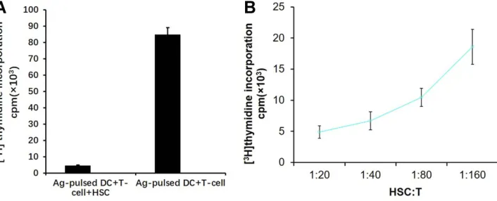
HSCs Inhibited T cell Proliferation
To evaluate the effects of HSCs on T cell proliferation, HSCs were added to an MLR culture containing splenic T cells stimulated by DCs pulsed with tumor antigens. HSCs
OncoTargets and Therapy downloaded from https://www.dovepress.com/ by 118.70.13.36 on 25-Aug-2020
inhibited T cell proliferation (P<0.05) (Figure 2A) in an
HSC:T cell ratio-dependent manner (Figure 2B).
HSCs Induced T Cell Apoptosis
We hypothesized that HSC-mediated inhibition of T cell pro-liferation may have resulted from apoptosis of activated Tcells. To test this hypothesis, splenic T cells were cultured for 3 days with tumor antigen-pulsed DCs in the presence or absence of HSCs. The cells were double-stained with anti-Annexin V
mAb and anti-CD3 mAb. As shown inFigure 3, the proportion
of cells that were double-positive for Annexin V and CD3 staining was higher in the group cultured with HSCs than that in the group cultured without HSCs (P<0.05). These
results confirmed that HSCs increased T cell apoptosis.
HSCs Promoted Treg Cell Expansion
To examine the effects of HSCs on Treg cell activity, Tregcells were quantified in MLRs in which splenic T cells were
cultured for 3 days with tumor antigen-pulsed DCs in the
presence of HSCs. The percentage of CD4+CD25+ FoxP3+ cells in MLR cultures with HSCs was higher than that in
MLR cultures without HSCs (P<0.05) (Figure 4B). This
result confirmed that HSCs promoted Treg cell expansion.
Cytokine Levels In Supernatants Of MLRs
Were Altered By HSCs
To evaluate the effects of HSCs on cytokine secretion, IL-2,
IL-4, IL-10, and IFN-γlevels were quantified in MLR culture
supernatants using ELISA. The results showed that the expres-sion levels of IL-2 and IL-4 in supernatants of MLRs with
HSCs were significantly lower than those in supernatants of
MLRs without HSCs (P<0.05) (Figure 5). No differences were
observed for IL-10 or IFN-γ expression in MLRs with or
without HSCs.
MLR Supernatants Promoted CT26 Cell
Proliferation And Migration
To determine the effects of MLR supernatants on CT26 cells, CCK8 assay was used to evaluate CT26 cell Figure 1DCs effectively took up tumor antigens. DCs that did not take up tumor antigens did not promote T cell proliferation. DCs able to effectively take up tumor antigens promoted T cell proliferation to a significantly greater extent than DCs that did not take up tumor antigens (P<0.05). The ratio of T cells to DCs was 20:1.
Figure 2(A) HSCs inhibited T lcell proliferation. The ratio of T cells to DCs to HSCs was 20:2:1. (B) In each group, 2×10⁵nylon wool-eluted spleen T cells from BALB/c mice were cultured with DCs in the presence or absence of HSCs. The ratio of T cells to DCs (10:1) was the same for all conditions, but the ratio of HSCs to T cells varied. HSCs inhibited T cell proliferation in a dose-dependent manner.
OncoTargets and Therapy downloaded from https://www.dovepress.com/ by 118.70.13.36 on 25-Aug-2020
Figure 3Nylon wool-eluted spleen T cells from BALB/c mice were cultured with DCs in the presence or absence of HSCs. The ratio of T cells to DCs to HSCs was 20:2:1. Apoptotic cells were double-stained with FITC-anti-CD3 and PE-anti-Annexin V. The number of apoptotic T cells in the group incubated with HSCs was markedly greater than that in the group without HSCs (P<0.05).
CD
25
Foxp3 100 101 102 103 104
FL3-H: CD4 PE-Cy5 0
50 100 150 200 250
t
h
gi
e
H-C
S
S:
H-C
S
S
62
100 101 102 103 104 FL1-H: Foxp3 FITC 100
101 102 103 104
E
P
5
2
D
C:
H-2
L
F
3 6.65
0.21 90.1
100 101 102 103 104 FL1-H: Foxp3 FITC 100
101 102 103 104
E
P
5
2
D
C:
H-2
L
F
1.12 14.9
1.17 82.8
Ag pulsed DC+T-cell Ag pulsed DC+T-cell+HSC
A
% CD25+Foxp3+
cells
in CD4+
cells
B
Figure 4HSCs induced expansion of Treg cells. T cells were co-cultured with DCs in the presence or absence of HSCs. (A) Gated on the CD4+ cell populations. (B) HSCs increased the number of CD4+CD25+FoxP3+ cells, and the percentage of Treg cells in the group with HSCs was higher than that in the group without HSCs (P<0.05).
OncoTargets and Therapy downloaded from https://www.dovepress.com/ by 118.70.13.36 on 25-Aug-2020
proliferation and transwell migration assay was used to
evaluate CT26 cell migration. As shown in Figure 6,
supernatants from MLRs with HSCs promoted prolifera-tion and migraprolifera-tion of CT26 cells to a greater extent than MLRs without HSCs (P<0.05).
HSCs Promoted Growth Of Liver
Metastatic Tumors In Vivo
To determine whether HSCs could promote CRC cell colo-nization in the liver in vivo, a murine model of CRC-derived liver metastasis was evaluated. In the control group, CT26
cells were injected via the portal vein. In the experimental group, a mixture of CT26 cells and HSCs was injected via the
portal vein. As shown in Figure 7A, the number of liver
metastatic tumors in the experimental group was greater than that in the control group (P<0.05). CT26 cell prolifera-tion was assessed using PCNA immunostaining of liver metastatic tumor samples. The number of PCNA-positive
cells was significantly greater in the experimental group
than in the control group (P<0.05) (Figure 7B).
Figure 5Supernatants were collected from MLRs and analyzed using ELISA. HSCs reduced the expression of IL-2 and IL-4 in the supernatants (P<0.05). However, HSCs did not alter the expression of IL-10 or IFN-γin the supernatants (P>0.05).
Figure 6(A) CCK8 assay was used to determine whether supernatants of MLRs containing HSCs promoted proliferation of CT26 cells. (B) Transwell migration assay was used to determine whether supernatants of MLRs containing HSCs increased CT26 cell migration.
Figure 7Effect of HSCs on liver metastasis in vivo. (A) The number of liver metastatic tumors in the experimental group was greater than that in the control group (P<0.05). (B) Immunohistochemical staining for PCNA in liver metastatic tumor samples. The number of PCNA-positive cells in the experimental group was significantly greater than that in the control group (P<0.05).
OncoTargets and Therapy downloaded from https://www.dovepress.com/ by 118.70.13.36 on 25-Aug-2020
Discussion
CRC is a common malignancy of the gastrointestinal tract. The liver, which is nourished by a rich blood supply from both arterial and portal venous systems, is the most com-mon location of CRC metastases. Liver metastasis is the
main cause of CRC-related death.14 A large body of
evi-dence has indicated that the liver microenvironment pro-vides autocrine and paracrine signals originating from both parenchymal and non-parenchymal cells that promote
development of hepatic metastases.15
HSCs are the main non-parenchymal cells in the liver. Quiescent HSCs can be activated by cytokines or by liver injury. Recent studies showed that activated HSCs play an
important role in liver fibrosis and exhibit
immunomodu-latory activities. Additionally, HSCs in the liver microen-vironment have been shown to promote proliferation and
infiltration of hepatoma cells.16,17 Platelet-derived growth
factor-C, which is secreted by colon carcinoma cells, induces proliferation of HSCs and promotes tumor
growth.18 However, the mechanisms responsible for the
effects of HSCs on development of CRC-derived liver metastases remain unclear.
HSCs exert potent immunosuppressive effects via
induction of activated T cell apoptosis.19 Furthermore,
HSCs can induce T cell hypo-responsiveness and promote Treg cell expansion, allowing hepatocellular carcinoma cells to escape immune detection, resulting in development
of liver carcinoma.20 We used MLRs to investigate the
effects of HSCs on T cells. Ourfindings demonstrated that
activated HSCs inhibited T cell proliferation and induced T cell apoptosis. In addition, HSCs promoted Treg cell expansion. Treg cells are a subgroup of CD4+ T cells that exhibit immunosuppressive activity. Transforming growth
factorβ(TGF-β) was one of thefirst cytokines discovered,
and plays an important role in differentiation of Treg
cells.21 Activated HSCs secrete TGF-β,11 which may
have been responsible for the increased percentage of CD4+CD25+ FoxP3+ cells in MLRs with HSCs. Based on this result, we hypothesized that HSCs may be respon-sible for creating an immunosuppressive liver microenvir-onment, resulting in an environment suitable for the growth of CRC metastases.
We also analyzed cytokine levels in MLR supernatants. IL-2 is primarily secreted by CD4+ and CD8+ T cells, resulting in T cell activation and enhanced NK cell
activity.22 Consistent with our results, Shimizu et al23
found that IL-2 production was significantly decreased in
patients with CRC and hepatic metastasis. IL-4 is primar-ily secreted by type 2 T helper cells, mast cells, and
basophilic granulocytes.24 Recent studies have shown
that binding of IL-4 to IL-4R promotes tumor cell
prolif-eration in breast and prostate cancer.25,26 However,
Morisaki et al27 found that IL-4/IL-4R prevents gastric
cancer cells from entering the G0/G1 phase, resulting in inhibition of cell proliferation. Our results suggest that IL-4 inhibits metastasis of CRC to the liver.
MLRs were used to simulate an immune microenvir-onment. HSCs, which are liver non-parenchymal cells, were added to MLR cultures to create a hepatic microen-vironment in vitro. CCK8 and transwell migration assays showed that supernatants from MLRs containing HSCs
promoted CT26 cell proliferation and migration, confi
rm-ing that HSCs in hepatic microenvironments could induce CRC cell proliferation and migration. Consistent with our in vitro results, we found that the presence of HSCs increased the number of liver metastases and increased proliferation of liver metastatic tumor cells in vivo.
Conclusion
Our study showed that HSCs may contribute significantly
to an immunosuppressive hepatic microenvironment, resulting in increased CRC cell proliferation and migra-tion. Furthermore, HSCs promoted CRC cell colonization
in the liver. Ourfindings indicate a potential strategy for
treatment of CRC liver metastases.
Acknowledgment
This study was supported by the “Belt and Road” Young
Scientist Communication International Cooperation Project (17410742100).
Disclosure
The authors report no conflicts of interest in this work.
References
1. Aran V, Victorino AP, Thuler LC, Ferreira CG. Colorectal cancer: epidemiology, Disease mechanisms and interventions to reduce onset and mortality.Clin Colorectal Cancer.2016;15:195–203. doi:10.1016/ j.clcc.2016.02.008
2. Nakagawa K, Tanaka K, Nojiri K, et al. The modified glasgow prog-nostic score as a predictor of survival after hepatectomy for colorectal liver metastases.Ann Surg Oncol.2014;21:1711–1718. doi:10.1245/ s10434-013-3342-6
3. Zimmitti G, Shindoh J, Mise Y, et al. RAS mutations predict radiologic and pathologic response in patients treated with chemotherapy before resection of colorectal liver metastases.Ann Surg Oncol.2015;22:834– 842. doi:10.1245/s10434-014-4042-6
OncoTargets and Therapy downloaded from https://www.dovepress.com/ by 118.70.13.36 on 25-Aug-2020
4. Kawada K, Hasegawa S, Murakami T, et al. Molecular mechanisms of liver metastasis.Int J Clin Oncol.2011;16:464–472. doi:10.1007/ s10147-011-0307-2
5. Ribatti D, Mangialardi G, Vacca A. Stephen paget and the‘seed and soil’theory of metastatic dissemination.Clin Exp Med.2006;6:145– 149. doi:10.1007/s10238-006-0117-4
6. Gu X, Wang Y, Xiang J, et al. Interferon-γtriggers hepatic stellate cell-mediated immune regulation through MEK/ERK signaling pathway. Clin Dev Immunol. 2013;2013:389807. doi:10.1155/ 2013/657424
7. Chou HS, Hsieh CC, Yang HR, et al. Hepatic stellate cells regulate immune response by way of induction of myeloid suppressor cells in mice.Hepatology.2011;53:1007–1019. doi:10.1002/hep.24162 8. Yang HR, Chou HS, Gu X, et al. Mechanistic insights into
immu-nomodulation by hepatic stellate cells in mice: a critical role of interferon-gamma signaling. Hepatology. 2009;50:1981–1991. doi:10.1002/hep.23202
9. Zhao W, Zhang L, Xu Y, et al. Hepatic stellate cells promote tumor progression by enhancement of immunosuppressive cells in an ortho-topic liver tumor mouse model. Lab Invest. 2014;94:182–191. doi:10.1038/labinvest.2013.139
10. Xu Y, Zhao W, Xu J, et al. Activated hepatic stellate cells promote liver cancer by induction of myeloid-derived suppressor cells through cyclooxygenase-2. Oncotarget. 2016;7:8866–8878. doi:10.18632/ oncotarget.6839
11. Yu MC, Chen CH, Liang X, et al. Inhibition of T-cell responses by hepatic stellate cells via B7-H1-mediated T-cell apoptosis in mice. Hepatology.2004;40:1312–1321. doi:10.1002/hep.20488
12. Gu X, Xiang J, Yao Y, Chen Z. Effects of RNA interference on CD80 and CD86 expression in bone marrow-derived murine dendritic cells.Scand J Immunol.2006;64:588–594. doi:10.1111/j.1365-3083.2006.01845.x 13. Kohashi S, Sato Y, Fukushima T, et al. Interferon-beta inhibits
liver metastases from murine colon 26 carcinoma and its highly metastatic variant. Surg Today. 2007;37:474–481. doi:10.1007/ s00595-006-3418-z
14. Kopetz S, Chang GJ, Overman MJ, et al. Improved survival in metastatic colorectal cancer is associated with adoption of hepatic resection and improved chemotherapy.J Clin Oncol.2009;27:3677– 3683. doi:10.1200/JCO.2008.20.5278
15. Milette S, Sicklick JK, Lowy AM, Brodt P. Molecular pathways: targeting the microenvironment of liver metastases. Clin Cancer Res.2017;23:6390–6399. doi:10.1158/1078-0432.CCR-15-1636
16. Amann T, Bataille F, Spruss T, et al. Activated hepatic stellate cells promote tumorigenicity of hepatocellular carcinoma. Cancer Sci.
2009;100:646–653. doi:10.1111/j.1349-7006.2009.01087.x
17. Antoine M, Tag CG, Gressner AM, Hellerbrand C, Kiefer P. Expression of E-selectin ligand-1 (CFR/ESL-1) on hepatic stellate cells: implications for leukocyte extravasation and liver metastasis. Oncol Rep.2009;21:357–362.
18. Bandapalli OR, Macher-Goeppinger S, Schirmacher P, Brand K. Paracrine signalling in colorectal liver metastases involving tumor cell-derived PDGF-C and hepatic stellate cell-derived PAK-2.Clin Exp Metastasis.2012;29:409–417. doi:10.1007/s10585-012-9459-3 19. Su YH, Shu KH, Hu C, et al. Hepatic stellate cells attenuate the immune
response in renal transplant recipients with chronic hepatitis.Transplant Proc.2012;44:725–729. doi:10.1016/j.transproceed.2011.11.049 20. Zhao W, Su W, Kuang P, et al. The role of hepatic stellate cells in the
regulation of T-cell function and the promotion of hepatocellular carcinoma.Int J Oncol.2012;41:457–464. doi:10.3892/ijo.2012.1497 21. Chen W, Jin W, Hardegen N, et al. Conversion of peripheral CD4 +CD25- naive T cells to CD4+CD25+ regulatory T cells by TGF-beta induction of transcription factor Foxp3.J Exp Med.2003;198:1875– 1886. doi:10.1084/jem.20030152
22. Al-Hakeim HK, Al-Rammahi DA, Al-Dujaili AH. IL-6, IL-18, sIL-2R, and TNFα proinflammatory markers in depression and schizo-phrenia patients who are free of overt inflammation.J Affect Disord.
2015;182:106–114. doi:10.1016/j.jad.2015.04.044
23. Shimizu H, Ito H, Kimura F, et al. Decreased cell-mediated immune status in colorectal cancer patients with hepatic metastasis. Hepatogastroenterology.2005;52:1106–1109.
24. Kelly-Welch AE, Hanson EM, Boothby MR, Keegan AD. Interleukin-4 and interleukin-13 signaling connections maps. Science.2003;300:1527–1528. doi:10.1126/science.1085458 25. Roca H, Craig MJ, Ying C, et al. IL-4 induces proliferation in
prostate cancer PC3 cells under nutrient-depletion stress through the activation of the JNK-pathway and survivin up-regulation. J Cell Biochem.2012;113:1569–1580. doi:10.1002/jcb.24025
26. Venmar KT, Kimmel DW, Cliffel DE, Fingleton B. IL4 receptorα mediates enhanced glucose and glutamine metabolism to support breast cancer growth.Biochim Biophys Acta.2015;1853:1219–1228. doi:10.1016/j.bbamcr.2015.02.020
27. Morisaki T, Yuzuki DH, Lin RT, Foshag LJ, Morton DL, Hoon DS. Interleukin 4 receptor expression and growth inhibition of gastric carcinoma cells by interleukin 4.Cancer Res.1992;52:6059–6065.
OncoTargets and Therapy
Dove
press
Publish your work in this journal
OncoTargets and Therapy is an international, peer-reviewed, open access journal focusing on the pathological basis of all cancers, potential targets for therapy and treatment protocols employed to improve the management of cancer patients. The journal also focuses on the impact of management programs and new therapeutic
agents and protocols on patient perspectives such as quality of life, adherence and satisfaction. The manuscript management system is completely online and includes a very quick and fair peer-review system, which is all easy to use. Visit http://www.dovepress.com/ testimonials.php to read real quotes from published authors.
Submit your manuscript here:https://www.dovepress.com/oncotargets-and-therapy-journal
OncoTargets and Therapy downloaded from https://www.dovepress.com/ by 118.70.13.36 on 25-Aug-2020