Research Article
CODEN: IJPNL6
STATISTICAL OPTIMIZATION OF PHYSIOCHEMICAL PARAMETERS FOR
ENHANCING THE ANTIMICROBIAL POTENTIAL OF LODHRA (SYMPLOCOS
RACEMOSA) BARK AND ITS BIOSAFETY EVALUATION
Henna Sood, Harpreet Kaur and Daljit Singh Arora
*Microbial Technology Laboratory, Department of Microbiology, Guru Nanak Dev University,
Amritsar-143005, India
*Corresponding author e-mail: [email protected]
ABSTRACT
Screening the aqueous extract of Symplocos racemosa bark demonstrated its broad spectrum antimicrobial potential with an average zone of inhibition 12–17mm. Using one-factor-at-a-time approach, 15% concentration, extraction for 20 min at 40 oC and a pH of 3.5 and filtration through Whatman filter paper, were found to be optimal for the best antimicrobial efficacy. Statistical optimization by response surface methodology enhanced the antimicrobial activity upto 1.3 folds. MIC values of the aqueous extract ranged from 5 –15 mg/ml. The viable cell count studies revealed P. aeruginosa to be the most sensitive and got killed instantaneously at 0 h. The aqueous extract was found to be reasonably thermostable at 100 °C for 1 h and had a better shelf life at refrigeration temperature (4 °C). Biosafety evaluation of the aqueous extract showed it to be neither cytotoxic nor mutagenic as revealed by Ames test and MTT assay, thus showing its promise for further pursuance.
Keywords: Symplocos racemosa, Antimicrobial, Minimum inhibitory concentration, Cytotoxicity.
INTRODUCTION
Antimicrobial resistance (AMR), a phenomenon which owes its emergence to overuse of antibiotics in humans and food- producing animals, is a global threat now a days and its spread is facilitated by suboptimal infection control measures. There is a surge in infectious diseases that are difficult and sometimes impossible to treat successfully due to multidrug resistant nature of the causal organisms .The stage is thus set for resistance altered ecology, not only in terms of resistant versus susceptible bacteria, but also in terms of the kinds of microorganisms surviving in the treated environment [1-2]. Hence , pharmacological studies are required that
facilitate the synthesis of more potent novel drugs with new targets, reduced toxicity and have the ability to overcome resistance mechanisms. The need of the hour is to screen the natural treasure for promising biological activities, which can check the spread of AMR posed by existing antibiotics [3].
Finding healing powers in nature’s lap, especially in plants, is an ancient idea. Plants have the ability to produce different bioactive compounds that serve as self-defence against different types of pathogens, and this shielding power can be exploited for human welfare. Interest in medicinal plants has been revived as a result of hardships associated with the use of antibiotics. This is the main reason why mainstream medicine is now becoming increasingly receptive to the use of antimicrobials and other drugs derived from plants. Scientists from divergent fields are investigating plants with an eye on their antimicrobial usefulness [4]. A large proportion of plant species are used for treatment of infectious diseases of microbial origin, which account for approximately one-half of the deaths in tropical countries [4]. This perspective holds value as many plant species still remain unexplored. Hence a large number of plants are constantly being screened for their antimicrobial potential [5-9]. Thus, it is clear that the plant kingdom harbors an inexhaustible source of active ingredients invaluable in the management of many intractable
International Journal of Pharmacy
diseases and its further exploration is required for betterment of mankind [3]. Symplocos racemosa is one such potential plant belonging to the family Symplocaceae in the order Ericales of Plant kingdom [10], commonly known as Lodh tree in English and
Lodhra in Sanskrit. It is a small, evergreen tree upto 6 m tall and 15 cm diameter. It is found in the plains and lower hills throughout north and east India up to 2,500 ft., from the terrain of Kumaon to Assam and Pegu, Chota Nagpur, Burma. It is one of the constituent of a plaster used to promote maturation of boils and other malignant growths [11]. Its bark is mainly used to treat various ailments like diarrhoea, dysentery, liver and bowel complaints, uterine disorders ,asthma, bronchitis and arthritis and is a specific remedy for ulcers ,eye, vaginal and menstrual disorders, diseases of blood, leprosy, dropsy, and menorraghia [12].Various biological activities such as anti-tumour [12-13], anti-cancer [14],
alpha-chymotrypsin inhibitor activity,
anti-androgenic effect [15], anti oxidant [12], analgesic and anti inflammatory [16], anti helminthic [8] etc have been scientifically explored. Antimicrobial activity of this plant has also been documented in literature [3, 17-18]. However, to the best of our knowledge, not much
extensive work on its antimicrobial properties has been done so far. In this light, the present study was designed to evaluate the antimicrobial activity of this
plant and optimal extraction methods were
established, both classically and statistically, which compliment maximal antimicrobial activity. In addition, thermostability, shelf life, minimal inhibitory concentration (MIC), viable cell count (VCC) studies and biosafety evaluation through Ames and MTT assay were performed, so as to work out its possibility for exploration as lead plant.
MATERIALS AND METHODS
Plant material and its extract preparation: The chemicals and standard antibiotics used in this work were purchased from Hi-Media, Mumbai, India. The plant Symplocos racemosa bark was procured from the local market of Amritsar, Punjab. The plant material was surface sterilized by first washing with autoclaved distilled water and soaking them in 1% mercuric chloride (HgCl₂) for 5 min followed by rinsing them 3 to 4 times using sterile distilled water and lastly drying at 40 °C overnight. The dried plant material was ground separately into powder form using an electric grinder. The aqueous extracts were prepared by weighing requisite amount of powdered plant material and suspending it in sterile distilled water under aseptic conditions. The suspension was thoroughly mixed and kept in a hot water bath for extraction at 40 °C for 20 min. The extracted material
was vacuum filtered through Whatman filter paper No.1 and the filtrate was subjected to antimicrobial testing by Agar well Diffusion Assay (ADA).
Test microorganisms: The reference strains of test bacteria and yeast, such as Enterococcus faecalis (MTCC 439), Staphylococcus aureus (MTCC 740) and Staphylococcus epidermidis (MTCC 435) as Gram positive bacteria ; Escherichia coli (MTCC 119), Klebsiella pneumoniae 1 (MTCC 109), K.
pneumoniae 2 (MTCC 530), Pseudomonas
aeruginosa (MTCC 741), Salmonella typhimurium 1
(MTCC 98), Salmonella typhimurium 2 (MTCC
1251) and Shigella flexneri (MTCC 1457) as Gram negative bacteria and yeast strains such as Candida albicans (MTCC 227) and Candida tropicalis (MTCC 230) were obtained from Microbial Type Culture Collection (MTCC), Institute of Microbial Technology (IMTECH), Chandigarh, India. These cultures were maintained on nutrient agar slants with regular subculturing; except Enterococcus faecalis, Candida tropicalis and Candida albicans which were maintained on trypticase soya agar (TSA), Sabouraud agar and yeast malt agar (YMA) respectively. Their glycerol stock was preserved at -80°C.
Inoculum preparation: The inoculum was prepared using a loopful of culture and inoculating it into 5 mL of suitable broth followed by 4 h incubation at 37 °C (bacterial cultures) and 25 °C (yeast strains). These actively growing cultures were then suitably diluted so as to obtain turbidity visually comparable to that of 0.5 McFarland standard [19].
Antimicrobial screening: Ten percent aqueous extract of the bark of Symplocos racemosa was tested for its antimicrobial activity against 4 Gram positive, 7 Gram negative and 2 yeast strains. For this 100µL of activated test organism (prepared as above) was inoculated onto suitable medium plates by spread plate method. Wells measuring 8 mm in diameter were cut out in the medium using sterilized stainless steel borer. Each well was filled with 0.1 mL of plant extract and kept for incubation in an upright position for 18 – 24 h. Sensitivity was measured in terms of diameter of the resultant zone of inhibition. Any organism with a clear zone of inhibition ≤ 12mm was considered to be resistant to the extract. The experiment was performed in duplicate and repeated twice.
optimized for obtaining best antimicrobial activity using Agar Diffusion Assay (ADA).
Effect of concentration: Different concentrations of the aqueous plant extract were assayed for their antimicrobial activity using Agar Diffusion Assay.
Effect of extraction temperature and time: In order to ascertain the optimum extraction temperature, the plant material at a concentration of 15%, was extracted in hot water bath at different temperatures (30 oC – 100 oC) for 20 min. The extracts were then filtered and subjected to antimicrobial screening. Similarly, to work out the suitable extraction time, 15% aqueous extract was prepared by extracting the samples at 40 oC for different time period ranging from 20 – 240 min. The extracts were vacuum filtered and tested for their antimicrobial potential.
Effect of pH: Optimum pH for maximum activity was determined by adjusting the pH of the 15% plant extract at 3(acidic), 7(Neutral) and 9 (alkaline) with 1N HCl or NaOH solution. The aqueous extract with unadjusted, natural pH (3.5) was also tested for comparison. Simultaneously, diluent with adjusted pH was tested as control.
Effect of different filtration strategies: To know whether the different filtration methods have any significant effect on the activity of the extract; 15% extract was filtered by using two different strategies , i.e., vacuum filtration (which included filtration through Whatman filter paper No.1, muslin cloth and normal filter paper) and centrifugation. These were then tested for their antimicrobial activity.
Statistical optimization of the physiochemical parameters by Response surface methodology (RSM) using Box-Behnken design: On the basis of earlier optimization studies carried out using one- factor-at- a- time (classical) method, parameters such as extraction temperature, extraction time and extract concentration were taken as independent variables for the optimization by RSM using box-behnken design of experiments [20]. Each variable was studied at three levels (-1, 0 ,+1); for temperature these were 40 °C, 50 °C and 60 °C; extraction time : 20 min, 40 min and 60 min, and concentration :10%, 15% and 20%. The experimental design included 17 tubes with five replicates having all the variables at their central coded values. The sensitivity of two Gram positive (S. aureus, MRSA) and two Gram negative (K. pneumoniae 1, E.coli ) bacteria in terms of zone of inhibition (in mm) was taken as responses G (1-4). The mathematical relationship of response G (for each parameter) and independent variable X (X1:
Concentration; X2: Time; X3: temperature) was calculated by the following quadratic model equation:
G (1–4) = β0 + β1X1 + β2X2 + β3X3 + β11X12 + β22X22+ β33X32 + β12X1X2 + β13X1X3 +β23X2X3
Where, G is the predicted response; β0, intercept; β1, β2, and β3, linear coefficients; β11, β22, and β33, squared coefficient and β12, β13, and β23 interaction coefficients. MINITAB statistical software was used to obtain optimal working conditions and generate response surface graphs. Statistical analysis of experimental data was also performed using this software.
Thermostability of the extract: Thermostability of 15% aqueous extract of the plant material was tested by exposing it to a range of temperature varying from 60 – 100 oC for 1 h. One set was kept as an untreated control. Antimicrobial activity of these sets was evaluated in terms of zone of inhibition and thereby percent loss at each temperature was calculated.
Shelf life study of the plant extract: In order to assess the stability of the aqueous extract under various storage conditions; two sets of the 15% extract were stored at refrigeration (4 °C) and at room temperature (28 °C). Antimicrobial activity of the two sets was checked against the reference strains at every 5 day interval upto a period of 30 days. The results were recorded as zone of inhibition and percent loss in activity, if any, was calculated.
Time kill assay of aqueous extract of Symplocos racemosa bark: Microbicidal activity of the plant extract was worked out using viable cell count method [5, 9], which is used to calculate the time taken (in hrs) by the extract to completely kill the pathogen. Stock solution of 10% aqueous extract was prepared for the study. Five ml of 4 h activated culture was serially diluted to 10-3 with suitable broth medium. Each diluted inoculum was mixed with equal amount of the extract under study and incubated at respective temperatures. Hundred microlitres of the mixed suspension was spread on suitable agar plates at different time intervals viz., 0, 2, 4, 6………. 24 h and incubated at respective suitable temperature for 24 hrs. The mean number of colonies were determined and compared with that of control in which the plant extract was replaced with sterilized distilled water. The experiment was performed in duplicates.
Biosafety evaluation of Symplocos racemosa bark Ames Mutagenicity test: The aqueous extract was subjected to Ames test by plate incorporation method to evaluate its mutagenicity [22-23]. The inoculum was activated overnight in nutrient broth at 37 °C and serially diluted upto 10-3 dilution. Mutagenicity testing was performed by adding 0.25 mL activated culture of Salmonella typhimurium (MTCC 1251, IMTECH, Chandigarh) and 0.25 mL extract equivalent to MIC (7.5mg/mL) to 5 ml of top agar containing 0.25 mL of 0.5 mM histidine–biotin mixture (1:1 ratio). The two were prepared separately and combined by first dissolving biotin in water at 60 °C and then adding histidine. The contents were mixed and poured onto glucose minimal agar plates immediately. Sodium azide (5 µL of 17.2 mg/mL) was used as a positive control while 0.25mL of sterilized distilled water was used as negative control. The plates were prepared in duplicate and incubated at 37 °C for 24 h. The number of visible revertant colonies was counted. The mutagenic potential of the extracts was determined on the basis of number of colonies as compared to positive control.
MTT assay: In order to check the level of cellular toxicity of the aqueous extract of the plant material, MTT [3-(4, 5-dimethylthiazol-2-yl)-2, 5-diphenyl tetrazolium bromide] assay was performed [23]. Ten milliliter sheep blood was taken into injection syringe containing 3 ml Alsever’s solution (anticoagulant) and transferred to sterile centrifuge tubes. The blood was centrifuged at 16,000 rpm at room temperature (25 °C) for 20 min so as to separate the plasma from the cells. The supernatant was discarded and 6 mL phosphate buffer saline (PBS) added which was again centrifuged. The blood cells were washed thrice with
PBS by centrifugation and the pellet was resuspended in 6 mL of PBS. Various dilutions of these cells were prepared using PBS and counted with the help of a hemocytometer under a light microscope so as to obtain cells equivalent to 1 × 10⁵ cells/ml. One hundred microlitre (100µL) of this diluted suspension was added in each well and incubated at 37 °C for overnight. The supernatant was removed carefully and 200 µL of the extract was added and incubated further for 24 h. Supernatant was removed again and added 20 µL MTT solution (5 mg/mL) to each well and incubated further for 3.5 h at 37 °C on orbital shaker at 60 rpm. After incubation, the supernatant was removed without disturbing the cells and 50 µL DMSO was added to each well to dissolve the formazan crystals. The absorbance was measured at 590 nm using an automated microplate reader (Biorad 680-XR, Japan). The wells with untreated cells served as control.
RESULTS
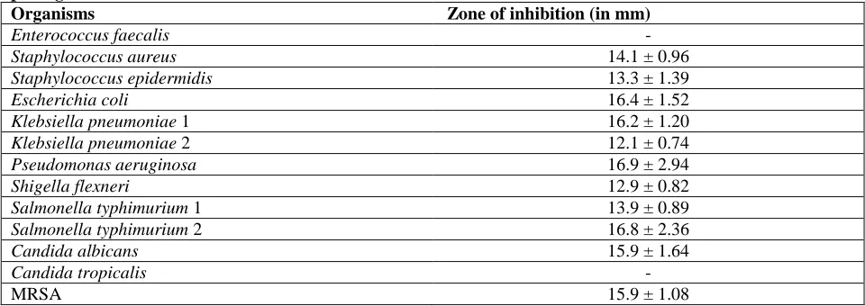
Preliminary screening of the aqueous extract for antimicrobial activity: Ten percent aqueous extract of Symplocos racemosa (Lodhra) bark was used for preliminary antimicrobial screening against some potential pathogens by Agar well Diffusion Assay (ADA). It exhibited a broad spectrum antimicrobial activity as it showed inhibitory action against 11 out of total 13 strains tested and its average zone of inhibition ranged from 12–17 mm (Table 1). Pseudomonas aeruginosa was the most sensitive bacteria (16.9 mm) closely followed by Salmonella typhimurium 2 > Escherichia coli > Klebsiella pneumoniae 1 > Candida albicans and MRSA. Staphylococcus aureus gave an inhibition zone of 14.1 ± 0.96mm followed by Salmonella typhimurium 1 > Staphylococcus epidermidis > Shigella flexneri >
Klebsiella pneumoniae 2. However, Candida
tropicalis and Enterococcus faecalis were found to be completely resistant and Klebsiella pneumoniae 2 (12.1mm) was least sensitive to the aqueous extract.
Therefore, Candida tropicalis and Klebsiella
pneumoniae 2 were not taken up for further studies. Based upon the above results, various
physicochemical parameters affecting the
Optimization of physiochemical parameters using one-factor-at-a-time (classical) approach
Effect of concentration: An increase in antimicrobial activity was observed with increase in
concentration. The aqueous extract at 5%
concentration was not effective against any organism except MRSA with an inhibition zone of 14.3 ± 0.58mm whereas Enterococcus faecalis did not respond at any concentration. Upto 15%, a sharp increase in activity was observed; though with further increase in concentration, the efficiency increased only marginally upto 30% thus suggesting 15% to be the optimum concentration which was selected for further studies. It was noted that at this concentration, C.albicans (20.3mm) was the most sensitive among all the pathogens tested. MRSA (19.6mm) among the Gram positive and E.coli (18.0mm), among the Gram negative bacteria were most sensitive to the extract (Fig. 1).
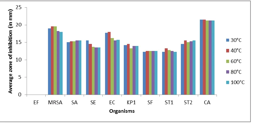
Effect of extraction temperature: In this study, a temperature range of 30 °C– 100 °C was used for extracting the plant material and at 40 °C, maximum antimicrobial activity against all the tested pathogens was observed. Thereafter an insignificant change in activity was observed upto 100 °C. Average zone of inhibition at 40 °C was 14.45mm which marginally decreased to 13.82mm at 100 °C. Therefore, 40 °C was chosen as optimum temperature for subsequent
experiments. At this optimum temperature,
C.albicans (21.5mm) was the most sensitive. Among the Gram positive and Gram negative; MRSA
(19.5mm) and E.coli (18.0mm) showed maximum
sensitivity, respectively (Fig. 2).
Effect of extraction time: The antimicrobial activity was best observed using 20min extracted plant material and thus 20min extraction period was selected for further experimentation. The activity slightly decreased with longer extraction time. C.albicans continued to be the most sensitive organism (23.25mm). Average zone of inhibition for Gram positive and Gram negative bacteria ranged from 14.75–17.5mm and 15.25–16.5mm respectively. MRSA (17.5mm) and E.coli (16.5mm) were the most sensitive. Enterococcus faecalis remained resistant at every tested time exposure. The average inhibition zone at 20 min extraction was 15.1mm, was reduced to 14.10 mm at 240 min exposure time which was significant (Fig. 3).
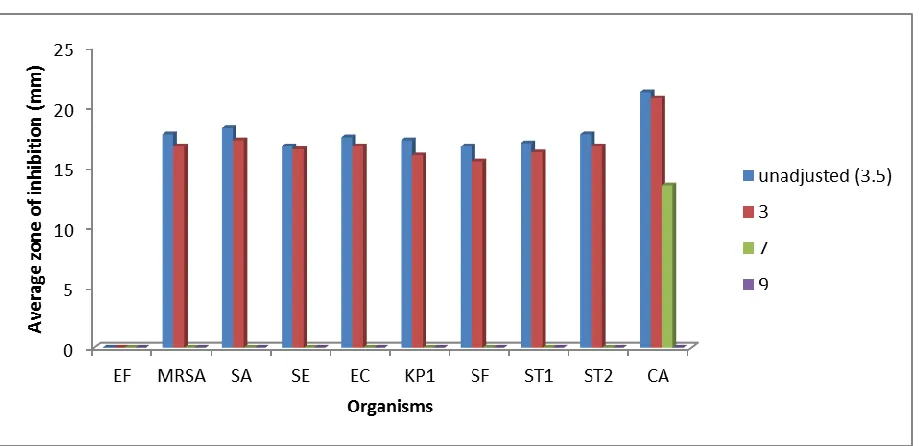
Effect of pH: The aqueous extract of the plant material; when tested at pH 3, 7, 9 and at natural pH (3.5); lost its potential at alkaline settings; pH 7 and 9, except C.albicans which was sensitive even at pH
7 but lost complete susceptibility at pH 9. A marginal decrease in activity was observed in all the organisms at pH 3 in comparison to natural or unadjusted pH (3.5). Thus, the natural pH was chosen as a supportive optimal in this study. At natural pH, the average zone of inhibition ranged from 16.75 mm -
21.5 mm. C.albicans (21.25mm) was the most
sensitive among all, whereas S.aureus (18.25mm) and S.typhimurium 2 (17.75mm) were most sensitive among the Gram positive and negative bacteria, respectively (Fig. 4).
Effect of different filtration strategies: The extract was subjected to different filtration techniques, i.e., Whatman filter paper No. 1, normal filter paper, muslin cloth and centrifugation. Though all the filtration strategies were found to be comparable, the extract filtered through Whatman filter paper no. 1 showed slightly better activity (14.54mm).The average zone of inhibition, as demonstrated by other methods, however, ranged from 14.09mm–14.54mm. Hence, Whatman filtration was taken as the method of choice for the rest of the study. Among the tested pathogens, Candida albicans was found to be most sensitive (18.87mm) in all the cases whereas Enterococcus faecalis remained resistant (Fig. 5).
Statistical optimization of the physiochemical parameters by Response surface methodology (RSM) using Box-Behnken design.
a) Fitting the model
The data obtained from the quadratic model equation was found to be significant. It was verified by F value and the analysis of variance (ANOVA) by fitting the data of all independent observations in response surface quadratic model (Table 2). The results for model F-value implies that the model is significant which indicate it to be suitable to represent
adequately the real relationship among the
parameters used. R2 value for all the responses ranged between 92–94.7% which showed suitable fitting of the model in the designed experiment. The final predictive equations for each response: S.aureus (G1), E.coli (G2), K. pneumoniae1 (G3), MRSA (G4) obtained are as follows:
G (1) = 15.4+ 1.50X1- 0.50X2+ 0X3- 1.07X1 2
b)Effect of different variables on S.aureus
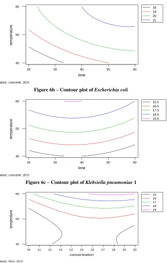
The linear effect of concentration (X1) was highly significant with P value ≤ 0.005. Similarly, the squared effect of concentration (X12) as well as interactive effect of concentration and time (X1X2) was found to be significant with P value ≤ 0.05. R2 value was 92.0%. The response surface graph showed that maximum predicted value of zone of inhibition for S.aureus (15.5mm) was obtained at 50°C with extraction time of 30 min at 15% concentration (Fig. 6a).
c)Effect of different variables on E.coli
The linear effect of concentration (X1) was highly significant with P value ≤ 0.005. Similarly, the linear effect of time (X2) and temperature (X3) was found to be significant with P value ≤ 0.05. R2 value was 92.8%.The maximum zone of inhibition for E.coli (21mm) was obtained at 55°C with extraction time of 50 min at 20% concentration (Fig. 6b).
d) Effect of different variables on K.pneumoniae 1 The linear effect of concentration (X1) and temperature (X3) was highly significant with P value ≤ 0.005. Whereas, the squared effect of concentration (X1
2
) and time (X2
2) was significant with P value ≤
0.05. R2 value was 93.0%.The response surface graph showed that the maximum predicted value of zone of inhibition for K.pneumoniae 1 is 19.5mm when 20% concentration was prepared at 60 °C for 35 min (Fig. 6c).
e) Effect of different variables on Methicillin- resistant S.aureus ( MRSA)
The linear effect of concentration (X1) as well as squared effect of temperature (X32) was obtained to be highly significant with P value ≤ 0.005. Even the interactive effect of time and temperature (X2X3) exhibited high level of significance with P value ≤ 0.005. Similarly, the linear effect of time (X2); squared effect of concentration (X12) and time (X22) plus the interactive effect of concentration and time (X1X2) was significant with P value ≤ 0.05. R2 value was 94.70%. Zone of inhibition (in mm) was highest (19mm) at 60 °C with extraction time of 20 min at 18% concentration (Fig. 6d).
Validation of Results: Thus from the overall assessment, temperature of 50–60 °C, concentration: 15–20 % and extraction time: 20–50 min can be
considered as the optimized conditions for
antimicrobial activity. The F value and R2 value showed that the model, correlated well with measured data and was statistically significant. To confirm the adequacy of the model, the verification experiments using optimum conditions were carried
out. This resulted in enhanced antimicrobial activity by 1.19 folds for S.aureus, 1.3 folds (E.coli and K.pneumoniae1) and 1.18 folds against MRSA.
Thermostability studies: The aqueous plant extract prepared at 40 °C was subjected to higher temperatures (60 – 100 °C) for 1 h so as to check its thermostability. One hour exposure of the extract to 60°C resulted in a maximum loss of 8.83% against
MRSA and minimum loss (1.75%) against
Salmonella typhimurium 1. Similarly, at higher exposure temperatures (70– 90 °C), the extract suffered a minimum loss of 2.86% and a maximum of 13.24% against various test organisms used. Exposure to boiling temperature resulted in a
maximium loss of 20.94% for C.albicans and
minimum of 13.4% for Salmonella typhimurium 1. This study thus shows the extract to be reasonably
thermostable. Upon comparison of the
thermostability profile derived from 1h exposure with a similar treatment given equivalent to extraction time (20 min), it was observed that exposure for a longer duration caused comparably more loss in activity. Exposure of the extract for 1h resulted in an average loss of 15.98% in its activity whereas exposure for 20min caused 11.85% reduction in the antimicrobial potential of the extract (Fig. 7).
Shelf life study: Shelf life studies revealed that the extract stored at refrigeration (4 °C) better retained its activity as compared to extract stored at room temperature (28 °C). Refrigerated extract suffered a loss in activity ranging from 6.28% – 35% over a period of 30 days whereas extract at ambient temperature suffered a minimum of 6.6% and a maximum of 40% loss in activity, when tested against various potential pathogens. It was also observed that the extract stored at room temperature started deteriorating at the 5th day of storage and suffered a higher loss than refrigerated extract; which was stable upto 20th day of storage.
Viable Cell Count (VCC) studies: On the basis of
VCC studies, P.aeruginosa reflected maximum
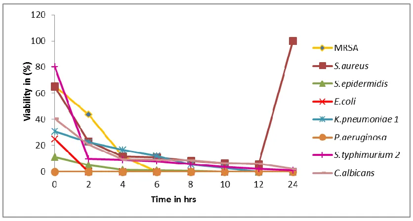
sensitivity to the plant extract and thus got killed instantaneously at 0 h. E.coli took 2 h for complete killing while MRSA was killed in 6 h. S.epidermidis also showed sensitivity with only 11.1% residual viability at the initial 0 h and finally got completely killed at 10h of incubation. K. pneumoniae 1, was however resistant and got killed at 12 h of incubation. In case of S.typhimurium 2 and C.albicans, 0.8% and 1.9% cells remained viable even after 24h of incubation, respectively. In this study, it was noted that S.aureus showed 65.4% viability at 0 h of incubation which reduced to 5.8% at 12h interval, however, its regrowth was observed after 24 h of incubation (Fig. 8).
Biosafety evaluation of Symplocos racemosa bark:
The aqueous extract was subjected to mutagenicity testing using Ames test. The number of colonies in the test plate was counted after 24 h of incubation at 37 °C and was compared to counts in positive control (Sodium azide) and distilled water as negative control. The number of revertant colonies in positive control (874) was much higher than the test extract (20) and control (18). Since the number of colonies in test were approximately similar to that of control and much less than positive control demonstrated non-mutagenicity of the aqueous extract. MTT assay was also carried out. Since, reduction of MTT can only occur in metabolically active cells, as MTT is converted into formazan crystals and hence the absorbance directly represents the viability of the cells. The extract was found to be non-cytotoxic, since the absorbance of test was comparable to that of untreated control and 98.13% viable cells were observed in the aqueous extract.
DISCUSSION
Antimicrobial resistance is one of the biggest challenges all over the world, generating interest among researchers to develop novel antimicrobial agents or to expand their spectrum. Natural products are worth promise to fight against various diseases caused by the resistant strains and many more. Among natural resources, lot of bioactive metabolites have been isolated from bacteria [24], fungi [25],
actinomycetes [26] which could be used as
antimicrobials, with some side effects or toxicity thus hampering the development of safe antimicrobials. Keeping this in mind our study has been directed towards the use of medicinal plants as safe antimicrobial agents. Symplocos racemosa (Lodhra) bark extract was screened for its antimicrobial activity which was found to be effective against 11 out of 13 reference strains of microorganisms tested,
indicating its importance as a broad spectrum antimicrobial agent, in accordance with the previous studies done by researchers on medicinal plants [27- 28]. An interesting observation recorded was about P.
aeruginosa, a fatal nosocomial pathogen being simultaneously resistant to many antibiotics, was found to be highly sensitive (16.9 mm) to the plant extract. Similarly drug- resistant organisms such as Methicillin- resistant Staphylococcus aureus (MRSA) also showed considerably good susceptibility (15.9 mm) to the plant extract thus highlighting its importance. Though, this plant had been used in the traditional medicine, however not much extensive scientific validation is available pertaining to its antimicrobial potential except a few reports [11, 17]. After screening against various microorganisms,
different physiochemical parameters such as
concentration, temperature, pH, and filtration were optimized for the best expression of antimicrobial activity.
The study thus gains importance as the resistant strains like MRSA showed the maximum sensitivity towards the plant extract. As the concentration increased, the antimicrobial activity also increased and reached a maximum at 15% and thereby remained more or less stable till 30% thus revealing 15% aqueous extract to be the best for antimicrobial activity. This concurs with the previous studies where 15- 25% aqueous extract of the plant material was found to be effective for antimicrobial activity [29-30]. In line with earlier studies, 40 °C extraction temperature was found to be the best for antimicrobial activity [9, 29] whereas optimal pH was its natural pH (3.5) in consonance with previous studies [29] which may be attributed to better metabolite production under such pH conditions. This shows that pH is also one of the determining factors for the biosynthesis of secondary metabolites. In consonance with earlier studies [9],different filtration strategies did not show much difference in the resulting antimicrobial activity; however the filtration through Whatman filter paper was considered to be the better choice and was pursued for rest of the studies.
from 92-94% which showed model to be fit. Apparently, to the best of our knowledge, no such studies have been reported for this plant extract. However, our results are in consonance with earlier studies on Streptomyces sp. where 2.73 folds increase in its antimicrobial activity was observed with an R2
value of 90% [33-34]. Minimum inhibitory
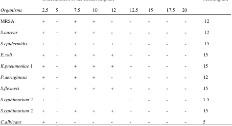
concentration of the aqueous plant extract ranged from 5- 15 mg/ml. Invariably, the results obtained from Agar Diffusion Assay (ADA) and MIC values are supportive of each other. MRSA, S.aureus and P.aeruginosa were relatively more sensitive and showed an MIC value of 12 mg/mL. Similarly, Sh. flexneri showing minimum inhibition zone of 12.9 mm was found to have higher MIC, i.e., 15 mg/ml which supported the data obtained from agar well diffusion assay. However, C.albicans gave the lowest MIC value of 5 mg/ml. Symplocos racemosa seems to have better antimicrobial potential supported by an MIC range of 5- 15 mg/ml in comparison to other medicinal plants such as Foeniculum vulgare, Antheum graveolens and Calotropis gigantean where MIC ranged from 20-60 mg/ml [35] and 1.5- 50 mg/ml [36]. Further, the plant extract was subjected to viable
cell count studies. Pseudomonas aeruginosa, a potent pathogen, could get completely killed at 0 h of incubation which gives further credence to this study. Similarly, E.coli took 2 h for complete killing and rest of the organisms tested took different time for complete killing depending upon their sensitivity to the plant extract. Our study on time kill assay found to be better than earlier viable cell count studies where P. aeruginosa and E.coli took 10 h and 12 h for complete killing respectively [35]. The aqueous plant extract was found to be relatively thermostable at 100 °C with minimum loss of 11% and maximum of 21% against E.coli and S.epidermidis respectively.
This property of being thermostable will be further helpful in purification purposes for the isolation of compound responsible for antimicrobial activity. Under refrigeration conditions, the extract was found to be more stable than at room temperature, suffering a maximum loss of 35% when observed at the 30th day of storage. Further, before developing the compound as a lead molecule, it needs to be tested for its cytotoxicity, and so was the aqueous extract of this plant. The plant extract used in this study was found to be non– mutagenic and non – cytotoxic. Thus, it can be said that medicinal plants needs to be explored further for development as lead drug molecules as proven by their safe cytotoxicity profile [23]. Many similar studies with different fungi were
also reported where the fungal extracts were non mutagenic and non cytotoxic [25, 37].
This study concluded the antimicrobial potential of the plant extract (Symplocos racemosa) against some medically important reference strains of bacteria including the most important MRSA. Moreover, the bioactivity was found to be thermostable and non-cytotoxic nature of the plant extract may help in its further exploitation as local medicine against some diseases. The purification of the compound/s responsible for antimicrobial activity will be of much significance for pharmaceutical applications.
ACKNOWLEDGMENTS
The work has been supported by financial assistance provided by Guru Nanak Dev University, Amritsar under UPE scheme. Henna Sood is thankful to University Grants Commission for UPE fellowship vide letter no. [8015/Estt./A-2 dated 16/04/2013].
.
Table 1: Antimicrobial activity of 10% Aqueous extract of Symplocos racemosa against some potential pathogens.
Organisms Zone of inhibition (in mm)
Enterococcus faecalis -
Staphylococcus aureus 14.1 ± 0.96
Staphylococcus epidermidis 13.3 ± 1.39
Escherichia coli 16.4 ± 1.52
Klebsiella pneumoniae 1 16.2 ± 1.20
Klebsiella pneumoniae 2 12.1 ± 0.74
Pseudomonas aeruginosa 16.9 ± 2.94
Shigella flexneri 12.9 ± 0.82
Salmonella typhimurium 1 13.9 ± 0.89
Salmonella typhimurium 2 16.8 ± 2.36
Candida albicans 15.9 ± 1.64
Candida tropicalis -
MRSA 15.9 ± 1.08
Table 2: Box-Behnken designs of different variables with their responses
Variables Zone of inhibition(mm)
RunOrder Concentration
(%)
Time (min)
Temperature (°C)
S.aureus E.coli K.pneumoniae 1 MRSA
1 10 20 50 13 15 14 14
2 15 40 50 16 18 16 16
3 15 40 50 15 20 16 17
4 15 60 60 13 19 16 14
5 15 40 50 15 19 16 16
6 15 40 50 16 18 16 16
7 20 40 60 15 21 20 19
8 15 60 40 14 18 14 17
9 15 20 60 15 18 16 19
10 15 20 40 15 16 14 16
11 20 60 50 16 21 17 16
12 20 40 40 15 18 15 17
13 10 40 60 13 17 17 15
14 20 20 50 15 19 17 15
15 15 40 50 15 19 16 16
16 10 40 40 12 15 14 15
17 10 60 50 11 16 16 12
Table 3: Minimum Inhibitory Concentration (MIC) of the aqueous extract
Organisms
Concentration of the extract (mg/ml) MIC(mg/ml)
2.5 5 7.5 10 12 12.5 15 17.5 20
MRSA + + + + ˗ ˗ ˗ ˗ ˗ 12
S.aureus + + + + ˗ ˗ ˗ ˗ ˗ 12
S.epidermidis + + + + + + ˗ ˗ ˗
15
E.coli + + + + + + ˗ ˗ ˗ 15
K.pneumoniae 1 + + + + + + ˗ ˗ ˗
15
P.aeruginosa + + + + ˗ ˗ ˗ ˗ ˗ 12
S.flexneri + + + + + + ˗ ˗ ˗ 15
S.typhimurium 2 + + - - - - ˗ ˗ ˗ 7.5
S.typhimurium 2 + + + + + + ˗ ˗ ˗ 15
Figure 1:- Effect of concentration on antimicrobial activity of Symplocos racemosa against some potential
pathogens.EF- Enterococcus faecalis; MRSA- Methicillin- Resistant Staphylococcus aureus ; SA- Staphylococcus
aureus ; SE- Staphylococcus epidermidis; EC- Escherichia coli; KP1- Klebsiella pneumoniae 1; SF- Shigella
flexneri; ST1- Salmonella typhimurium 1; ST2- Salmonella typhimurium 2; CA- Candida albicans.
Figure 2:- Effect of extraction temperature on antimicrobial activity of Symplocos racemosa against some
potential pathogens: EF- Enterococcus faecalis; MRSA- Methicillin- Resistant Staphylococcus aureus ; SA-
Staphylococcus aureus ; SE- Staphylococcus epidermidis; EC- Escherichia coli; KP1- Klebsiella pneumoniae 1;
Figure 3:- Effect of extraction time on antimicrobial activity of Symplocos racemosa against some
potential pathogens: EF- Enterococcus faecalis; MRSA- Methicillin- Resistant Staphylococcus aureus ; SA-
Staphylococcus aureus ; SE- Staphylococcus epidermidis; EC- Escherichia coli; KP1- Klebsiella pneumoniae
1; SF- Shigella flexneri; ST1- Salmonella typhimurium 1; ST2- Salmonella typhimurium 2; CA- Candida
albicans.
Figure 4:- Effect of pH on antimicrobial activity of Symplocos racemosa against some potential pathogens.
EF- Enterococcus faecalis; MRSA- Methicillin- Resistant Staphylococcus aureus ; SA- Staphylococcus aureus
; SE- Staphylococcus epidermidis; EC- Escherichia coli; KP1- Klebsiella pneumoniae 1; SF- Shigella flexneri;
Figure 5:- Effect of different filtration strategies ( 1- Whatman filter paper No. 1; 2- centrifuge; 3-
muslin cloth; 4- normal filter paper) on antimicrobial activity of aqueous extract of Symplocos racemosa
against some potential pathogens. EF- Enterococcus faecalis; MRSA- Methicillin- Resistant Staphylococcus
aureus ; SA- Staphylococcus aureus ; SE- Staphylococcus epidermidis; EC- Escherichia coli; KP1- Klebsiella
pneumoniae 1; SF- Shigella flexneri; ST1- Salmonella typhimurium 1; ST2- Salmonella typhimurium 2; CA-
Candida albicans.
14.0 14.5 15.0 15.5
60 50
40 30
20 60
50
40
time
tem
perat
ure
Contour Plot of S.aureus
Hold values: concentr: 15.0
18 19 20 21
60 50
40 30
20 60
50
40
time
tem
perat
ure
Contour Plot of E.coli
Hold values: concentr: 20.0
Figure 6b – Contour plot of Escherichia coli
15.5 16.5 17.5 18.5 19.5
60 50
40 30
20 60
50
40
time
tem
perat
ure
Contour Plot of K.pneumo
Hold values: concentr: 20.0
Figure 6c – Contour plot of Klebsiella pneumoniae 1
15 16 17 18 19
20 19 18 17 16 15 14 13 12 11 10 60
50
40
concentration
tem
perat
ure
Contour Plot of MRSA
Hold values: time: 20.0
Figure 7: Comparison of the thermostability profile after 20min and 1h exposure
Figure 8: Viable cell count studies of aqueous extract of Symplocos racemosa bark.
REFERENCES:
1. Levy SB. J Antimicrob Chemother, 2002; 49: 25–30.
2. Huttner A, Harbarth S, Carlet J, Cosgrove S, Goossens H, Holmes A, Jarlier V, Voss A, Pittet D. Antimicrob Resist Infect Control, 2013;2: 31.
3. Menghani E, Ojha CK, Negi RS, Agarwal Y, Pareek, A. Global J Sci Front Res, 2011; 11: 1. 4. Sintayehu B, Asres K, Mazumder A. Int J Pharm Sci Res, 2012; 3: 1523–27.
5. Arora DS, Kaur J. Int J Antimicrob Ag, 1999; 12: 57– 62. 6. Arora DS, Kaur GJ, Kaur H. Int J Food prop, 2009; 12: 286–94.
7. Kan A, Ozcelik B, Kartal M, Ozdemir, ZA, Ozgen S. Trop J Pharm Res, 2010; 9: 475–81.
8. Narsimha Rao R, Bhavya B, Pavani K, Swapna A, Prasoona CH. Int J Pharm Biol Sci, 2011; 1: 198– 30. 9. Onsare JG, Kaur H, Arora DS. Acad J Med Plants, 2013; 1: 80–91.
10. Bhusnar HU, Nagore DH, Nipanikar SU. Pharmacol, 2014; 5: 76–83. 11. Devmurari VP. Int J PharmTech Res , 2010;2: 1359–63.
14. Bhuvan RP, Jignesh PD, Bhavik PA, Ashok GL. Rom J Biol - Plant Biol, 2009; 54: 135– 40. 15. Jadhav M, Menon S, Shailajan S. J Coast Life Med, 2013; 1: 309– 14.
16. Kumar SS, Sharma SS, Saini V, Mohapatra S. Int Res J Pharmacy, 2013; 4: 136– 39. 17. Deattu N, Suseela L, Narayanan N. J Drug Deliv Ther, 2012; 2: 53-55.
18. Kumar GS, Jayaveera KN, Kumar CK, Sanjay UP, Swamy BM, Kumar DV. Trop J Pharm Res, 2007;6:
717- 23.
19. Arora DS, Onsare JG. World J Pharm Res, 2014; 3: 2772–88. 20. Chandra P, Arora DS. Free Rad Antiox, 2011; 1: 48-55. 21. Wiegand I, Hilpert K, Hancock REW. Nature, 2008; 3: 163–75. 22. Mortelmans K, Zeiger E. Mutat Res, 2000; 455: 29–60. 23. Arora DS, Onsare JG. Ind Crop prod, 2014; 52: 125–35.
24. Asha Devi NK, Rajendran R, Sundaram SK. Indian J Nat Prod Resour, 2011; 2: 59– 64. 25. Kaur H, Arora DS, Sharma V. App Biochem Biotech , 2014; 173: 1963–76.
26. Narayana KJP, Prabhakar P, Vijayalakshmi M, Venkateshwari Y, Krishna PSJ. Pol J Microbiol , 2008; 57: 35– 39.
27. Chandra M. Int J Biotech Bioeng Res, 2014; 7: 653–58.
28. Sharma A, Chandraker S, Patel VK, Ramteke P. Indian J Pharm Sci, 2009;71: 136–39. 29. Arora DS, Kaur GJ. J Nat Med, 2007; 61: 313–17.
30. Rawani A, Pal S, Chandra G. Asian Pac J Trop Biomed, 2011; 1: 71– 5.
31. Katapodis P, Christakopoulou V, Kekos D, Christakopoulos P. Biochem Eng J, 2007;35: 136–41. 32. Wu QL, Chen T, Gan Y, Chen X, Zhao, XM. Appl Microbiol Biotechnol, 2007; 76: 783–94. 33. Wang Y, Fang X, An F, Wang G, Zhang X. Microb Cell Fact, 2011; 10: 98.
34. Zhengyan G, Ling S, Zhiqin J, Wenjun W. Int J Mol Sci, 2012;13: 5230–41. 35. Kaur GJ, Arora DS. BMC Compl Alternative Med, 2009; 9: 30.