CASE REPORT OPEN ACCESS
Diffuse large B cell lymphoma with central pontine
myelinolysis
Sumaira Shafi, Cristian Papazoglu, Prasanta Basak,
Stephen Jesmajian
ABSTRACT
Introduction: Diffuse large B cell lymphoma is the most common type of non-Hodgkin, lymphoma among adults, with an annual incidence of 7–8 cases per 100, 000 people per year. Case Report: We report a case of diffuse large B cell lymphoma with isolated splenomegaly, complicated by central pontine myelinolysis. Solitary splenic non-Hodgkin’s lymphoma is rare; with an incidence less than 1%. Treatment of diffuse large B cell lymphoma led to the resolution of the pontine lesion. The association of central pontine myelinolysis with diffuse large B cell lymphoma is exceedingly rare, with only one case report published till date. Conclusion: Diffuse large B cell lymphoma presenting as isolated splenomegaly is rare and can be a diagnostic challenge. The association of central pontine myelinolysis with diffuse large B cell lymphoma is rare and the possibility that central pontine myelinolysis could be an early manifestation of lymphoma or a paraneoplastic syndrome needs to be further explored.
Sumaira Shafi1, Cristian Papazoglu1, Prasanta Basak2, Stephen Jesmajian3
Affiliations: 1Resident, Department of Internal Medicine, Albert Einstein College of Medicine and Montefiore New Rochelle Hospital, New Rochelle, NY, USA; 2Attending Physician, Department of Internal Medicine, Albert Einstein College of Medicine and Montefiore New Rochelle Hospital, New Rochelle, NY, USA; 3Program Director, Department of Internal Medicine, Albert Einstein College of Medicine and Montefiore New Rochelle Hospital, New Rochelle, NY, USA. Corresponding Author: Sumaira Shafi, 16 Guion Place, New Rochelle, NY, USA, 10802; Ph: 1-914-6891659; Fax: 1-914-365- 5489; Email: sshafi@montefiore.org
Received: 29 June 2015 Accepted: 30 July 2015 Published: 08 December 2015
Keywords: Diffuse large B cell lymphoma, Central pontine myelinolysis
How to cite this article
Shafi S, Papazoglu C, Basak P, Jesmajian S. Diffuse large B cell lymphoma with central pontine myelinolysis. J Case Rep Images Oncology 2015;1:10– 14.
Article ID: 100003Z10SS2015
*********
doi:10.5348/Z10-2015-3-CR-3
INTRODUCTION
Many hematological diseases are associated with splenomegaly. Primary splenic lymphoma is rare, accounting for approximately 1% of all lymphoma cases [1]. Splenomegaly is the predominant, and sometimes the only manifestation in these cases [2]. Diffuse large B cell lymphoma (DLBCL) is the most common subtype of non-Hodgkin’s lymphoma (NHL), usually presenting with lymphadenopathy. We report a case of DLBCL presenting initially with isolated splenomegaly and associated with central pontine myelinolysis (CPM).
The CPM, now known as osmotic demyelination syndrome, is an acquired demyelinating lesion of the pons that typically occurs after rapid correction of hyponatremia [3]. Central pontine myelinolysis is also found in patients with chronic alcoholism, malnutrition, hyponatremia, liver disease and infections [4]. The association of DLBCL with asymptomatic CPM is rarely seen.
CASE REPORT
A 58-year-old male presented with a 10-month history of fever, weight loss and upper abdominal discomfort. He had intermittent fever, mostly at night, associated with night sweats. He had an unintentional weight loss of 20 lbs in the last six months. He had recently completed treatment for latent tuberculosis with six-month of isoniazid. On admission he was afebrile, and physical examination was significant for pallor and a palpable spleen. No lymph nodes were palpable. Laboratory data included hemoglobin 8.2 g/dL, white blood cell count 8.6x10³/µL with a normal differential and platelet count 273x103/µL. Peripheral blood smear showed microcytosis and anisocytosis with no parasites identified on thick and thin smears. Sodium was 127 mEq/L. Other laboratory data included total bilirubin 0.7 mg/ dL, albumin 2.9 g/dL, AST 72 IU/L, ALT 24 IU/ L, LDH 1, 047 U/L, β2 microglobulin 4.2 mg/L, ESR 50 mm/hr and CRP 95.4 mg/L. Computed tomography scan of the chest, abdomen and pelvis showed homogenous splenomegaly with enlargement of the liver, without any intra-abdominal, pelvic, or retroperitoneal lymphadenopathy (Figure 1).
Serology done at another facility before current admission was negative for Lyme disease, tularemia, HIV and hepatitis. Echocardiogram was normal with no evidence of vegetations. Bone marrow biopsy showed a normocellular marrow with trilineage hematopoiesis, with no abnormal cells.
During the second day of hospitalization, the patient complained of neck pain. MRI of cervical spine showed abnormal signal in the pons. Brain MRI, T2-weighted and Fluid Attenuated Inversion Recovery (FLAIR) both revealed a hyperintense lesion in the pons. Diffusion weighted image showed hyper-intensity within the central pons with restricted diffusion on ADC (Figure 2A– D). Findings were consistent with CPM. The sodium level was 130 mmol/L essentially excluding rapid correction of hyponatremia as a cause for CPM.
Endoscopic gastroduodenoscopy and colonoscopy were negative. Since the diagnosis remained unclear, an elective splenectomy was planned and the patient was sent home.
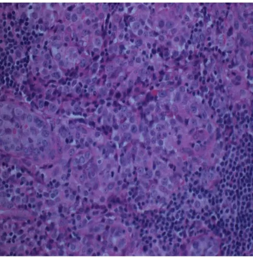
He was lost to follow-up and two months later when he showed up, a 2-cm firm non-tender lymph node was detected in the right axilla. An excisional lymph node biopsy revealed large atypical lymphoma cells located within sub-capsular and para-cortical regions (Figure 3). Immunohistochemistry indicated positive staining for CD20, Bcl-2 and Bcl-6. CD3, CD30, S-100, CD31, CD34 were negative (Figure 4). The diagnosis of DLBCL was established.
He was started on chemotherapy with lenalidomide (revlimid), rituximab, cyclophosphamide, doxorubicin, vincristine, and prednisone ( R2-CHOP). Three months later his symptoms had improved. A repeat MRI scan of the brain after four cycles of chemotherapy showed complete resolution of the pontine lesion (Figure 5).
Figure 1: Computed tomography scan of abdomen showing hepatosplenomegaly.
Figure 2: Brain MRI: Axial diffusion weighted image (A) showing hyper-intensity within the central pons with low signal on ADC (B) in keeping with restricted diffusion. Axial T2 weighted (C) and FLAIR (D) sequences demonstrate increased signal with the central pons.
DISCUSSION
Diffuse large B cell lymphoma is the most frequent (31%) of all NHLs with an aggressive clinical course [5].
As a secondary lymphoid organ, the spleen is usually involved as part of the systemic illness. However, the spleen is rarely the exclusive site of the disease burden. DLBCL of the spleen accounts for about one-third of all primary splenic lymphomas [6]. The common age of occurrence is in the sixth and seventh decade. Both primary and secondary DLBCL can present with a single large tumor mass, occupying more than 50% of the spleen. These patients usually present with low grade fever, night sweats and left upper quadrant pain related to splenomegaly.
The most common laboratory finding seen in these patients is anemia, increased ESR, and high LDH levels [5]. Splenectomy is usually needed for the diagnosis of isolated splenic lymphoma [5]. The detection of a late appearing palpable lymph node in our patient, helped establish the diagnosis of DLBCL without undergoing a splenectomy.
The suggested chemotherapy for splenic DLBCL is R-CHOP [7]. Our patient was identified with an activated B cell variant of DLBCL, and thus was treated with R2-CHOP. The use of lenalidomide (Revlimid) for DLBCL is emerging as a new therapeutic strategy.
The CPM was first described by Adams et al. in 1959 as a disease affecting alcoholics and malnourished [8]. In 1976, a link between CPM and the rapid correction of sodium in hyponatremic patients was suggested [9], and by 1982 the link was firmly established [7]. CPM has also been reported in adults with a variety of serious illnesses and after certain surgical procedures.
The CPM is a symmetric, sharply demarcated lesion in the basis pontis with a characteristic radiographic appearance [3]. Similar radiographic appearance was observed in our patient. Usually, the severity of the lesion does not correlate with the severity of the symptoms but the pontine lesion decreases in size and intensity as the symptoms subside [3]. Microscopically, CPM is characterized by loss of oligodendrocytes, dissolution of the myelin sheaths and sparing of nerve axons within the central aspect of the basis pontis [3]. Clinical and laboratory studies indicated that myelinolysis is the result of the cellular stress caused by fluctuating osmotic forces and ion shifts that trigger changes in neuronal volume and neuronal cell membrane function. Other studies have suggested that myelinolysis may be caused by compression of myelin by edematous elements [4, 10]. Hyperintense lesions located in central pons, similar to those seen in pontine osmotic demyelination syndrome have been reported in patients with large B-cell lymphoma [11–13]. In two of the previous published articles, large B cell lymphoma with pontine involvement was described [11, 12]. Treatment in those cases included intrathecal or high dose methotrexate in addition to systemic chemotherapy. Resolution of pontine lesions was observed following treatment. There is only one previously reported case, where DLBCL was associated with CPM [13]. Our case is unique with presentation of
Figure 4: Lymph node biopsy, immunohistochemistry showing CD 20 positive B cells (x200).
DLBCL as isolated splenomegaly and then having a rarely reported association of DLBCL with CPM.
Other risk factors for developing CPM in our patient included a history of chronic alcohol abuse, and mild hyponatremia. However, published evidence strongly suggests that there could be a correlation between CPM and some histological sub-types of lymphoma [13, 14]. In contrast to symptomatic CPM cases, the asymptomatic pontine lesions are only found by chance and therefore their incidence might be underestimated.
CONCLUSION
Diffuse large B cell lymphoma (DLBCL) presenting as isolated splenomegaly is rare. In the absence of lymphadenopathy, isolated splenomegaly can be a diagnostic challenge. With positive B symptoms and splenomegaly, primary splenic lymphoma should be considered as a differential, and splenic biopsy may be indicated. The association of central pontine myelinolysis (CPM) with DLBCL is an even rarer finding. Although this association is intriguing, the possibility that CPM could be an early manifestation of certain types of lymphomas or a paraneoplastic syndrome remains to be explored.
*********
Author Contributions
Sumaira Shafi – Substantial contributions to conception and design, Acquisition of the data, Drafting the article, Revising it critically for important intellectual content, Final approval of the version to be published
Cristian Papazoglu – Substantial contributions to conception and design, Drafting the article, Final approval of the version to be published.
Prasanta Basak – Substantial contributions to conception and design, Revising it critically for important intellectual content, Final approval of the version to be published Stephen Jesmajian – Substantial contributions to conception and design, Revising it critically for important intellectual content, Final approval of the version to be published
Guarantor
The corresponding author is the guarantor of submission.
Conflict of Interest
Authors declare no conflict of interest.
Copyright
© 2015 Sumaira Shafi et al. This article is distributed under the terms of Creative Commons Attribution License which permits unrestricted use, distribution and reproduction in any medium provided the original author(s) and original publisher are properly credited. Please see the copyright policy on the journal website for more information.
REFERENCES
1. Brox A, Bishinsky JI, Berry G. Primary non-Hodgkin lymphoma of the spleen. Am J Hematol 1991 Oct;38(2):95–100.
2. Grosskreutz C, Troy K, Cuttner J. Primary splenic lymphoma: report of 10 cases using the REAL classification. Cancer Invest 2002;20(5-6):749–53. 3. Hurley RA, Filley CM, Taber KH. Central pontine
myelinolysis: a metabolic disorder of myelin. J Neuropsychiatry Clin Neurosci 2011 Fall;23(4):369– 74.
4. Lupato A, Fazio P, Fainardi E, Cesnik E, Casetta I, Granieri E. A case of asymptomatic pontine myelinolysis. Neurol Sci 2010 Jun;31(3):361–4. 5. Gurumurthy R, Mohapatra RK, Easow JM, Mohan
S. Cutaneous B cell lymphomas: Report of two interesting cases. Indian J Dermatol 2015 Mar-Apr;60(2):176–8.
6. Kattepur AK, Rohith S, Shivaswamy BS, Babu R, Santhosh CS. Primary splenic lymphoma: a case report. Indian J Surg Oncol 2013 Sep;4(3):287–90. 7. Iannitto E, Tripodo C. How I diagnose and treat
splenic lymphomas. Blood 2011 Mar 3;117(9):2585– 95.
8. Adams RD, Victor M, Mancall EL. Central pontine myelinolysis: a hitherto undescribed disease occurring in alcoholic and malnourished patients. AMA Arch Neurol Psychiatry 1959 Feb;81(2):154–72.
9. Tomlinson BE, Pierides AM, Bradley WG. Central pontine myelinolysis. Two cases with associated electrolyte disturbance. Q J Med 1976 Jul;45(179):373–86.
10. Cramer SC, Stegbauer KC, Schneider A, Mukai J, Maravilla KR. Decreased diffusion in central pontine myelinolysis. AJNR Am J Neuroradiol 2001 Sep;22(8):1476–9.
11. Yamamoto A, Kikuchi Y, Homma K, O’uchi T, Furui S. Characteristics of intravascular large B-cell lymphoma on cerebral MR imaging. AJNR Am J Neuroradiol 2012 Feb;33(2):292–6.
12. Sekiguchi Y, Shimada A, Imai H, et al. Intravascular large B-cell lymphoma with pontine involvement successfully treated with R-CHOP therapy and intrathecal administration: a case report and review of literature. Int J Clin Exp Pathol 2014 May 15;7(6):3363–9.
13. Zhou AY, Barnes C, Razzaq R. Central pontine myelinolysis in a patient with non-Hodgkin lymphoma. Br J Haematol 2013 Apr;161(2):156. 14. Chintagumpala MM, Mahoney DH Jr, McClain K, et
al. Hodgkin’s disease associated with central pontine myelinolysis. Med Pediatr Oncol 1993;21(4):311–4.
SUGGESTED READING
• A clinical evaluation of the International Lymphoma Study Group classification of non-Hodgkin’s lymphoma. The Non-Hodgkin’s Lymphoma Classification Project”. Blood. 1997;89:3909–18.
sub-type: a report from the Haematological Malignancy Research Network.Br J Cancer. 2011;105:1684–92.
• Bairey O, Shvidel L, Perry C, Dann E J, Ruchlemer R, Tadmor T and Goldschmidt N. Characteristics of primary splenic diffuse large B-cell lymphoma and role of splenectomy in improving survival. Cancer. 2015; doi:10.1002/cncr.29487.
• Epperla N, Landeck J, Sabbagh S. Osmotic
demyelination syndrome. WMJ.
Access full text article on