R E S E A R C H A R T I C L E
Open Access
Methotrexate resistance in relation to treatment
outcome in childhood acute lymphoblastic
leukemia
Anna Wojtuszkiewicz
1, Godefridus J. Peters
3, Nicole L. van Woerden
1, Boas Dubbelman
1, Gabriele Escherich
6,
Kjeld Schmiegelow
7,8, Edwin Sonneveld
9, Rob Pieters
9,10, Peter M. van de Ven
4, Gerrit Jansen
5, Yehuda G. Assaraf
11,
Gertjan J. L. Kaspers
1and Jacqueline Cloos
1,2*Abstract
Background:Methotrexate (MTX) eradicates leukemic cells by disrupting de novo nucleotide biosynthesis and DNA replication, resulting in cell death. Since its introduction in 1947, MTX-containing chemotherapeutic regimens have proven instrumental in achieving curative effects in acute lymphoblastic leukemia (ALL). However, drug resistance phenomena pose major obstacles to efficacious ALL chemotherapy. Moreover, clinically relevant molecular mechanisms underlying chemoresistance remain largely obscure. Several alterations in MTX metabolism, leading to impaired accumulation of this cytotoxic agent in tumor cells, have been classified as determinants of MTX resistance. However, the relation between MTX resistance and long-term clinical outcome of ALL has not been shown previously.
Methods:We have collected clinical data for 235 childhood ALL patients, for whom samples taken at the time of diagnosis were also broadly characterized with respect to MTX resistance. This included measurement of concentrations of MTX polyglutamates in leukemic cells, mRNA expression of enzymes involved in MTX metabolism (FPGS, FPGH, RFC, DHFR, and TS), MTX sensitivity as determined by the TS inhibition assay, and FPGS activity.
Results:Herein we demonstrate that higher accumulation of long-chain polyglutamates of MTX is strongly associated with better overall (10-year OS: 90.6 vs 64.1 %,P= 0.008) and event-free survival (10-year EFS: 81.2 vs 57.6 %,P= 0.029) of ALL patients. In addition, we assessed both the association of several MTX resistance-related parameters determined in vitro with treatment outcome as well as clinical characteristics of pediatric ALL patients treated with MTX-containing combination chemotherapy. High MTX sensitivity was associated with DNA hyperdiploid ALL (P< 0.001), which was linked with increased MTX accumulation (P= 0.03) and elevated reduced folate carrier (RFC) expression (P= 0.049) in this subset of ALL patients.TEL-AML1fusion was associated with increased MTX resistance (P= 0.023). Moreover, a low accumulation of MTX polyglutamates was observed inMLL-rearranged andTEL-AML1rearranged ALL (P< 0.05).
Conclusions:These findings emphasize the central role of MTX in ALL treatment thereby expanding our understanding of the molecular basis of clinical differences in treatment response between ALL individuals. In particular, the identification of patients that are potentially resistant to MTX at diagnosis may allow for tailoring novel treatment strategies to
individual leukemia patients.
Keywords:Acute lymphoblastic leukemia, Antifolate, Methotrexate, Drug resistance, Hyperdiploid, FPGS, DHFR, RFC, TS
* Correspondence:j.cloos@vumc.nl
1Department of Pediatric Oncology/Hematology, VUmc Cancer Center
Amsterdam, VU University Medical Center, Room CCA 4.28, De Boelelaan 1117, 1081HV Amsterdam, The Netherlands
2
Department of Hematology, VU University Medical Center, Amsterdam, The Netherlands
Full list of author information is available at the end of the article
Background
The treatment outcome of pediatric acute lymphoblastic leukemia (ALL) has greatly improved over the past seven decades with the current regimens resulting in a 5-year event-free survival (EFS) of around 80 % [1–3]. This impressive improvement has been largely attributed to novel prognostic factors, including cytogenetic abnor-malities such asTEL-AML1andE2A-PBX1gene fusions associated with good prognosis as well as MLL gene re-arrangements that confer unfavorable prognosis [4, 5]. Yet, the high cure rates achieved with current treatment protocols are still paralleled by approximately 20 % risk of relapse, which is consequently associated with poor prognosis [2, 3]. The emergence of relapse is largely at-tributable to drug resistance phenomena of leukemic cells. Thus, further advances in understanding the mo-lecular basis underlying these drug resistance phenomena as well as accurate prediction of chemotherapy resistance prior to drug treatment may pave the way to overcoming chemoresistance.
Historically, one of the backbones of contemporary ALL treatment is the folate antimetabolite—methotrexate (MTX) [6]. Folates are essential enzyme cofactors involved in one-carbon metabolism, which includes several cellular biosynthetic pathways including thymidylate and de novo purine biosynthesis, amino acid metabolism, and mito-chondrial protein synthesis [7]. Antifolates potently inhibit several folate-dependent enzymes engaged in nucleotide biosynthesis, which leads to cessation of DNA replication eventually resulting in cell death [6]. High-dose (HD)-MTX is used as part of the central nervous system (CNS)-directed therapy (intrathecal MTX), and MTX is an essen-tial component of the maintenance treatment [1]. MTX is predominantly taken up into cells via the reduced folate carrier (RFC/SLC19A1), and in the case of HD-MTX treatment, also by passive diffusion across cellular mem-branes, at least to some extent [8, 9]. Upon entry into the cytoplasm, MTX undergoes polyglutamylation—a unique metabolic conversion catalyzed by folylpolyglutamate syn-thetase (FPGS) [9]. This polyglutamylation, which is based on the sequential addition of multiple glutamate residues to theγ-carboxyl group of both folates and MTX, ensures efficient intracellular retention as well as sustains and en-hances target enzyme inhibition [10–12]. The main targets of polyglutamylated MTX are dihydrofolate reductase (DHFR—also inhibited by MTX monoglutamates), thy-midylate synthase (TS), and several enzymes involved in purine synthesis [9]. On the other hand, a lysosomal glycoprotein—folylpolyglutamate hydrolase (FPGH)—can counteract polyglutamylation, thereby increasing the ef-flux of MTX by the efef-flux transporters of the ATP-binding cassette superfamily including for example ABCC1 and ABCG2 [9, 13, 14]. Overall, the intracellular accumulation of MTX polyglutamates in leukemic cells
proved to be an important determinant of the antileukemic activity of MTX in childhood ALL patients in vivo [15–17]. At the same time, high concentration of long-chain but not total MTX polyglutamates was associated with inhibition of de novo purine synthesis [15]. Consequently, a spectrum of alterations in MTX metabolism resulting in its decreased cellular accumulation has been identified to induce MTX resistance and compromise its curative effect. MTX resistance has been attributed to inactivating mutations or down-regulation affecting the RFC gene as well as increased levels of DHFR and TS enzymes together with mutations that decrease their affinity for antifo-lates [8, 9, 18–21]. In addition, different polymorphisms in RFC,TS, andDHFR were previously reported, several of which were related to increased risk of relapse [22–24]. The cytotoxic effect elicited by MTX is also largely influ-enced by FPGS activity. Consequently, loss of FPGS func-tion is a well established mechanism of resistance to MTX and other polyglutamylation-dependent antifolates in leukemic cells [17, 25–28]. Differential MTX sensitivity was shown to be associated with several cytogenetic ab-normalities. Precursor B cell ALL displaying TEL-AML1 or E2A-PBX1 gene fusions were characterized by de-creased levels of MTX polyglutamates as compared to precursor B cell ALL with normal karyotype [16], while patients with hyperdiploid karyotype were highly sensitive to MTX [29, 30]. Next, to its own cytotoxic effect, MTX is also important in the metabolism of other chemothera-peutics, such as mercaptopurine, used routinely in ALL treatment. MTX was shown to promote the conversion of mercaptopurine to one of its active metabolites— thiogua-nine nucleotides [31, 32]—of which high concentration in leukemic cells was associated with increased EFS in leukemia patients [33]. Therefore, it is imperative to characterize the extent of resistance to this important chemotherapeutic as well as the mechanisms underlying this phenomenon.
The aim of the current study was therefore to deter-mine which parameters of MTX resistance are related to the long-term clinical outcome in childhood ALL pa-tients treated with combination chemotherapy. Towards this goal, we have determined a range of in vitro param-eters associated with MTX resistance in a large cohort of pediatric ALL patients and subsequently assessed their relation with treatment outcome as well as with clinical characteristics.
Results
The association of ex vivo MTX resistance and the clinical outcome
the cellular concentration of total (1–6 glutamate residues) as well as long-chain (4–6 glutamate residues) MTX polyglutamates and the extent of TS inhibition (thymi-dylate synthase inhibition assay (TSIA)). For the TSIA parameter, we used both a continuous (TSIA cont.) and a short-term (TSIA short) exposure to MTX, followed by a drug-free period. TSIA cont. represents the overall ability of cells to accumulate MTX, while TSIA short enables the efflux of MTX during the drug-free period and hence rather points to the polyglutamylation cap-acity of the cells. This approach reliably reflects MTX sensitivity of leukemic blasts [34]. In addition, FPGS
activity and mRNA expression of enzymes and proteins involved in MTX metabolism and transport were deter-mined includingDHFR,TS,FPGS,FPGH, andRFC. The abovementioned parameters were determined in a co-hort of 235 samples derived from pediatric ALL patients at diagnosis. The patients under study were enrolled in several treatment protocols. The proportion of patients assigned to specific protocols along with their clinical characteristics is listed in Additional file 1: Table S1. Due to logistic challenges and leukemic blast number limitations, none of the MTX-related parameters could be measured in all the patients in our cohort. Therefore,
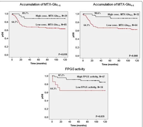
Fig. 1Kaplan–Meier analysis of the accumulation of MTX polyglutamates and FPGS activity in relation to overall survival (Plogrank) of total ALL patients. Each survival curve is labeled with the OS rates at 10 years and the number of patients included in each subgroup. The cut-off values used were: 1935 pmol/109cells for total accumulation of MTX polyglutamates, 1000 pmol/109cells for long-chain MTX polyglutamates and 0.35
the number of cases included in the statistical analysis varied considerably for the different parameters and the number of patients used for each measurement as listed in the various tables.
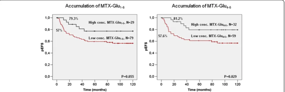
The prognostic value of MTX resistance associated pa-rameters was assessed with respect to long-term overall survival (OS) as well as EFS of childhood ALL patients. We identified three MTX-related variables that were sig-nificantly associated with OS in Kaplan–Meier analysis (Fig. 1). Higher levels of both the total concentration of cellular MTX as well as long-chain MTX polyglutamates were predictive of a better outcome. A 10-year OS reached 89.7 % in patients with high concentrations of total MTX polyglutamates and 90.6 % in case of long-chain MTX polyglutamates, compared to 64 % in patients with low levels of these MTX metabolites (P = 0.018 for total concentration and P = 0.008 for long-chain MTX polyglutamates, respectively). Consistently, higher FPGS activity was also predictive of a better OS (P= 0.039). For EFS, only the accumulation of long-chain MTX polygluta-mates was a significant predictor of a better outcome (Fig. 2; P = 0.029). The total concentration of MTX showed a similar trend, although it did not reach signifi-cance (Fig. 2,P= 0.055). The univariate Cox regression model further confirmed the concentration of both long-chain (EFS: HR = 2.61, P = 0.035, OS: HR = 4.44,
P = 0.015) and total MTX polyglutamates (EFS: HR = 2.29,P = 0.062, OS: HR = 3.8, P = 0.028) as the stron-gest prognostic variables (Table 1 and Additional file 1: Table S2). Due to the large heterogeneity of treatment protocols applied in therapy of the patients under study, we performed our analyses both with and with-out the correction for the treatment protocol. In this analysis, the concentration of the long-chain MTX polyglutamates was still significantly associated with OS (HR = 3.82,P = 0.03) but not with EFS (HR = 2.23,
P = 0.084), whereas the levels of total MTX polygluta-mates were not significant for both the EFS and OS (EFS: HR = 1.73, P = 0.232, OS: HR = 2.57, P = 0.125). Intri-guingly, lowRFCmRNA expression was a significant pre-dictor of a longer EFS when corrected for the treatment protocol (HR = 0.15,P= 0.009). Trends observed for the concentration of both the total as well as long-chain MTX polyglutamates in the whole patient cohort were also ap-parent when precursor B cell ALL patients were analyzed separately (data not shown), while the numbers of T cell ALL patients were too low for further analysis.
Next, the independent prognostic values of the con-centration of total as well as long-chain MTX polygluta-mates were examined in the multivariate Cox regression model, in combination with clinical factors significantly associated with the EFS (white blood cell count—WBC, age, and CNS involvement—see Additional file 1: Tables S3 and S4) and OS (WBC, age, CNS involvement, lineage, and DNA index). Neither of the parameters tested had independent prognostic value in the multi-variate analysis of EFS (Additional file 1: Table S5), both with (HR = 1.65,P= 0.292 for the total MTX polygluta-mates, HR = 2.16, P = 0.112 for the long-chain MTX polyglutamates) and without the correction for treat-ment protocol (HR = 1.9, P = 0.170 for the total MTX polyglutamates, HR = 2.33,P= 0.073 for the long-chain MTX polyglutamates).RFC mRNA expression showed a significant association with OS in the multivariate analysis both with (HR = 0.18, P= 0.025) and without (HR = 0.13,P= 0.026) the correction for the treatment protocol. As in the univariate analysis, low RFC ex-pression was predictive of a better OS which is the op-posite of what was expected. Neither the accumulation of the total nor the long-chain MTX polyglutamates showed significant association with the OS (Additional file 1: Table S6).
Ex vivo MTX resistance in pediatric ALL patient specimens
To substantiate the associations found in the clinical outcome analysis, we performed a more in-detail characterization of MTX resistance in the patient cohort under study. Since, precursor B cell ALL and T cell ALL have a different MTX response, we first compared the distribution of the MTX-resistance parameters between these two subtypes of ALL (Table 2). Indeed, our results support previously reported findings in a patient group partially overlapping with the currently analyzed cohort
[17, 34, 35]. Accordingly, precursor B cell ALL patient specimens showed higher MTX sensitivity than T cell ALL samples, as determined by lower median TSI50values
for the short-term exposure (3.9-fold difference in the median, P < 0.001), as well as higher accumulation of both total and long-chain MTX polyglutamates (1.9-fold and 3.3-fold, respectively, P < 0.001). These differences were paralleled by higher mRNA expression (2.6-fold,
P = 0.008) and enzyme activity (5.6-fold, P< 0.001) of FPGS in precursor B cell ALL as well as elevated levels
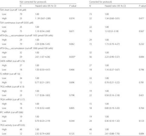
Table 1Results of univariate Cox regression model of MTX resistance-related variables in relation to event-free survival Not corrected for protocols Corrected for protocols
N Hazard ratio (95 % CI) Pvalue N Hazard ratio (95 % CI) Pvalue
TSIA short (cut-off 1.44μM)
Low 79 1.00 73 1.00
High 23 1. 39 (0.67–2.89) 0.374 22 1.34 (0.60–3.01) 0.477
TSIA continuous (cut-off 0.035μM)
Low 25 1.00 22 1.00
High 75 1.19 (0.54–2.60) 0.671 70 1.3 (0.53–3.18) 0.567
MTX-Glu1–6accumulation (cut-off 1935 pmol/109 cells)
High 29 1.00 29 1.00
Low 79 2.29 (0.96–5.45) 0.062 72 1.73 (0.70–4.27) 0.232
MTX-Glu4–6accumulation (cut-off 1000 pmol/109 cells)
High 32 1.00 32 1.00
Low 59 2.61 (1.07–6.36) 0.035* 56 2.23 (0.90–5.57) 0.084
DHFR mRNA (cut-off 3.74)
High 27 1.00 27 1.00
Low 18 1.50 (0.50–4.47) 0.466 18 1.16 (0.37–3.67) 0.796
TS mRNA (cut-off 10)
Low 34 1.00 33 1.00
High 12 0.77 (0.21–2.81) 0.696 12 0.78 (0.21–3.22) 0.781
FPGS mRNA (cut-off 4.13)
High 19 1.00 19 1.00
Low 23 1.17 (0.36–3.83) 0.798 22 0.58 (0.16–2.18) 0.421
FPGH mRNA (cut-off 3.17)
High 16 1.00 15 1.00
Low 18 1.18 (0.32–4.40) 0.805 18 0.80 (0.19–3.33) 0.764
RFC mRNA (cut-off 0.86)
High 19 1.00 19 1.00
Low 25 0.70 (0.23–2.19) 0.544 24 0.36 (0.10–1.32) 0.123
FPGS activity (cut-off 0.35)
High 48 1.00 48 1.00
Low 12 2.32 (0.79–6.80) 0.125 11 2.61 (0.88–7.76) 0.084
TSIA continuous and short-term exposure (TSIA short) are expressed as the concentration of MTX (inμM) necessary to inhibit 50 % of the TS activity (TSI50) compared to the controls incubated without MTX (in triplicate); the concentration of MTX polyglutamates (MTX-Glu1–6and MTX-Glu4–6) is expressed as pmol MTX-Glun/10
9
cells; mRNA expression of folate enzymes is expressed as ratio normalized toβ-actin; FPGS activity is expressed as pmol MTX-Glu2formed/h/10 6
of DHFR and TS mRNA in T cell ALL (3.2-fold and 3.9-fold, respectively, bothP< 0.001).
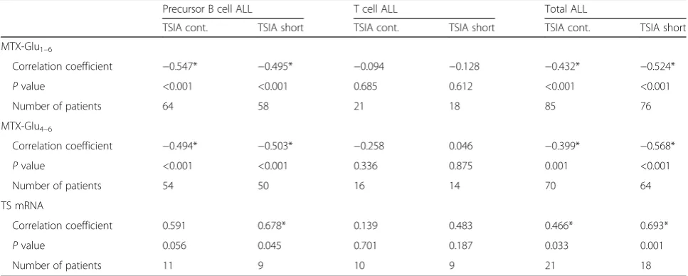
Next, in order to determine which variables strongly influence MTX resistance in ALL patients, we exam-ined the correlation between cellular levels of MTX polyglutamates, activities, and mRNA expression levels of MTX metabolic enzymes with in vitro MTX sensi-tivity as determined by the TSIA. In line with previous
findings [34], both the TSIA results based on continuous and the short-term MTX exposure correlated with con-centrations of total (TSIA continuous: R =−0.432 and TSIA short-term:R=−0.524,P< 0.001) and long-chain MTX polyglutamates (Table 3, R = −0.399, P = 0.001 and R = −0.568, P < 0.001) as well as with the TS mRNA expression (TSIA continuous: R = 0.466, P = 0.033 and TSIA short-term:R= 0.693,P= 0.001). Simi-lar correlations were obtained when precursor B cell patients were examined separately. Weaker and non-significant correlations were found in T cell ALL, sug-gesting that there might be a substantial difference between the factors determining MTX resistance in precursor B cell and T cell leukemia. To formally test whether the strength of association between TSIA and MTX polyglutamates orTSmRNA expression differed between B cell and T cell leukemia, we used a linear re-gression model that included an interaction between the level of MTX polyglutamates and the lineage. No significant difference in association between the con-centration of MTX polyglutamylates and both the continuous and short-term TSIA was found between the precursor B cell and T cell ALL (data not shown).
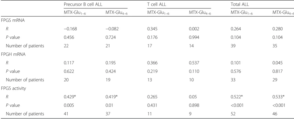
Intriguingly, the accumulation of both total and long-chain MTX polyglutamates correlated with FPGS activity but not with its mRNA levels (Table 4). This association was observed for the whole cohort as well as for precursor B cell ALL alone. Consequently,FPGSmRNA expression did not correlate with FPGS activity (P = 0.125 for the whole cohort, P = 0.75 for precursor B cell ALL, and P = 0.544 for T cell ALL).
Table 2MTX resistance-related variables in precursor B and T cell ALL
Precursor B cell ALL T cell ALL
N Median (range) N Median (range) Pvalue TSIA cont. 85 0.058 (0.01–0.76) 30 0.062 (0.01–0.22) 0.595
TSIA short 87 0.37 (0.16–12.97) 30 1.44 (0.16–40.0) <0.001
MTX-Glu1–6 97 1210 (120–4838) 31 630 (314–1943) <0.001
MTX-Glu4–6 83 856 (80–3190) 25 260 (0–843) <0.001
DHFR mRNA 37 3.86 (0.27–55.06) 17 12.42 (1.48–34.11) <0.001
TS mRNA 37 2.64 (0.24–23.04) 18 10.26 (1.64–25.04) <0.001
FPGS mRNA 32 6.26 (1.12–39.64) 20 2.39 (0.52–17.20) 0.008
FPGH mRNA 27 2.12 (0.16–18.00) 16 4.83 (0.18–27.65) 0.196
RFC mRNA 35 0.89 (0.24–6.90) 20 0.79 (0.23–4.79) 0.668
FPGS activity 57 1.01 (0.03–11.33) 16 0.18 (0.02–1.57) <0.001
Pvalue is determined by the Mann–WhitneyUtest; TSIA continuous (TSIA cont.) and short-term exposure (TSIA short) are expressed as the concentration of MTX (inμM) necessary to inhibit 50 % of the TS activity (TSI50) compared to the controls incubated without MTX (in triplicate); the concentration of MTX polyglutamates (MTX-Glu1–6and MTX-Glu4–6) are expressed as pmol MTX-Glun/10
9
cells; mRNA expression of folate enzymes is expressed as ra-tio normalized toβ-actin; FPGS activity is expressed as pmol MTX-Glu2 formed/h/106
cells
Table 3Correlation of the TSIA assay (continuous and short exposure) with accumulation of MTX polyglutamates and TS mRNA expression
Precursor B cell ALL T cell ALL Total ALL
TSIA cont. TSIA short TSIA cont. TSIA short TSIA cont. TSIA short
MTX-Glu1–6
Correlation coefficient −0.547* −0.495* −0.094 −0.128 −0.432* −0.524*
Pvalue <0.001 <0.001 0.685 0.612 <0.001 <0.001
Number of patients 64 58 21 18 85 76
MTX-Glu4–6
Correlation coefficient −0.494* −0.503* −0.258 0.046 −0.399* −0.568*
Pvalue <0.001 <0.001 0.336 0.875 0.001 <0.001
Number of patients 54 50 16 14 70 64
TS mRNA
Correlation coefficient 0.591 0.678* 0.139 0.483 0.466* 0.693*
Pvalue 0.056 0.045 0.701 0.187 0.033 0.001
Number of patients 11 9 10 9 21 18
Ex vivo MTX resistance in association with chromosomal abnormalities
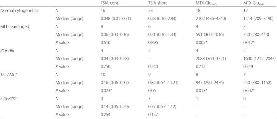
Next, we examined the possible association of MTX re-sistance with the hyperdiploid karyotype as well as three other recurrent cytogenetic abnormalities seen in ALL:
MLL rearrangements, TEL-AML1, and E2A-PBX1. We found that blasts of hyperdiploid ALL patients were significantly more sensitive to MTX than blasts from non-hyperdiploid ALL patients (Fig. 3), both in the con-tinuous and the short-term TSIA (P< 0.001). Consistent with previous reports [30, 36], the leukemic blasts of these patients accumulated significantly higher levels of total MTX and in the same time displayed elevatedRFC mRNA levels, confirming that increased uptake of MTX contributes to MTX hypersensitivity observed in this group.
Similarly, the analyzed cytogenetic subtypes of ALL showed several associations with MTX resistance-related parameters (Table 5). In line with findings from others [16], we established that TEL-AML1 fusion results in lower accumulation of both total and long-chain MTX polyglutamates (2.2-fold,P= 0.013 and 2.8-fold,P= 0.007, respectively). In addition, these patients showed higher TSI50values for both continuous and short-term exposure
(3.3-fold, P = 0.023 and 2.9-fold P = 0.06, respectively). Interestingly, ALL patients withMLLrearrangements also displayed lower accumulation of total and long-chain MTX polyglutamates (3.6-fold,P = 0.005 and 4.3-fold,
P = 0.012, respectively), however this was not paralleled by elevated TSI50values. No significant correlations were
observed for patients harboring the E2A-PBX1nor
BCR-ABLgene fusions, which can partially be explained by the small patient numbers in both groups.
Discussion
In the current study, which used multiple parameters as-sociated with MTX sensitivity, we demonstrate that the cellular level of MTX-induced cytotoxicity is an import-ant determinimport-ant of long-term treatment outcome in childhood ALL. We showed that accumulation of long-chain MTX polyglutamates was the strongest MTX resistance-associated variable, as reflected by its signifi-cant association with both OS and EFS in the analyzed patient cohort. Our data support and further extend pre-vious studies documenting better antileukemic effect of MTX as well as favorable 5-year EFS in childhood ALL patients, whose leukemic cells accumulated higher levels of MTX polyglutamates [15, 29, 37]. On the other hand, a study of the Children’s Oncology Group (COG) sug-gested that the association between the concentration of MTX polyglutamates and EFS is abrogated by higher dose MTX therapy [38]. However, this study involved a different mode of MTX administration and cut-off levels for high/low polyglutamate accumulation, which might have influenced the different outcome of the analysis. When analyzed in multivariate Cox regression with cor-rection for established clinical factors (including WBC, age, CNS involvement, lineage, and DNA index) as co-variates, the cellular level of long-chain MTX polygluta-mates was not an independent prognostic factor. This indicates that despite the important role in ALL treat-ment response, MTX polyglutamates may not have additional predictive value to the already existing models. Surprisingly, low levels of RFC were predictive of a better overall survival in both univariate and multi-variate analysis, contradicting previous reports [9, 39]. The relation betweenRFCmRNA expression and overall
Table 4Correlation of cellular MTX polyglutamate levels with mRNA expression of FPGS and FPGH as well as FPGS activity
Precursor B cell ALL T cell ALL Total ALL
MTX-Glu1–6 MTX-Glu4–6 MTX-Glu1–6 MTX-Glu4–6 MTX-Glu1–6 MTX-Glu4–6
FPGS mRNA
R −0.168 −0.082 0.345 0.002 0.264 0.280
Pvalue 0.456 0.724 0.176 0.994 0.104 0.104
Number of patients 22 21 17 14 39 35
FPGH mRNA
R 0.117 0.195 0.366 0.537 0.101 0.045
Pvalue 0.622 0.424 0.219 0.110 0.576 0.817
Number of patients 20 19 13 10 33 29
FPGS activity
R 0.429* 0.419* 0.265 0.05 0.522* 0.533*
Pvalue 0.005 0.01 0.431 0.898 <0.001 <0.001
Number of patients 41 37 11 9 52 46
RandPvalues were estimated using Spearman’s Rho test; the concentration of MTX polyglutamates (MTX-Glu1–6and MTX-Glu4–6) is expressed as pmol
survival was the strongest when corrected for the treat-ment protocol, suggesting that it might be linked to high doses of MTX used in some treatment protocols. When examined within each treatment protocol separately, this association was only observed in patients treated at DCOG ALL8 protocol, on which patients depending on the risk group were administered MTX at 2 g/m2or 5 g/ m2 [40]. In contrast, on COALL protocol ALL-97, for which this relation was absent, MTX was administered at 1 g/m2[2, 41]. This discrepancy may be explained by the limited number of patients treated with 1 g/m2 in this cohort (6 patients for COALL-97 and 16 for DCOG ALL8). Decreased RFCexpression as well as mutations in theRFCgene resulting in less efficient uptake of anti-folates by tumor cells, have been associated with MTX resistance [9, 39]. On the other hand, some of the muta-tions found in theRFCgene were previously reported to increase the affinity of RFC to folates, resulting in in-creased intracellular accumulation and competition of fol-ate with MTX [42]. It is therefore possible that extremely high RFC expression results in high concentration of folates in the cells, leading to decreased efficacy of MTX and worse treatment outcome. Unfortunately, the num-bers of patients treated on the other protocols for which
RFC mRNA expression data were available were too low to allow this analysis.
Moreover, we show that the lineage as well as a num-ber of chromosomal abnormalities are associated with distinct levels of MTX sensitivity. In agreement with previous analysis in a partially overlapping patient co-hort as well as in other studies [17, 34, 35], we observed here elevated accumulation of MTX polyglutamates together with decreased expression of DHFR and TS mRNA and consequently higher MTX sensitivity in pre-cursor B cell ALL, as compared to T cell ALL. The dif-ference in the accumulation of the long-chain MTX polyglutamates between these two distinct subtypes of ALL exceeded that obtained for the total MTX polyglu-tamates. This observation, together with the FPGS activ-ity being significantly increased in the precursor B cell ALL, suggests that differences in MTX response be-tween the precursor B cell and T cell leukemia are highly dependent on FPGS activity and consequent MTX poly-glutamylation. Indeed, a previous study comparing lineage-based differences in MTX sensitivity in leukemia cell lines also pointed to the involvement of FPGS activ-ity and DHFR levels [43]. The importance of the cellular accumulation of MTX polyglutamates was further cor-roborated by its association with the TSIA, which was previously shown to be a good determinant of MTX sen-sitivity, as reflected by the correlation between the TSIA results and MTX cytotoxicity [34]. This association was
found in both the entire patient cohort as well as in pre-cursor B cell ALL alone but not in T cell ALL when ana-lyzed separately. The linear regression analysis did not show that the association of cellular MTX polyglutamate levels with the TSIA is actually different between precur-sor B cell and T cell leukemia. The apparent differences between the correlations may be due to a large uncer-tainty in the estimates for the correlations of the T cell ALL, which may have been caused by the small sample size for this subgroup. Taken together, our data indicate that despite the differences in concentration of MTX polyglutamates and MTX sensitivity profiles between precursor B cell and T cell ALL, the accumulation of MTX polyglutamates is likely an indicator of MTX re-sistance as determined by the TSIA in both of these ALL subtypes.
Intriguingly, in parallel to differences in both FPGS mRNA levels and activity observed between precursor B cell and T cell ALL, we found no correlation between
FPGSmRNA level and enzyme activity or the concentra-tion of MTX polyglutamates. This suggests that the level of FPGS mRNA might not directly translate to its en-zymatic activity. A strong correlation was previously re-ported between FPGS mRNA expression and activity in leukemic cell lines [43]. In fact, our previous finding of the high propensity of humanFPGS gene in ALL speci-mens to undergo impaired splicing is consistent with the fact that transcript levels may not correlate at all with FPGS catalytic activity [28, 44], Wojtuszkiewicz et al. un-published observations. Moreover, decreased rates of
FPGS mRNA translation affecting its activity were
previously detected in murine leukemic cell lines se-lected with another polyglutamylation-dependent antifo-late—edatrexate [45]. In addition, we have reported that aberrant FPGSsplicing is a potential contributing factor to the loss of FPGS function, as various FPGS mRNA splicing alterations were detected in MTX-resistant leukemic cell lines devoid of FPGS activity as well as in adult ALL patient samples [28].
MTX sensitivity in the patient cohort under study differed between several genetic subtypes of precursor B cell ALL. This included the hyperdiploid patients, dis-playing high MTX sensitivity associated with increased
RFC expression as well as TEL-AML1 fusion and MLL rearrangements displaying an increased MTX resistance. Elevated RFC expression might be caused by an additional copy of RFC gene-carrying chromosome 21, which is often observed in hyperdiploid ALL. Our re-sults support previous findings showing that hyperdi-ploid patients accumulate higher levels of MTX polyglutamates associated with elevated RFCexpression [29, 30]. Consequently, this subset of patients responded favorably to MTX-containing chemotherapy as com-pared to patients with a non-hyperdiploid karyotype [37]. Although the remarkable sensitivity of hyperdiploid ALL cannot be entirely explained by high MTX sensitiv-ity, the current results as well as observations of others [29, 30, 37] suggest that it is an important biological fea-ture contributing to sensitivity of this ALL subtype. Pre-cursor B cell ALL withTEL-AML1 fusion as well as with rearrangedMLLgene were associated with lower accumu-lation of MTX polyglutamates, which in the case of
TEL-Table 5MTX sensitivity and accumulation of MTX polyglutamates in relation to cytogenetic abnormalities
TSIA cont. TSIA short MTX-Glu1–6 MTX-Glu4–6
Normal cytogenetics N 16 23 18 17
Median (range) 0.048 (0.01–0.71) 0.28 (0.16–2.84) 2102 (436–4240) 1514 (209–3190)
MLL-rearranged N 8 6 4 3
Median (range) 0.06 (0.03–0.16) 0.21 (0.16–1.33) 591 (360–1016) 350 (280–443)
Pvalue 0.610 0.896 0.005* 0.012*
BCR-ABL N 4 2 4 2
Median (range) 0.04 (0.03–0.28) – 2088 (360–3721) 1630 (1212–2047)
Pvalue 0.750 0.240 0.712 0.749
TEL-AML1 N 10 9 8 7
Median (range) 0.16 (0.06–0.37) 0.82 (0.54–11.21) 945 (290–2476) 550 (380–1152)
Pvalue 0.023* 0.06 0.013* 0.007*
E2A-PBX1 N 3 3 1 0
Median (range) 0.14 (0.05–0.29) 0.77 (0.57–1.12) – –
Pvalue 0.254 0.157 – –
First, the Kruskal–Wallis analysis of variance was used to compare all the four groups, and variables with thePvalue above 0.1 were further examined in detail. The Mann–WhitneyUtest was used to compare MTX-related variables between each subgroup of ALL patients carrying certain cytogenetic abnormality and patients displaying normal karyotype as a reference group; TSIA continuous (TSIA cont.) and short-term exposure (TSIA short) are expressed as the concentration of MTX (inμM) necessary to inhibit 50 % of the TS activity (TSI50) compared to the controls incubated without MTX (in triplicate); the concentration of MTX polyglutamates (MTX-Glu1–6and MTX-Glu4–6) is expressed as pmol MTX-Glun/10
9
AML1 was accompanied by increased MTX resistance as reflected by the TSIA. Precursor B cell ALL carrying either
TEL-AML1 or E2A-PBX1 gene fusions was previously shown to accumulate decreased levels of MTX polygluta-mates as compared to precursor B cell ALL which are devoid of these cytogenetic aberrations [16]. This was associated with diminished expression of RFC in
E2A-PBX1ALL and elevated expression of ABCG2—an MTX efflux transporter—in ALL patients with TEL-AML1 fu-sion [16]. Decreased RFC activity resulting from transcrip-tional silencing as well as mutations and allele loss was previously reported as the cause of antifolate resistance in several tumor types, including leukemia, breast cancer, and osteosarcoma [19–21]. Similar overexpression of ABC transporters is an established mechanism of antifolate re-sistance [9, 13, 46]. Moreover, another study suggests that bothTEL-AML1 andE2A-PBX1 gene fusions downregu-late FPGS expression by interacting with its promoter re-gion [47, 48]. Interestingly,TEL-AML1andE2A-PBX1are generally associated with a relatively good prognosis, as opposed to patients harboring rearrangedMLL gene [4], which can be explained by the differences in sensitivity to other chemotherapeutics between these ALL subtypes [5]. The numbers of ALL patients displaying these cytogenetic abnormalities for which MTX sensitivity-associated vari-ables were measured was very limited in this study. Hence, we were not able to evaluate the role of particular triso-mies (i.e., trisomy 21) across the patients with hyperdi-ploid karyotype or the analyzed cytogenetic abnormalities. Therefore, these associations should be further addressed in large future studies. However, the current data suggest that TEL-AML1, E2A-PBX1, and MLL-rearranged ALL may benefit from courses with high-dose MTX, partly overcoming the polyglutamylation and transport defects [16, 18].
One of the major limitations of the current study was the low numbers of patients for which particular param-eters were recorded. This especially influenced analyses in various ALL groups, such as cytogenetic subtypes, where the patient numbers were extremely low. More-over, patients included in this study were treated on di-verse protocols with distinct MTX administration doses. This issue, however, was addressed in the statistical ana-lysis by including the treatment protocol as a covariate. Finally, the lack of MTX-related toxicity data limited our insight into the associations of particular MTX resistance-related parameters and the clinical outcome of ALL patients. These issues should be carefully considered in future studies.
Conclusions
Taken together, our study clearly shows that the low cellu-lar level of long-chain polyglutamates of MTX is an im-portant predictor of MTX resistance and is associated
with dismal therapeutic outcome. Its additional prognostic value warrants further investigation in larger studies using more up-to-date treatment regimens. As MTX re-mains one of the re-mainstays of contemporary ALL treat-ments, expanding our understanding of its contribution to the treatment outcome is of supreme therapeutic value. In particular, the identification of patients that are potentially resistant to MTX at start of the treat-ment may allow for tailoring novel treattreat-ment strategies to individual leukemia patients in the context of com-bination chemotherapy.
Materials and methods
ALL patients
We analyzed a total of 235 newly diagnosed (see Additional file 1: Table S1 for patient characteristics), untreated pediatric ALL patients treated on Dutch Childhood Oncology Group (DCOGN= 125) protocols ALL6–ALL9 [40, 49], German Co-operative ALL study group (COALL
N = 93) protocols 92–97 [2, 41] and Nordic Society of Paediatric Haematology and Oncology (NOPHON= 17) protocols ALL-92–ALL-2000 [50]. All patients (or parents or legal guardians of patients) have provided a written in-formed consent.
Immunophenotyping and DNA index flow cytometry were performed at reference laboratories of the partici-pating groups; patient characteristics were collected by the study centers.
For all patients, leukemic cells freshly obtained from the bone marrow or peripheral blood of ALL patients at diagnosis were isolated within 48 h of sampling as described previously [51]. When necessary, contaminat-ing normal cells were removed by monoclonal anti-bodies linked to magnetic beads as described previously [52]. All samples contained >80 % leukemic blasts with the majority of samples reaching blast percentages around 90 % (only 11 out of 235 patient samples con-tained <85 % blasts), as determined by cytospin prepa-rations stained with May-Grunwald-Giemsa (Merck, Darmstadt, Germany).
This study was approved by the Local Ethics Committee VUmc. Date of approval: December 5, 2000 (file number TJFS/bz2568a).
In situ thymidylate synthase inhibition assay
The TSIA was used to measure in vitro efficacy of MTX to inhibit TS, which is known to determine cytotoxicity in MTX exposed cells. Inhibition of TS was determined in whole cells by measuring the TS-catalyzed conversion of 3H-dUMP to dTMP and 3H2O, as described
of MTX necessary to inhibit 50 % of the TS activity (TSI50)
compared to the controls incubated without MTX (in triplicate) [34, 53].
MTX accumulation and polyglutamylation
Ten million leukemic cells were incubated for 24 h with 1 μM [3′, 5′, 7-3H]-MTX (Moravek Biochemicals; final specific activity 2 Ci/mmol) in a 5-ml culture medium, as described previously [17]. After washing, samples were counted for radioactivity, cell number, and viability. The remaining suspension was centrifuged, and the cell pellet was used for measurement of polyglutamates using high-performance liquid chromatography on an anion exchange column as described previously [17, 53]. The data are expressed as pmol MTX-Glun/109 cells.
Total MTX polyglutamates included MTX associated with 1 up to 6 glutamate residues, while long-chain MTX polyglutamates corresponded to MTX associated with 4 up to 6 glutamate residues.
RNA extraction, reverse transcription, and quantitative PCR assay
Freshly isolated cells were washed twice in RPMI (Gibco) containing 2 % fetal calf serum, followed by lysis in 1 ml RNAzol. Next, the samples were frozen in liquid nitrogen and stored at−80 °C until further pro-cessing. RNA extraction was performed according to manufacturer’s instructions with previously described adjustments [35]. Competitive template PCR was per-formed as described previously [35].
FPGS activity assay
The FPGS activity assay was performed as described previously [17]. Briefly, the reaction was carried out in crude cell extracts employing a 2-h incubation with a final concentration of 250μM MTX and 4 mM [3 H]-L-glutamic acid at 37 °C, which was next terminated with ice-cold glutamic acid. The resulting MTX-[3H]-Glu2
was separated from unreacted [3H]-L-glutamic acid by reverse-phase column chromatography and quantified as described previously [17].
Statistical analysis
All statistical analyses were performed using the IBM SPSS Statistics 20 software. The Mann–WhitneyU test was used to compare the levels of MTX resistance-related variables between different subgroups of ALL patients, while Spearman’s Rho test and linear regression were applied to assess correlations. Kaplan–Meier ana-lysis and Cox regression were used in the anaana-lysis of EFS as well as OS of the patients. The cut-off values for sur-vival analysis were selected based on literature (clinical factors) [2, 40, 41, 49, 50]. For MTX-related variables, if the distribution was bimodal, we chose the cut-off value
midway between the two modes of the distributions; otherwise, we used the median as a cut-off. Kaplan– Meier analysis and Cox regression were used for testing associations between MTX resistance-related variables and event-free (defined as time from complete remission to an event) as well as overall survival of the patients. Events considered in event-free survival were relapse and death. MTX resistance-related variables which showed significant impact on survival in the univariate analysis (P< 0.05 at two-sided testing) were added to a multivari-ate Cox regression model with identified prognostic clin-ical factors in order to assess their added prognostic value. As different protocols were used with different patients, additional analyses were performed in which we corrected for the protocol used by including protocol as an add-itional independent variable in the Cox regression model.
Additional file
Additional file 1:Patient characteristics and supplemental survival analysis. Table S1.Clinical characteristics of pediatric ALL patients included in this study.Table S2.Results of univariate Cox regression model of MTX resistance related variables in relation to overall survival. Table S3.Treatment outcome (EFS) in the whole cohort according to presenting features (not corrected for the treatment protocol).Table S4. Treatment outcome (EFS) in the whole cohort according to presenting features (corrected for the treatment protocol).Table S5.Results of multivariate Cox regression model of MTX resistance related variables in relation to event-free survival.Table S6.Results of multivariate Cox regression model of MTX resistance related variables in relation to overall survival.
Competing interests
The authors declare that they have no competing interests.
Authors’contributions
JC was the principal investigator and takes primary responsibility for the paper; GE, KS, and ES provided patient samples and clinical data; AW, NLW, PMV, and BD performed the research; JC, GJLK, GJ, GJP, and YGA designed the research; AW wrote the paper; JC, GJLK, GJ, GJP, GE, KS, ES, NLW, PMV, BD, YGA, and RP edited the paper. All authors read and approved the final manuscript.
Acknowledgements
The authors would like to thank Marianne Rots and Michael Dworzak for their data contributions. This study was supported by KiKa (Children cancer-free). Y. G. Assaraf is a recipient of a visiting professor fellowship of the Royal Netherlands Academy of Arts and Sciences.
Author details 1
Department of Pediatric Oncology/Hematology, VUmc Cancer Center Amsterdam, VU University Medical Center, Room CCA 4.28, De Boelelaan 1117, 1081HV Amsterdam, The Netherlands.2Department of Hematology, VU University Medical Center, Amsterdam, The Netherlands.3Department of
Medical Oncology, VU University Medical Center, Amsterdam, The Netherlands.4Department of Epidemiology and Biostatistics, VU University
Medical Center, Amsterdam, The Netherlands.5Department of
Rheumatology, VU University Medical Center, Amsterdam, The Netherlands.
6
Department of Pediatric Hematology/Oncology, University Medical Center Hamburg-Eppendorf, Hamburg, Germany.7Institute of Clinical Medicine,
University of Copenhagen, Copenhagen, Denmark.8Department of Pediatrics and Adolescent Medicine, University Hospital Rigshospitalet, Copenhagen, Denmark.9Dutch Childhood Oncology Group (DCOG), The Hague, The Netherlands.10Princess Máxima Center for Pediatric Oncology, Utrecht, The
Received: 9 March 2015 Accepted: 19 May 2015
References
1. Pui CH, Mullighan CG, Evans WE, Relling MV. Pediatric acute lymphoblastic leukemia: where are we going and how do we get there? Blood. 2012;120:1165–74.
2. Escherich G, Horstmann MA, Zimmermann M, Janka-Schaub GE. Cooperative study group for childhood acute lymphoblastic leukaemia (COALL): long-term results of trials 82,85,89,92 and 97. Leukemia. 2010;24:298–308.
3. Einsiedel HG, Von Stackelberg A, Hartmann R, Fengler R, Schrappe M, Janka-Schaub G, et al. Long-term outcome in children with relapsed ALL by risk-stratified salvage therapy: results of trial acute lymphoblastic leukemia-relapse study of the Berlin-Frankfurt-Munster Group 87. J Clin Oncol. 2005;23:7942–50.
4. Pui CH, Relling MV, Downing JR. Acute lymphoblastic leukemia. N Engl J Med. 2004;350:1535–48.
5. Meijerink JPP, den Boer ML, Pieters R. New genetic abnormalities and treatment response in acute lymphoblastic leukemia. Semin Hematol. 2009;46:16–23. 6. Bertino J. Karnofsky memorial lecture. Ode to methotrexate. J Clin Oncol.
1993;11:5–14.
7. Stokstad ELR. Historical perspectives on key advances in the biochemistry and physiology of folates. In: Picciano MF, Stokstad ELR, Gregory JF, editors. Evaluation of Folic Acid Metabolism in Health and Disease. New York, NY: Wiley-Liss; 1990. p. 1–21.
8. Cheok MH, Lugthart S, Evans WE. Pharmacogenomics of acute leukemia. Annu Rev Pharmacol Toxicol. 2006;46:317–53.
9. Assaraf YG. Molecular basis of antifolate resistance. Cancer Metastasis Rev. 2007;26:153–81.
10. Allegra CJ, Hoang K, Yeh GC, Drake JC, Baram J. Evidence for direct inhibition of de novo purine synthesis in human MCF-7 breast cells as a principal mode of metabolic inhibition by methotrexate. J Biol Chem. 1987;262:13520–6.
11. Allegra CJ, Chabner BA, Drake JC, Lutz R, Rodbard D, Jolivet J. Enhanced inhibition of thymidylate synthase by methotrexate polyglutamates. J Biol Chem. 1985;260:9720–6.
12. Baggott JE, Vaughn WH, Hudson BB. Inhibition of 5-aminoimidazole-4-carboxamide ribotide transformylase, adenosine deaminase and 5′-adenylate deaminase by polyglutamates of methotrexate and oxidized folates and by 5-aminoimidazole-4-carboxamide riboside and ribotide. Biochem J. 1986;236:193–200.
13. Gonen N, Assaraf YG. Antifolates in cancer therapy: structure, activity and mechanisms of drug resistance. Drug Resist Updat. 2012;15:183–210. 14. Hooijberg JH, Broxterman HJ, Kool M, Assaraf YG, Peters GJ, Noordhuis P,
et al. Antifolate resistance mediated by the multidrug resistance proteins MRP1. Cancer Res. 1999;59:2532–5.
15. Masson E, Relling MV, Synold TW, Liu Q, Schuetz JD, Sandlund JT, et al. Accumulation of methotrexate polyglutamates in lymphoblasts is a determinant of antileukemic effects in vivo. J Clin Invest. 1996;97:73–80. 16. Kager L, Cheok M, Yang W, Zaza G, Cheng Q, Panetta JC, et al. Folate
pathway gene expression differs in subtypes of acute lymphoblastic leukemia and influences methotrexate pharmacodynamics. J Clin Invest. 2005;115:110–7.
17. Rots MG, Pieters R, Peters GJ, Noordhuis P, van Zantwijk CH, Kaspers GJ, et al. Role of folylpolyglutamate synthetase and folylpolyglutamate hydrolase in methotrexate accumulation and polyglutamylation in childhood leukemia. Blood. 1999;93:1677–83.
18. Cheok MH, Pottier N, Kager L, Evans WE. Pharmacogenetics in acute lymphoblastic leukemia. Semin Hematol. 2009;46:39–51.
19. Rothem L, Aronheim A, Assaraf YG. Alterations in the expression of transcription factors and the reduced folate carrier as a novel mechanism of antifolate resistance in human leukemia cells. J Biol Chem.
2003;278:8935–41.
20. Rothem L, Stark M, Kaufman Y, Mayo L, Assaraf YG. Reduced folate carrier gene silencing in multiple antifolate-resistant tumor cell lines is due to a simultaneous loss of function of multiple transcription factors but not promoter methylation. J Biol Chem. 2004;279:374–84.
21. Kaufman Y, Ifergan I, Rothem L, Jansen G, Assaraf YG. Coexistence of multiple mechanisms of PT523 resistance in human leukemia cells harboring 3 reduced folate carrier alleles: transcriptional silencing, inactivating mutations, and allele loss. Blood. 2006;107:3288–94.
22. Schmiegelow K. Advances in individual prediction of methotrexate toxicity: a review. Br J Haematol. 2009;146:489–503.
23. De Jonge R, Hooijberg JH, van Zelst BD, Jansen G, van Zantwijk CH, Kaspers GJL, et al. Effect of polymorphisms in folate-related genes on in vitro methotrexate sensitivity in pediatric acute lymphoblastic leukemia. Blood. 2005;106:717–20.
24. Gregers J, Christensen IJ, Dalhoff K, Lausen B, Schroeder H, Rosthoej S, et al. The association of reduced folate carrier 80G> A polymorphism to outcome in childhood acute lymphoblastic leukemia interacts with chromosome 21 copy number. Blood. 2010;115:4671–7.
25. Mauritz R, Peters GJ, Priest DG, Assaraf YG, Drori S, Kathmann I, et al. Multiple mechanisms of resistance to methotrexate and novel antifolates in human CCRF-CEM leukemia cells and their implications for folate homeostasis. Biochem Pharmacol. 2002;63:105–15.
26. Zhao R, Titus S, Gao F, Moran RG, Goldman ID. Molecular analysis of murine leukemia cell lines resistant to 5, 10-dideazatetrahydrofolate identifies several amino acids critical to the function of folylpolyglutamate synthetase. J Biol Chem. 2000;275:26599–606.
27. Liani E, Rothem L, Bunni MA, Smith CA, Jansen G, Assaraf YG. Loss of folylpoly-gamma-glutamate synthetase activity is a dominant mechanism of resistance to polyglutamylation-dependent novel antifolates in multiple human leukemia sublines. Int J cancer. 2003;103:587–99.
28. Stark M, Wichman C, Avivi I, Assaraf YG. Aberrant splicing of
folylpolyglutamate synthetase as a novel mechanism of antifolate resistance in leukemia. Blood. 2009;113:4362–9.
29. Whitehead VM, Vuchich MJ, Lauer SJ, Mahoney D, Carroll AJ, Shuster JJ, et al. Accumulation of high levels of methotrexate polyglutamates in lymphoblasts from children with hyperdiploid (greater than 50 chromosomes) B-lineage acute lymphoblastic leukemia: a Pediatric Oncology Group study. Blood. 1992;80:1316–23.
30. Belkov VM, Krynetski EY, Schuetz JD, Yanishevski Y, Masson E, Raimondi S, et al. Mechanism for ploidy but not lineage differences in methotrexate mechanism for ploidy but not lineage differences in methotrexate accumulation. Blood. 1999;93:1643–50.
31. Dervieux T, Hancock M, Evans W, Pui C, Relling M. Effect of methotrexate polyglutamates on thioguanine nucleotide concentrations during continuation therapy of acute lymphoblastic leukemia with mercaptopurine. Leukemia. 2002;16:209–12.
32. Schmiegelow K, Nielsen S, Frandsen TL, Nersting J. Mercaptopurine/ methotrexate maintenance therapy of childhood acute lymphoblastic leukemia: clinical facts and fiction. J Pediatr Hematol Oncol.
2014;36:503–17.
33. Lennard L, Lilleyman JS, Van Loon J, Weinshilboum RM. Genetic variation in response to 6-mercaptopurine for childhood acute lymphoblastic leukaemia. Lance. 1990;336:225–9.
34. Rots MG, Pieters R, Kaspers GJ, van Zantwijk CH, Noordhuis P, Mauritz R, et al. Differential methotrexate resistance in childhood T- versus com-mon/preB-acute lymphoblastic leukemia can be measured by an in situ thymidylate synthase inhibition assay, but not by the MTT assay. Blood. 1999;93:1067–74.
35. Rots M, Willey J, Jansen G, van Zantwijk CH, Noordhuis P, DeMuth J, et al. mRNA expression levels of methotrexate resistance-related proteins in childhood leukemia as determined by a standardized competitive template-based RT-PCR method. Leukemia. 2000;14:2166–75. 36. Synold TW, Relling MV, Boyett JM, Rivera GK, Sandlund JT, Mahmoud H,
et al. Blast cell methotrexate-polyglutamate accumulation in vivo differs by lineage, ploidy, and methotrexate dose in acute lymphoblastic leukemia. J Clin Invest. 1994;94:1996–2001.
37. Whitehead VM, Rosenblatt DS, Vuchich MJ, Shuster JJ, Witte A, Beaulieu D. Accumulation of methotrexate and methotrexate polyglutamates in lymphoblasts at diagnosis of childhood acute lymphoblastic leukemia: a pilot prognostic factor analysis. Blood. 1990;76:44–9.
38. Whitehead VM, Shuster JJ, Vuchich MJ, Mahoney DH, Lauer SJ, Payment C, et al. Accumulation of methotrexate and methotrexate polyglutamates in lymphoblasts and treatment outcome in children with B-progenitor-cell acute lymphoblastic leukemia: a Pediatric Oncology Group study. Leukemia. 2005;19:533–6.
40. Kamps WA, van der Pal-de Bruin KM, Veerman AJP, Fiocco M, Bierings M, Pieters R. Long-term results of Dutch Childhood Oncology Group studies for children with acute lymphoblastic leukemia from 1984 to 2004. Leukemia. 2010;24:309–19.
41. Harms DO, Janka-Schaub GE. Co-operative study group for childhood acute lymphoblastic leukemia (COALL): long-term follow-up of trials 82, 85, 89 and 92. Leukemia. 2000;14:2234–9.
42. Jansen G, Mauritz R, Drori S, Sprecher H, Kathmann I, Bunni M, et al. A structurally altered human reduced folate carrier with increased folic acid transport mediates a novel mechanism of antifolate resistance. J Biol Chem. 1998;273:30189–98.
43. Galpin AJ, Schuetz JD, Masson E, Yanishevski Y, Synold TW, Barredo JC, et al. Differences in folylpolyglutamate synthetase and dihydrofolate reductase expression in human B-lineage versus T-lineage leukemic lymphoblasts: mechanisms for lineage differences in methotrexate polyglutamylation and cytotoxicity. Mol Pharmacol. 1997;52:155–63.
44. Wojtuszkiewicz A, Assaraf YG, Maas M, Kaspers G, Jansen G, Cloos J. Pre-mRNA splicing in cancer: the relevance in oncogenesis, treatment and drug resistance. Expert Opin Drug Metab Toxicol. 2015;11:673–89.
45. Roy K, Egan M, Sirlin S, Sirotnak F. Posttranscriptionally mediated decreases in folylpolyglutamate synthetase gene expression in some folate analogue-resistant variants of the L1210 Cell. J Biol Chem. 1997;272:6903–8. 46. Assaraf YG. The role of multidrug resistance efflux transporters in antifolate
resistance and folate homeostasis. Drug Resist Updat. 2006;9:227–46. 47. Leclerc GJ, Sanderson C, Hunger S, Devidas M, Barredo JC.
Folylpolyglutamate synthetase gene transcription is regulated by a multiprotein complex that binds the TEL-AML1 fusion in acute lymphoblastic leukemia. Leuk Res. 2010;34:1601–9.
48. Leclerc GJ, Mou C, Leclerc GM, Mian AM, Barredo JC. Histone deacetylase inhibitors induce FPGS mRNA expression and intracellular accumulation of long-chain methotrexate polyglutamates in childhood acute lymphoblastic leukemia: implications for combination therapy. Leukemia. 2010;24:552–62. 49. Veerman AJ, Kamps WA, van den Berg H, van den Berg E, Bökkerink JPM, Bruin
MCA, et al. Dexamethasone-based therapy for childhood acute lymphoblastic leukaemia: results of the prospective Dutch Childhood Oncology Group (DCOG) protocol ALL-9 (1997–2004). Lancet Oncol. 2009;10:957–66. 50. Schmiegelow K, Forestier E, Hellebostad M, Heyman M, Kristinsson J,
Söderhäll S, et al. Long-term results of NOPHO ALL-92 and ALL-2000 studies of childhood acute lymphoblastic leukemia. Leukemia. 2010;24:345–54. 51. Pieters R, Loonen AH, Huismans DR, Broekema G, Dirven M, Heyenbrok M,
et al. In vitro drug sensitivity of cells from children with leukemia using the MTT assay with improved culture conditions. Blood. 1990;76:2327–36. 52. Kaspers GJ, Veerman AJ, Pieters R, Broekema GJ, Huismans DR, Kazemier KM,
et al. Mononuclear cells contaminating acute lymphoblastic leukaemic samples tested for cellular drug resistance using the methyl-thiazol-tetrazolium assay. Br J Cancer. 1994;70:1047–52.
53. Mauritz R, Bekkenk M, Rots M, Pieters R, Mini E, van Zantwijk CH, et al. Ex vivo activity of methotrexate versus novel antifolate inhibitors of dihydrofolate reductase and thymidylate synthase against childhood leukemia cells. Clin Cancer Res. 1998;4:2399–410.
Submit your next manuscript to BioMed Central and take full advantage of:
• Convenient online submission
• Thorough peer review
• No space constraints or color figure charges
• Immediate publication on acceptance
• Inclusion in PubMed, CAS, Scopus and Google Scholar
• Research which is freely available for redistribution