Open Access
Review
Aorto-ventricular tunnel
Roxane McKay
Address: Division of Cardiovascular Surgery, Le Bonheur Children's Hospital, Memphis, TN 38103, USA Email: Roxane McKay - [email protected]
Abstract
Aorto-ventricular tunnel is a congenital, extracardiac channel which connects the ascending aorta above the sinutubular junction to the cavity of the left, or (less commonly) right ventricle. The exact incidence is unknown, estimates ranging from 0.5% of fetal cardiac malformations to less than 0.1% of congenitally malformed hearts in clinico-pathological series. Approximately 130 cases have been reported in the literature, about twice as many cases in males as in females. Associated defects, usually involving the proximal coronary arteries, or the aortic or pulmonary valves, are present in nearly half the cases. Occasional patients present with an asymptomatic heart murmur and cardiac enlargement, but most suffer heart failure in the first year of life. The etiology of aorto-ventricular tunnel is uncertain. It appears to result from a combination of maldevelopment of the cushions which give rise to the pulmonary and aortic roots, and abnormal separation of these structures. Echocardiography is the diagnostic investigation of choice. Antenatal diagnosis by fetal echocardiography is reliable after 18 weeks gestation. Aorto-ventricular tunnel must be distinguished from other lesions which cause rapid run-off of blood from the aorta and produce cardiac failure. Optimal management of symptomatic aorto-ventricular tunnel consists of diagnosis by echocardiography, complimented with cardiac catheterization as needed to elucidate coronary arterial origins or associated defects, and prompt surgical repair. Observation of the exceedingly rare, asymptomatic patient with a small tunnel may be justified by occasional spontaneous closure. All patients require life-long follow-up for recurrence of the tunnel, aortic valve incompetence, left ventricular function, and aneurysmal enlargement of the ascending aorta.
Disease name and synonyms
In their original description of aorto – left ventricular tun-nel, Edwards and Burchell [1] considered the malforma-tion a "separamalforma-tion between the aorta and the heart", or type of aneurysm which "lay against the outflow tract of the right ventricle and origin of the pulmonary trunk". The term "aortico-left ventricular tunnel" was used subse-quent to Levy's publication in 1963 [2], and "aorto-left ventricular tunnel" was introduced about ten years later by Ross and colleagues [3]. Recognizing that the tunnel may extend to either the left or the right ventricular cavity, the more general designation "aorto-ventricular tunnels"
has recently been applied to this group of malformations [4]. The defect is not a component of any described genetic syndrome, although cystic medial degeneration has been observed in an ascending aortic aneurysm resected fifteen years after tunnel repair [5].
Definition and diagnostic criteria
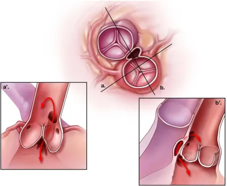
An aorto-ventricular tunnel is an extracardiac channel which connects the ascending aorta above the sinutubular junction to the cavity of the left or right ventricle. Among 130 cases reported in the literature, more than 90% com-municated with the left ventricle (Figure 1). It differs from
Published: 8 October 2007
Orphanet Journal of Rare Diseases 2007, 2:41 doi:10.1186/1750-1172-2-41
Received: 25 July 2007 Accepted: 8 October 2007
This article is available from: http://www.OJRD.com/content/2/1/41 © 2007 McKay; licensee BioMed Central Ltd.
a ruptured sinus of Valsalva aneurysm (sinus of Valsalva fistula) in having its vascular orifice in the tubular aorta, rather than a sinus of the aortic valve, and in passing out-side the heart into the tissue plane between the muscular subpulmonary infundibulum and the aortic valvar sinuses. The aortic opening of most tunnels lies above the right coronary sinus of Valsalva. In these cases, the tunnel virtually always communicates with the left ventricle in the fibrous triangle beneath the left – right coronary com-missure, or the right ventricle immediately above or below the subpulmonary infundibulum. In aorto-left
ven-tricular tunnel, the right coronary aortic leaflet is thus unsupported for a variable portion of its hinge-point and may appear to arise from a bar of fibrous tissue spanning the aortic root [5]. Tunnels lying above the left sinus of Valsalva or the intercoronary commissure have less uni-form morphology and may enter the left ventricle further away from the aortic valve, apparently through infoldings of fibrous tissue. It is extremely rarely, if ever, that an aorto-ventricular tunnel passes through intracardiac myo-cardium to reach the cavity of the ventricle, a feature
Schematic representation of the most common type of aorto-left ventricular tunnel Figure 1
which serves to differentiate it from coronary-cameral fis-tula [4].
The ostium of a coronary artery may lie within an aorto-ventricular tunnel, and absence of the origin (atresia) of the left [6,7] or right [3,8-13] have both been observed with this anomaly. In one reported case, there was a fistula between the right coronary artery and the distal segment of a tunnel to the left ventricle [11]. Associated lesions of the aortic valve occur in about 20% of patients, ranging from two-leaflet valves without obstruction [1,2] to severe dysplasia or atresia [11,14-16]. In addition, older patients may acquire leaflet perforation [17,18] or aortic incompe-tence [19] as the result of hydrodynamic trauma to the unsupported right coronary cusp or progressive aortic dil-atation. Stenosis of the pulmonary valve [20,21] occurs less frequently (around 5% of reported cases), while com-pression of the right ventricular outflow tract by the tun-nel may produce subpulmonary obstruction [22]. Rarely, both semilunar valves are stenotic [2,23].
Histologically, the arterial end of the tunnel resembles the aorta with fibrous tissue, elastic fibers and smooth muscle cells, while the ventricular end contains hyalinized colla-gen and muscle. This reflects that the "walls" of tunnels incorporate the structures through which they pass. Within the tunnel itself, there may be a well-defined junc-tion between ventricular and arterial components, in addition to cystic or membranous structures reminiscent of cardiac valve leaflets [4,24].
Epidemiology
The incidence of aorto-left ventricular tunnel has been variably estimated to be around 0.1% of congenitally mal-formed hearts from review of clinical and pathological material [25], 0.05% among patients undergoing cardiac catheterization during a 35-year interval at the Children's Hospital in Boston [11], and 0.46% of cardiac malforma-tions identified by fetal echocardiography [5]. About twice as many cases have been reported in males as in females, but it is seldom seen in patients of Asian, Orien-tal, or African descent. Although extremely rare, aorto-ventricular tunnel is the most common cause of abnormal blood flow from the aorta to a ventricle in infancy.
Clinical description
A loud "to-and-fro" murmur, usually with systolic and diastolic thrills, invariably radiates over the entire precor-dium in aorto-ventricular tunnel, and bounding pulses indicate rapid aortic run-off. In older patients, these signs may suggest aortic valve stenosis with incompetence, but the second heart sound should have a normal aortic com-ponent in uncomplicated aorto-ventricular tunnel. Although spontaneous closure has been documented by echocardiography in a single case of aorto-left ventricular
tunnel [11], most patients develop symptoms of heart failure during the first year of life. The onset, severity and progression of heart failure is, however, quite variable, and ranges from many years of asymptomatic compensa-tion [19,26-28] to rapid decompensacompensa-tion [8,29], sudden death [30], or death in utero [16], This spectrum may reflect variable compression of coronary arteries, associ-ated left ventricular outflow obstruction, or obstruction to the right ventricular outflow tract, although it has not, in general, been possible to correlate clinical course with specific morphology of the tunnel. The exceptions to this generalization are aorto-right ventricular tunnel with pul-monary stenosis, and tunnels with severe associated aortic valve obstruction. In the former, the onset of heart failure is delayed [31], while in the latter group, congestive heart failure, with or without low cardiac output, supervenes early, nearly one third of reported cases having died before birth or on the first day of life.
Etiology
While the etiology of aorto-ventricular tunnel is unknown, the substrate for its formation and that of the associated valvar and coronary arterial lesions may be inferred from developmental anatomy [32-34]. The cush-ions which form the facing aortic and pulmonary sinuses with their respective valvar leaflets normally become sep-arated by an extracardiac tissue plane, due to regression of surrounding muscle. The coronary arteries, also initially encased by this cuff of myocardium, grow through it to connect with the aortic sinuses. Failure of this tissue plane to develop normally might then result in a tunnel above one of the facing aortic sinuses and explain also the poten-tial involvement of the proximal coronary arteries and valve leaflets. This produces one of the few congenital malformations which may simultaneously involve both the pulmonary and aortic valves.
Diagnostic methods
is now indicated only when associated lesions or coronary arterial origins cannot be evaluated with certainty on non-invasive studies.
Differential diagnosis
Aorto-ventricular tunnel must be distinguished from other lesions which cause rapid run-off of blood from the aorta and produce cardiac failure. These include sinus of Valsalva fistula, common arterial trunk with valvar regur-gitation, aorto-pulmonary window, ventricular septal defect with aortic regurgitation, persistent patency of the arterial duct, coronary-cameral fistula, valvar aortic steno-sis and regurgitation, and cerebral arterio-venous malfor-mation. Because of its "to-and-fro murmur", tetralogy of Fallot with absent pulmonary valve can also mimic aorto-ventricular tunnel with associated right aorto-ventricular out-flow obstruction.
Antenatal diagnosis
It is possible to reliably diagnose aorto-ventricular tunnel on fetal echocardiography after 18 weeks gestation. Hypertrophy and dilatation of the left ventricle with pro-gressive reduction of its shortening fraction are consistent features, and there is often disproportionate dilatation of the aortic root with apparent incompetence of the valve. Using color flow Doppler imaging, blood flow around the aortic valve has been demonstrated [16,42], as well as flow specifically within the tunnel itself [40,41]. There are no known molecular markers for aorto-ventricular tunnel at present, and it is not associated with any recognized genetic syndrome. However, the recent finding of cystic medial necrosis within the wall of an ascending aortic aneurysm resected fifteen years after repair of aortico-left ventricular tunnel in early childhood [43] raises the possi-bility that markers of an associated or underlying connec-tive tissue disorder may emerge.
Management
In general, surgical correction of a tunnel carrying signifi-cant blood flow should be undertaken without delay, even in asymptomatic patients, as only those repaired in the first six months of life have been shown to have sub-sequent normalization of left ventricular size and func-tion [9]. Based on a single report of spontaneous closure over a two-to-three year period, it has been suggested that observation may be appropriate for the occasional asymp-tomatic patient with a very small (2 millimeter) aorto-left ventricular tunnel [11]. In this particular case, however, critical valvar aortic stenosis was relieved by balloon val-vuloplasty at the time of diagnosis on the first day of life, so extrapolation to other situations should be done with caution.
Repair consists of closing the tunnel such that the aortic valve is supported, the coronary circulation is not
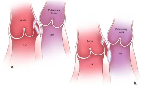
com-promised, and left or right ventricular outflow obstruction is prevented or relieved. In most cases of aorto-left ven-tricular tunnel, this has been accomplished by transaortic patch closure of the aortic end, and placement of a second patch through the tunnel itself to close the ventricular ori-fice and support the aortic valve (Figure 2a). Alternatively, the tunnel wall itself can be used to achieve an equivalent anatomical result [44]. Closure of the aortic orifice by direct suture also has sometimes given good results [11,45,46], but more often, the tunnel recurs or progres-sive aortic regurgitation through an unsupported or dis-torted right coronary leaflet leads to subsequent valve replacement. If the ventricular end of an aorto-left ven-tricular tunnel is not closed, residual high pressure in the blind-ending pouch may compress the right ventricular outflow [22]. In tunnels communicating with a low-pres-sure right ventricle, it is less certain that the ventricular ori-fice need be closed, although this has been done through a right ventricular incision in most reported cases (Figure 2b).
When the ostium of a coronary artery arises proximally within a tunnel, the patch is deviated distally to conserve perfusion from the aorta. More distal origin of a coronary artery from a tunnel above the right aortic sinus is man-aged by resection of the orifice and reattachment to the ascending aorta [12,44,47]. Distal coronary origin in a tunnel arising above the left aortic sinus is more difficult to manage, because it lies behind the heart. As these gen-erally have been associated with tunnels to the right ven-tricle, however, closure of just the ventricular end is an option to maintain coronary perfusion [31]. Patch angi-oplasty using autologous pericardium or saphenous vein has been successful in restoring flow to a right coronary artery whose ostium was atretic [7].
Associated lesions of the aortic valve are treated as indi-cated either separately or at the time of tunnel repair. This has included balloon valvuloplasty [11], open commis-surotomy [48-50], homograft root replacement [51], or aortoventriculoplasty [15] for stenosis or atresia in neonates or small infants, as well as repair or replacement of the valve in older patients. Obstruction of the pulmo-nary valve has been successfully managed by percuta-neious valvuloplasty preoperatively [21] or open valvotomy at the time of surgery [23,31]. However, attempted percutaneous balloon dilation did not relieve the obstruction on one occasion [31].
ven-tricular function. Given the desirability of supporting the aortic leaflet and the variable origins of coronary arteries in this malformation, however, it is questionable if percu-taneous interventions can achieve long-term outcomes equivalent to those of current surgical techniques, for which operative mortality approaches zero [9,11].
Unresolved questions
While follow-up extending to 35 years has now docu-mented that mild aortic regurgitation may remain stable for a considerable period of time in postoperative patients [11,54], the very long-term results of two-patch repair in the modern era are awaited, as are elucidation of the molecular or genetic basis of the anomaly. The natural history of disproportionate ascending aortic enlargement which occurs early in life with this malformation is also uncertain and may eventually emerge as the ultimate determinant of outcome.
References
1. Edwards JE, Burchell HB: The pathological anatomy of deficien-cies between the aortic root and the heart, including aortic sinus aneurysms. Thorax 1957, 12:125-39.
2. Levy MJ, Lillehei CW, Anderson RC, Amplatz K, Edwards JE: Aor-tico-left ventricular tunnel. Circulation 1963, 27:841-53. 3. Sommerville J, English T, Ross DN: Aorto-left ventricular tunnel.
Clinical features and surgical management. Br Heart J 1974,
36:321-8.
4. McKay R, Anderson RH, Cook AC: The aorto-ventricular tun-nels. Cardiol Young 2002, 12:563-80.
5. Cook AC, Fagg NKL, Ho SY, Groves AMM, Sharland GK, Anderson RH, Allen LD: Echocardiographic-anatomical correlations in aorto-left ventricular tunnel. Br Heart J 1995, 74:443-8. 6. Saylam A, Tuncali T, Ikizler C, Aytaç A: Aorto-right ventricular
tunnel. A new concept in congenital cardiac malformations. Ann Thorac Surg 1974, 18:634-7.
7. Bonnet D, Bonhoeffer P, Sidi D, Kachaner J, Acar P, Villain E, Vouhé PR: Surgical angioplasty of the main coronary arteries in chil-dren. J Thorac Cardiovasc Surg 1999, 117:352-7.
8. Bove KE, Schwartz DC: Aortico-left ventricular tunnel. A new concept. Am J Cardiol 1967, 19:696-709.
9. Horváth P, Balaji S, Škovránek S, Hucin B, de Leval MR, Stark J: Sur-gical treatment of aortico-left ventricular tunnel. Eur J Cardi-othorac Surg 1991, 5:113-7.
10. Hovaguimian H, Cobanoglu A, Starr A: Aortico-left ventricular tunnel: a clinical review and new surgical classification. Ann Thorac Surg 1988, 45:106-12.
Surgical repair of aorto-left ventricular tunnel (a) and aorto-right ventricular tunnel (b) Figure 2
11. Martins JD, Sherwood MC, Mayer JE Jr, Keane JF: Aortico-left ven-tricular tunnel: 35 – year experience. J Am Coll Cardiol 2004,
44:446-50.
12. Rauzier JM, Bonnet D, Zniber L, Sidi D, Aggoun Y, Acar P, Kachaner J, Vouhe P: Aortic-ventricular tunnel with right coronary artery atresia. Arch Mal Coeur Vaiss 1997, 90:725-7.
13. Rosengart TK, Redel DA, Stark JF: Surgical repair of aorto-right ventricular tunnel in an infant. Ann Thorac Surg 1993, 55:520-2. 14. Bitar FF, Smith FC, Kavey R-EW, Kveselis DA, Byrum CJ, Brandt B, Gaum WE: Aortico-left ventricular tunnel with aortic atresia in the newborn. Am Heart J 1993, 126:1480-2.
15. Guyton RA, Michalik RE, McIntyre AB, Plauth WH Jr, Nugent EW, Hatcher CR Jr, Williams WH: Aortic atresia and aortico-left ventricular tunnel: successful surgical management by Konno aortoventriculoplasty in a neonate. J Thorac Cardiovasc Surg 1986, 92:1099-1105.
16. Sousa-Uva M, Touchot A, Fermont L, Piot D, Delezoide AL, Serraf A, Lacour-Gayet F, Roussin R, Bruniaux J, Planché C: Aortico-left ven-tricular tunnel in fetuses and infants. Ann Thorac Surg 1996,
61:1805-10.
17. Meldrum-Hanna W, Schroff R, Ross DN: Aortico-left ventricular tunnel: late follow-up. Ann Thorac Surg 1986, 42:304-6.
18. Warnke H, Bartel J, Blumenthal-Barby Ch: Aortico-ventricular tunnel. Thorac Cardiovasc Surg 1988, 36:86-8.
19. Akalin H, Erol Ç, Oral D, Çorapçioglu T, Uçanok K, Özyurda Ü, Ulu-soy V: Aortico-left ventricular tunnel: successful diagnostic and surgical approach to the oldest patient in the literature. J Thorac Cardiovasc Surg 1989, 97:804-5.
20. Jureidini SB, de Mello D, Norui S, Kanter K: Aortico-right ven-tricular tunnel and critical pulmonary stenosis: diagnosis by two-dimensional and Doppler echocardiography and angiog-raphy. Pediatr Cardiol 1989, 10:99-103.
21. Martin Jimenez J, Gonzales Diegues CC, Quero Jimenez C, Rico Gomez F, Bermudez Canete R, Quero Jimenez M: Aortico-left ven-tricular tunnel associated with pulmonary valve stenosis. Rev Esp Cardiol 1996, 49:921-4.
22. Knott-Craig CJ, van der Merwe PL, Kalis NN, Hunter J: Repair of aortico-left ventricular tunnel associated with subpulmonary obstruction. Ann Thorac Surg 1992, 54:557-9.
23. Turley K, Silverman NH, Teitel D, Mavroudis C, Snider R, Rudolph A:
Repair of aortico-left ventricular tunnel in the neonate: sur-gical, anatomic and echocardiographic considerations. Circu-lation 1982, 65:1015-20.
24. Kleikamp G, Minami K, Thies W-R, Dohmann R, Raute-Kreisen U, Meyer H, Körfer R: Aotra-right ventricular tunnel with a rudi-mentary valve and an anomalous origin of the left coronary artery. J Thorac Cardiovasc Surg 1992, 104:1759-60.
25. Okoroma EO, Perry LW, Scott LP III, McClenathan JE: Aortico-left ventricular tunnel. Clinical profile, diagnostic features and surgical considerations. J Thorac Cardiovasc Surg 1976, 71:238-44. 26. Kafka H, Chan KL, Leach AJ: Asymptomatic aortico-left
ven-tricular tunnel in adulthood. Am J Cardiol 1989, 63:1021-2. 27. Ribeiro P, Bun-Tan LB, Oakley CM: Management of aortic left
ventricular tunnel. Br Heart J 1985, 54:333-6.
28. Serino W, Andrade JL, Ross D, de Leval M, Sommerville J: Aorto-left ventricular communication after closure. Late postopera-tive problems. Br Heart J 1983, 49:501-6.
29. Palacio J, Perretta A, Sanchez B, Alperovich M: Intrapericardial congenital supravalvular aortic aneurysm communicating with the outflow-tract of the left ventricle. Hypoplasia of the aortic orifice and ascending aorta. J Cardiovasc Surg (Torino)
1964, 5:401-407.
30. Roberts WC, Morrow AG: Aortico-left ventricular tunnel. A cause of massive aortic regurgitation and of intracardiac aneurysm. Am J Med 1965, 39:662-7.
31. Hruda J, Hazekamp MG, Sobotka-Plojhar MA, Ottenkamp J: Repair of aorto-right ventricular tunnel with pulmonary stenosis and an anomalous origin of the left coronary artery. Eur J Car-diothorac Surg 2002, 21:1123-5.
32. Bernanke DH, Velkey JM: Development of the coronary blood supply: Changing concepts and current ideas. Anat Rec 2002,
269(4):198-208.
33. Bogers AJ, Gittenberger-de Groot AC, Dubbledam JA, Huysmans HA: The inadequacy of existing theories on development of the proximal coronary arteries and their connexions with the arterial trunks. Int J Cardiol 1998, 20(1):117-123.
34. Ya J, van den Hoff MJ, de Boer PA, Tesink-Taekema S, Franco D, Moorman AF, Lamers WH: Normal development of the outflow tract in the rat. Circ Res 1998, 82:464-72.
35. Bash SE, Huhta JC, Nihill MR, Vargo TA, Hallman GL: Aortico-left ventricular tunnel with ventricular septal defect: two-dimen-sional/Doppler echocardiographic diagnosis. J Am Coll Cardiol
1985, 5:757-60.
36. Grant P, Abrams LD, De Giovanni JV, Shah KJ, Silove ED: Aortico-left ventricular tunnel arising from the Aortico-left aortic sinus. Am J Cardiol 1985, 55:1657-8.
37. Humes RA, Hagler DJ, Julsrud PR, Levy JM, Feldt RH, Schaff HV: Aor-tico-left ventricular tunnel: diagnosis based on two-dimen-sional echocardiography, color flow Doppler imaging, and magnetic resonance imaging. Mayo Clin Proc 1986, 61:901-7. 38. Perry JC, Nanda NC, Kicks DG, Harris JP: Two-dimensional
echocardiographic identification of aortico-left ventricular tunnel. Am J Cardiol 1983, 52:913-4.
39. Sreeram N, Franks R, Arnold R, Walsh K: Aortico-left ventricular tunnel: long-term outcome after surgical repair. J Am Coll Car-diol 1991, 17:950-5.
40. Biffanti R, Reffo E, Sanders SP, Maschietto N, Stellin G, Milanesi O:
Two-dimensional and real-time three-dimensional echocar-diographic fetal diagnosis of aorto-ventricular tunnel. Circula-tion 2005, 111:e367-8.
41. Grab D, Paulus WE, Terinde R, Lange D: Prenatal diagnosis of an aortico-left ventricular tunnel. Ultrasound Obstet Gynecol 2000,
15:435-8.
42. Siepe M, Dittrich S, Beyersdorf F, Schlensak C: Aortic atresia with aortico-left ventricular tunnel mimicking severe aortic incompetence in utero. Eur J Cardiothorac Surg 2006,
29(5):845-847.
43. Sakurai M, Takahara Y, Takeuchi S, Mogi K: Ascending aortic aneu-rysm following aortico-ventricular tunnel repair. Jpn J Thorac Cardiovasc Surg 2006, 54:182-4.
44. Grünenfelder J, Zünd G, Prêtre R, Schmidli J, Vogt PR, Turina MI:
Right coronary artery from aorto-left ventricular tunnel: case report of a new surgical approach. J Thorac Cardiovasc Surg
1998, 116:363-5.
45. Norwicki ER, Abderdeen E, Friedman S, Rashkind WJ: Congenital left aortic sinus-left ventricle fistula and review of aortocar-diac fistulas. Ann Thorac Surg 1997, 23(4):378-388.
46. Spooner EW, Dunn JM, Behrendt DM: Aortico-left ventricular tunnel and sinus of Valsalva aneurysm. Case report with operative repair. J Thorac Cardiovasc Surg 1978, 75:232-6. 47. Hucin B, Horvath P, Skovránek J, Reich O, Samánek M: Correction
of aortico-left ventricular tunnel during the first day of life. Ann Thorac Surg 1989, 47:254-6.
48. Diamant S, Luber JM Jr, Gootman N: Successful repair of aortico-left ventricular tunnel associated with severe aortic stenosis in a newborn. Pediatr Cardiol 1985, 6:171-3.
49. Villani M, Tirboschi R, Marino A, De Tommasi M, Velitti F, Giani PC, Parenzan L: Aortico-left ventricular tunnel in infancy. Two sur-gical cases. Scand J Thorac Cardiovasc Surg 1980, 14(2):169-175. 50. Webber S, Johnston B, LeBlanc J, Patterson M: Aortic-left
ventricu-lar tunnel associated with critical aortic stenosis in the new-born. Pediatr Cardiol 1991, 12:237-40.
51. Weldner P, Dhillon R, Taylor JF, de Leval MR: An alternative method for repair of aortico-left ventricular tunnel associ-ated with severe aortic stenosis presenting in a newborn. Eur J Cardiothorac Surg 1996, 10:380-2.
52. Chessa M, Chaudhari M, De Giovanni JV: Aorto-left ventricular tunnel: transcatheter closure using an Amplatzer duct occluder device. Am J Cardiol 2000, 86:253-4.
53. Vijayalakshmi IB, Chitra N, Prabhu Deva AN: Use of an Amplatzer duct occluder for closing an aortico-left ventricular tunnel in a case of noncompaction of the left ventricle. Pediatr Cardiol
2004, 25:77-9.