©2016 JNAS Journal-2016-5-6/237-241 ISSN 2322-5149 ©2016 JNAS
Adenosarcoma of the Uterus: A Case Report
Fatemeh Montazer
1*, Anahita Nosrati
1and Mahboobeh Majlesi
21- Assistant Professor of pathology, Department of pathology, Gastrointestinal Cancer Register Center , Imam khomaini Hospital, Mazandaran University of Medical Sciences, Sari, Iran.
2- Resident of pathology, Department of pathology,Imam khomaini Hospital, Mazandaran University of Medical Sciences, Sari, Iran.
Corresponding author:
Fatemeh Montazer
ABSTRACT: Adenosarcoma is a biphasic tumor with benign epithelial elements and a sarcomatous stroma. Typically it presents as a solitary large polypoid mass arising from the uterine fundus. In this case report we presented a 64 year-old woman with postmenopausal bleeding and endometrial polypoid lesion in sonographic report. Pathologic examination of the curetting specimen revealed a mixed epithelial-mesenchymal tumor that it was diagnosed as adenosarcoma.
Keywords: adenosarcoma, postmenopausal bleeding.
INTRODUCTION
Adenosarcoma is a biphasic tumor with benign epithelial elements and a sarcomatous stroma(1). This entity was originally described by Clement and Scully in 1974 as Müllerian adenosarcoma(2). Although it usually arises in the endometrium, it can arise in the cervix, the myometrium, fallopian tubes, and ovaries. (1). Adenosarcoma is a rarely observed mullerian tumor of the uterus and represents 8% of uterine sarcomatous tumors(3). Occurs in women of all ages. The median age is 50 years, with a range of 13-83years (4). Although adenosarcoma is typically low grade tumor, recurrences have been reported in up to 30–40% of patients while 20–25% of women die from their tumors (1).
Typically it presents as a solitary large polypoid mass arising from the uterine fundus and fills the endometrial cavity and protrudes from the uterine cervix(1). The most common presenting symptom is abnormal vaginal bleeding .The diagnosis can be made with endometrial curettage(5).
The aim of our experience is to show the difficulty of the diagnosis with endometrial biopsies and difficulty of the diagnosis from other Mixed epithelial-mesenchymal tumors such as adenofibromas or carcinosarcomas.
Case presentation:
238
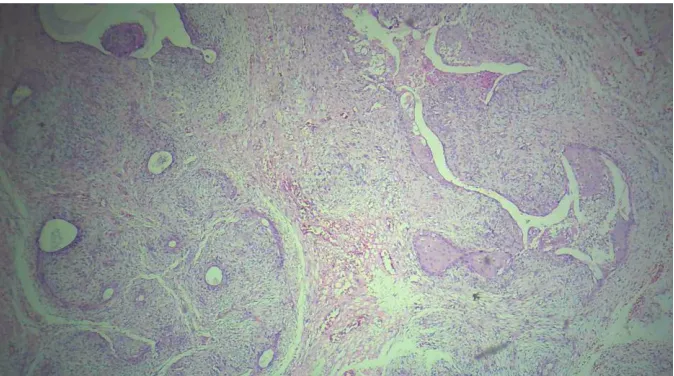
Fig 1. mixed epithelial-mesenchymal tumor with cleft like spaces(phyllodes tumor-like)Fig 2. mixed epithelial-mesenchymal tumor with prominent squamous epithelial component
239
Fig 4. Increased cellularity of stroma around the epithelial elements forming a cambium layer.Fig 5. Mesenchymal cells were showed mild nuclear atypia and mitotic feature.
Laparatomy was planned on the basis of this diagnosis. After the peritoneal washing, a total abdominal hysterectomy-bilateral salpingo-oophorectomy was performed along with extirpation of the parametrium and pelvic-para-aortic lymph node sampling. At laparotomy, the uterus was fist size. On opening in lower segment of uterus a small polypoid lesion measuring 2×0.5×0.5 cm was seen. On microscopic examination the tumor showed glandular epithelium with little atypia and proliferation of atypical mesenchymal cells. Mitosis exceeded 2 per 10 high power fields. No myometrial invasion or lymph node metastasis was seen. The lesion was confined to the uterus. no serosal invasion was observed. Both ovaries were intact. Peritoneal cytology revealed no malignant cells. Histopathological final diagnosis was adenosarcoma.
Discussion:
240
Microscopically, the glands were lined by benign or atypical glandular epithelium, together with sarcomatous stromal cells which showed characteristic structures of ‘periglandular cuff’ of increased cellularity and ‘intraglandular polypoid projections. (9)The epithelium typically is cytologically bland, but hyperplastic and even atypical hyperplastic epithelium is occasionally noted (10). Increased cellularity of stroma around the epithelial elements forming a cambium layer could be a useful clue suggesting in diagnosis of adenosarcoma (8). Using the World Health Organization definition, stromal mitotic activity of 2 or more per 10 high-power fields is required for a diagnosis of adenosarcoma but in practice the diagnosis is made with stromal mitotic activity less than this if the characteristic architecture and cambium layer is present (11).
The diagnosis could be sometimes only made or confirmed after multiple pathologic examinations.In some cases, many endometrial samples are needed in order to find uterine adenosarcoma (12). In fact, in our case, the diagnosis of uterine adenosarcoma was difficult and only suspected after multiple histologic analysis. Because of suspected lesion, hysterectomy was performed and permitted to confirm histologically the diagnosis of uterine adenosarcoma. Adenosarcomas are characterized by weaker malignancy potential, late-onset local recurrence (25%),and rare metastasis. recurrence is observed more frequently in the vagina and pelvis (60%) (5).
Unfavourable prognostic factors are sarcomatous overgrowth, deep myometrial invasion, presence of heterologous elements and extrauterine spread (13).
The epithelial component of adenosarcoma is keratin positive, and it usually stains for estrogen and progesterone receptors.
The mesenchymal cells in adenosarcoma typically show cytoplasmic staining for CD10, and there is nuclear staining for estrogen and progesterone receptors and for WT-l (14).
Adenofibroma is a rare benign tumor of the cervix or endometrium. Over time the diagnostic criteria for adenosarcoma have been broadened to the point that some authors now doubt the existence of adenofibromas (4).The overall architecture is similar to adenosarcoma, but the stroma is predominantly fibrous and less cellular than that of an adenosarcoma, with no condensation of stromal cells around the epithelial elements. The stromal cells are bland, and MF are absent or difficult to find. Some adenosarcomas contain bland areas similar in appearance to an adenofibroma, so the diagnosis of an adenofibroma generally requires hysterectomy so that the entire tumor can be evaluated microscopically(15).
Adenosarcoma is usually treated by hysterectomy and bilateral salpingo-oophorectomy. Metastasis to lymph nodes is rare, although occasional patients whose tumors show sarcomatous overgrowth have pelvic lymph node metastases (16). Adenosarcomas without sarcomatous overgrowth do not require adjuvant therapy of any type. Hormonal therapy may be an option in rare cases with advanced or recurrent disease. adenosarcomas with sarcomatous overgrowth should be treated according to recommendations for high-grade sarcomas.(17)
REFERENCES
1- Taga S, Sawada M, Nagai A, Yamamoto D, Hayase R. A case of adenosarcoma of the uterus. Case Rep Obstet Gynecol. 2014;2014:342187.
2- Clement PB, Scully RE. Müllerian adenosarcoma of the uterus. A clinicopathologic analysis of ten cases of a distinctive type of müllerian mixed tumor. Cancer. 1974 Oct;34(4):1138-49.
3- Piura B, Rabinovich A, Meirovitz M, Yanai-Inbar I. Mullerian adenosarcoma of the uterus: case report and review of literature. Eur J Gynaecol Oncol. 2000;21(4):387-90.
4- Gallardo A, Prat J. Mullerian adenosarcoma: a clinicopathologic an immunohistochemical study of 55 cases challenging the existence of adenofibroma. Am J Surg Pathol. 2009 Feb;33(2):278-88.
5- Tinar S, Sehirali S, Inal MM, Yildrim Y, Celik E, Yigit S. Adenosarcoma of the uterus: a case report. MedGenMed. 2004 Jan 12;6(1):51.
6- Fait T, Zivny J, Freitag P, Kuzel D. Adenosarcoma of the uterine body in a 19-year-old woman - three year survival: case report. Eur J Gynaecol Oncol. 2001, 22: 61-63.
7- Fatnassi R, Amri F. Adenosarcoma of the uterus: a case report. J Gynecol Obstet Biol Reprod (Paris). 2005 May;34(3 Pt 1):270-2.
8- Sakr R, Marzouk P, Bricou A, Demaria F, Cortez A, Benifla JL. Uterine adenosarcoma associated to lymphovascular
emboli: a case report.Cases J. 2009 May 18;2:7515.
9- Shi Y, Liu Z, Peng Z, Liu H, Yang K, Yao X. The diagnosis and treatment of Mullerian adenosarcoma of the uterus. Aust N Z J Obstet Gynaecol. 2008 Dec;48(6):596-600
10- Gallardo A, Prat J. Mullerian adenosarcoma: a clinicopathologic and immunohistochemical study of 55 cases challenging the existence of adenofibroma. Am J Surg Pathol. 2009 Feb;33(2):278-88.
241
12- McCluggage WG. My approach to the interpretation of endometrial biopsies and curettings. J Clin Pathol.2006;59:801–812.
13- Sinha A, Phukan JP, Sengupta S, Guha P. Mullerian adenosarcoma of uterus with sarcomatous overgrowth and heterologous component associated with stromal deposit in omentum: a case report and review of the literature. Case Rep Med. 2012;2012:820378.
14- Amant F, Schurmans K, Steenkiste E, Verbist L, Abeler VM, Tulunay G,et al.Immunohistochemical determination of
estrogen and progesterone receptor positivity in uterine adenosarcoma. Gynecol Oncol. 2004 Jun;93(3):680-5 15- Clement PB, Scully RE. Mullerian adenosarcoma of the uterus: a clinicopathologic analysis of 100 cases with a
review of the literature. Hum Pathol. 1990 Apr;21(4):363-81.
16- Kaku T, Silverberg SG, Major FJ, Miller A, Fetter B, Brady MF.Adenosarcoma of the uterus: a Gynecologic Oncology Group clinicopathologic study of 31 cases. Int J Gynecol Pathol. 1992;11(2):75-88.