OncoTargets and Therapy
Dove
press
O r i g i n a l r e s e a r c h
open access to scientific and medical research
Open access Full Text article
crenolanib, a PDgFr inhibitor, suppresses
lung cancer cell proliferation and inhibits
tumor growth in vivo
Ping Wang1
liqiang song2
hui ge1
Pule Jin1
Yifang Jiang1
Wenxia hu1
nan geng1
1Department of respiratory
Medicine, Fourth hospital of hebei Medical University, shijiazhuang, hebei, People’s republic of china;
2Department of Pathology, school of
Basic Medical sciences, Peking Union Medical college, Beijing, People’s republic of china
correspondence: Ping Wang
12 Jiankang rd, Qiaodong, shijiazhuang, hebei 050011, People’s republic of china Tel +86 139 3113 0696
email pingwang1393@gmail.com
Abstract: Platelet-derived growth factor (PDGF) and its receptors (PDGFR), including PDGFRα and PDGFRβ, play important roles in tumorigenesis, tumor progression, and the regulation of stromal cell function. Constitutive activation of PDGFR signaling, gene rear-rangement, and activating mutations of PDGFR have been identified in various types of human tumors and malignancies. PDGFRα and PDGFRβ belong to the family of type III receptor tyrosine kinases and, upon stimulation, activate downstream signaling cascades. Crenolanib is a specific tyrosine kinase inhibitor that targets and inhibits the kinase activity of PDGFR and the FMS-related tyrosine kinase 3. Its clinical efficacy in several human tumors is currently under investigation in Phase II clinical trials. In this study, we examined the potential role of crenolanib in the treatment of non-small-cell lung cancer (NSCLC). Using A549 cells as a model system, we have shown that crenolanib is capable of suppressing proliferation and inducing apoptosis in a dose-dependent manner. Crenolanib-treated cells have reduced migratory activity in response to inducers of chemotaxis. Furthermore, the in vivo antitumor activity of crenolanib was confirmed in an NSCLC xenograft tumor model. Injection of crenolanib significantly inhib-ited the growth of tumor mass by inducing apoptosis in tumor cells. Our results provide strong evidence supporting the use of crenolanib as a potential therapeutic agent in treating NSCLC. This work sets a foundation for further development of targeted and personalized therapeutics for lung cancer.
Keywords: platelet-derived growth factor receptor signaling, receptor tyrosine kinase, tyrosine kinase inhibitor, non-small-cell lung cancer, chemotherapy, targeted therapy
Introduction
Platelet-derived growth factor receptors (PDGFRs), including PDGFRα and PDGFRβ,
belong to the family of cell surface type III receptor tyrosine kinases (RTKs).1,2 Upon binding of the ligands, platelet-derived growth factors (PDGFs), the receptor complex is activated and the cytosolic domains serve as docking sites for coactivators and sub-sequently initiate downstream signaling cascades such as MAPK, PI3K, and STAT3 pathways.3–6 PDGFR signaling regulates a variety of biological processes, including cellular growth, cellular differentiation, cell migration, and angiogenesis.1,7 Deregulated PDGFR signaling has been implicated in the pathogenesis of several human diseases and malignancies.8–11 For example, in patients with gastrointestinal stromal tumors, chronic myelomonocytic leukemia, and glioblastoma multiforme, mutations have been identi-fied in the genes encoding PDGFR, which results in constitutive activation of the kinase activity, overstimulation of signal transduction, interaction with adjacent stroma and vasculature, and eventually autocrine regulation of tumor cell growth.12–15 Therefore, the
OncoTargets and Therapy downloaded from https://www.dovepress.com/ by 118.70.13.36 on 26-Aug-2020
For personal use only.
Number of times this article has been viewed
This article was published in the following Dove Press journal: OncoTargets and Therapy
Dovepress
Wang et al
PDGFR pathway represents a potential therapeutic target in patients harboring activating mutations of PDGFR.
With PDGFR as a promising target in cancer therapeutics, efforts have been made to develop specific kinase inhibitors that disrupt the receptor activation of PDGFR. Crenolanib is a spe-cific tyrosine kinase inhibitor that targets and inhibits the kinase activity of PDGFR and the FMS-related tyrosine kinase 3 (FLT3).16–18 In a completed Phase I clinical trial, its pharma-cokinetics and safety were evaluated and the recommended dosage was well tolerated by patients.16 The clinical efficacy of crenolanib in several human tumors is currently under inves-tigation in multiple Phase II clinical trials. Unlike other RTK inhibitors such as imatinib mesylate and motesanib, which have been previously studied and tested in clinical trials for various types of cancer, crenolanib preferentially targets the phospho-rylated, thus active, form of PDGFR that is often derived from constitutively active mutations of genes encoding PDGFR.17,19,20 Therefore, crenolanib may provide targeted therapy to patients with PDGFR mutations and have reduced toxicity compared with other broad-spectrum multikinase inhibitors.
In this study, we aim to test the potential efficacy of crenolanib in treating non-small-cell lung cancer (NSCLC), a collective type of epithelial lung carcinoma. Previous research has suggested a strong correlation between
deregu-lated PDGFR signaling and NSCLC tumor progression.21–24
Expression and activity of PDGF and PDGFR have been used as prognostic indicators in lung carcinoma. The traditional treatment for NSCLCs involves surgical resection followed by applications of antiangiogenic agents such as targeted cytotoxic drugs.25,26 A549 cells were derived from human
lung carcinoma and express PDGFRα in high levels.27 By
inhibiting the PDGFR signaling, our data demonstrated that crenolanib treatment suppressed the proliferation and migra-tion of A549 cells and inhibited tumor growth in an NSCLC xenograft tumor model. Our study provides strong evidence supporting the use of crenolanib as a personalized and effec-tive second-line therapy for NSCLC patients.
Materials and methods
reagents, cells, and mice
Crenolanib (CP-868596) was purchased from Selleck Chemicals LLC (Houston, TX, USA). Human NSCLC cell line A549 was purchased from the American Type Culture Collection (Rockville, MD, USA). A549 cells were maintained in RPMI medium supplemented with 10% inactivated fetal bovine serum, 2 mM L-glutamine, and 100 U/ mL penicillin–streptomycin in a humidified incubator at 37°C in 5% CO2. The athymic nude mice were purchased from Slack Company (Shanghai, People’s Republic of China). Care and
treatment of mice were in accordance with the Animal Care and Use Committee guidelines of Hebei Medical University (Shijiazhuang, Hebei, People’s Republic of China).
cell viability assay
Cells were seeded in 96-well plates in triplicate and treated with different doses of crenolanib for 24, 48, or 72 hours as indicated in Figure 1. Cell viability at the end of each treat-ment was measured using CellTiter-Glo Assay Kit (Promega, Madison, WI, USA) according to the manufacturer’s instruc-tions. Briefly, an equal volume of the CellTiter-Glo reagent (100 µL) was added directly to the cell culture in 96-well plates and the plates were placed on an orbital shaker for 2 minutes followed by incubation at room temperature for 10 minutes. Luminescence was read in a luminometer (Molecular Devices, Sunnyvale, CA, USA).
Flow cytomery analysis
Cells treated with different concentrations of crenolanib were harvested, fixed, and stained with propidium iodide using a BD Cycletest Plus DNA Reagent Kit (Becton Dickinson, Franklin lakes, NJ, USA). Cells were collected and analyzed for deoxyribonucleic acid (DNA) content using FACSCalibur (BD Biosciences, San Jose, CA, USA) and CellQuest Pro flow cytometry analytic software (Becton Dickinson).
nuclei staining
Cells were seeded on polylysine-coated glass coverslips and treated with different concentrations of crenolanib as indicated in Figure 2. After 48 hours, cells were fixed with 4% paraform-aldehyde for 10 minutes and stained with Hoechst (Sigma-Aldrich, St Louis, MO, USA) for 10 minutes. Images were obtained and analyzed using an Olympus BX51 fluorescent microscope (Olympus America Inc., Melville, NY, USA).
Transwell assay
1×10⁵ cells in RPMI media supplemented with 1% fetal bovine serum were seeded into the upper compartments of the 24-transwell Boyden chamber (Costar, Bedford, MA, USA). Different concentrations of crenolanib were added to cells
as indicated in Figure 3. Medium (650 µL) supplemented
with 10% fetal bovine serum was loaded into the lower chambers to be used as a chemoattractant. After incubation for 12 hours, the nonmigrating cells were wiped off from the upper surface, and the migrated cells on the lower side were fixed, stained with 0.1% crystal violet, and photographed using an Eclipse LV150L microscope (Nikon Corporation, Tokyo, Japan). For absorbance measurement, the migrated cells on the lower side of the chamber were lysed with 10% acetic acid and measured at 595 nm.
OncoTargets and Therapy downloaded from https://www.dovepress.com/ by 118.70.13.36 on 26-Aug-2020
Dovepress crenolanib inhibits lung cancer cell growth
Efficacy studies in a mouse
xenograft model in vivo
A549 cells were injected into the axillary regions of mice (2×10⁶ cells/mouse). When the tumor volumes reached
70 mm3, the mice were randomly allocated to the control
group, low-dose crenolanib group (10 mg/kg), or high-dose crenolanib group (20 mg/kg) (n=6 per group). The vehicle for crenolanib treatment consists of 10% 1-methyl-2-pyrrolidinone and 90% polyethylene glycol 300 Aldrich). The tumor size and mouse body weight were measured every other day for about 2 weeks. The tumor volume was calculated as follows: (mm3) = (width × width × length)/2. After treatment, the mice were euthanized using carbon dioxide, and the tumors were harvested and analyzed.
immunohistochemistry analysis
Tumor samples were harvested from mice, trimmed
into 5 mm3 pieces, and immediately fixed in 10% neutral
buffered formalin. After fixation, the samples were trans-ferred to 70% ethanol, embedded into paraffin blocks, and sectioned in 5 mm thicknesses. Terminal deoxynucleotidyl transferase dUTP nick end labeling (TUNEL) assay was per-formed using FragEL kit (Calbiochem, Darmstadt, Germany) according to the manufacturer’s instructions. Briefly, slides were stained with TUNEL reagent for 1 hour followed by incubation with an Alexa Fluor 488-conjugated antibody for 30 minutes. The nuclei were stained with Hoechst for 20 minutes. The slides were photographed using an Olympus BX51 fluorescent microscope (Olympus America Inc.).
statistics
Student’s t-test and analysis of variance were performed using StatView (SAS Institute, Cary, NC, USA). The data shown are the mean values of triplicate measurements with error bars corresponding to standard deviation. P,0.05 was considered statistically significant.
Results
crenolanib decreases nsclc
cell viability
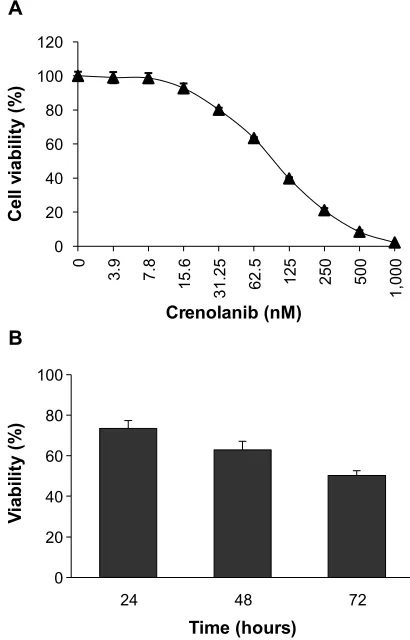
PDGFR signaling promotes cell survival through activation of various downstream signal transduction cascades, including MAPK, PI3K, and STAT3 pathways. To determine whether inhibition of PDGFR signaling affects cellular proliferation, the A549 NSCLC cells were treated with crenolanib with a series of concentrations for 72 hours, and the cell viability was measured. We observed a dose-dependent inhibition of
cancer cell viability (Figure 1A). To find out the onset of action for crenolanib, A549 cells were treated with crenolanib (50 nM) for 24, 48, or 72 hours, followed by measurement of cell viability. The inhibitory effect of crenolanib was observed as early as 24 hours after treatment (Figure 1B).
crenolanib induces apoptosis
in nsclc cells
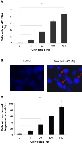
PDGF is a potent mitogenic factor that enhances pro-liferation and antagonizes apoptosis. We further tested whether treatment of crenolanib was capable of inducing apoptosis in cancer cells. After treatment with crenolanib for 48 hours, the sub-G1 DNA content of A549 cells was measured as an indicator of late stage apoptosis.28 Similar to the previous observation in cell viability, cre-nolanib caused increased sub-G1 DNA content in a dose-dependent manner, indicating that inhibition of PDGFR signaling promoted cells to undergo apoptosis (Figure 2A). Apoptosis-induced condensed and fragmented DNA is visualized in Figure 2B. Consistently, the number of cells
0 20 40 60 80 100 120
0
3.
9
7.
8
15
.6
31.25 62
.5
125 250 500
1,000
Ce
ll
viabil
it
y
(%
)
Crenolanib (nM)
0 20 40 60 80 100
24 48 72
V
iabil
it
y
(%
)
Time (hours) A
B
Figure 1 crenolanib decreases non-small-cell lung cancer cell viability.
Notes: (A) a549 cells were treated with crenolanib (3.9∼1,000 nM) for 72 hours followed by measurement with cell viability assay. (B) a549 cells were treated with crenolanib (50 nM) for 24, 48, or 72 hours followed by measurement with cell viability assay. in both experiments, viability of cells without crenolanib treatment was set to 100%.
OncoTargets and Therapy downloaded from https://www.dovepress.com/ by 118.70.13.36 on 26-Aug-2020
Dovepress
Wang et al
A
B
C 100
80
60
Cells with sub-G1 DNA
(%)
Crenolanib (nM)
Crenolanib (nM)
Crenolanib (500 nM) Control
** **
Cells with condensed/ fragmented nuclei (%)
40
20
0
100
80
60
40
20
0
0 4 20 100 500
0 4 20 100 500
Figure 2 crenolanib induces apoptosis in non-small-cell lung cancer cells.
Notes: (A) A549 cells were treated with crenolanib at the indicated concentrations for 48 hours followed by propidium iodide staining and flow cytometry analysis for sub-g1 deoxyribonucleic acid (Dna) content. (B) a549 cells were incubated with crenolanib (500 nM) for 48 hours. The nuclei were stained with hoechst and analyzed using a fluorescent microscope. The representative images are shown. Red arrows indicate apoptotic cells with condensed or fragmented DNA. (C) The number of cells with condensed/fragmented nuclei was quantitated by counting in five random fields. Percentage was calculated and the averaged results are shown.
Note: **P,0.01.
with condensed/fragmented DNA increased proportionally with crenolanib treatment (Figure 2C).
crenolanib inhibits cell migration
in nsclc cells
PDGF is a critical chemoattractant that binds to its cell surface receptor and regulates cell proliferation and chemotaxis.29
Malignant tumor cells are capable of migrating and invading to a second site for tumorigenesis. PDGFR signaling plays an important role in this process and the following angiogenesis to contribute to the metastasis of tumors. Therefore, PDGF/ PDGFR becomes an attractive target for cancer therapeutics.
We next investigated the effect of PDGFR inhibition on cancer cell migration. As shown in Figure 3A and B, treatment
OncoTargets and Therapy downloaded from https://www.dovepress.com/ by 118.70.13.36 on 26-Aug-2020
Dovepress crenolanib inhibits lung cancer cell growth
120
100
80
60
40
20
0
12.5 25 50
Crenolanib (nM)
Inhibition (%)
A Control Crenolanib (12.5 nM) Crenolanib (25 nM) Crenolanib (50 nM)
B **
Figure 3 crenolanib inhibits cell migration in non-small-cell lung cancer cells.
Notes: (A) a549 cells were treated with crenolanib (12.5, 25, or 50 nM) for 10 hours. The nonmigrated cells on the upper chamber of the transwell were removed and the migrated cells on the lower side were stained and photographed. The representative images are shown. (B) The migrated cells were lysed and colorimetric determination was made at 595 nm. summary of the quantitation of the inhibition from transwell assay is shown.
Note: **P,0.01.
with crenolanib inhibited the migration of A549 cells in a dose-dependent manner. This inhibition is unlikely due to the cytotoxicity of crenolanib, as the duration of treat-ment was significantly shorter and the concentrations were significantly lower compared with the conditions used in Figure 1.
crenolanib suppresses nsclc
tumor growth in vivo
We then went on to investigate the antitumor effect of cre-nolanib in vivo. A549 cells were injected into the axillary regions of athymic nude mice. After successful inoculation, crenolanib (10 mg/kg and 20 mg/kg) was administered, and the tumor volumes were measured every other day. Crenolanib significantly inhibited the growth of tumor mass, and the strongest inhibitory effect was observed with 20 mg/kg treatment (Figure 4A). Consistent with the in vitro studies, crenolanib induced massive apoptosis in tumor cells, which was demonstrated by significantly increased DNA fragmentation detected in both TUNEL assay and nuclei staining (Figure 4B). Furthermore, the dosage of crenolanib applied was well tolerated by recipient mice. No weight loss was observed during the course of treatment (Figure 4C). Taken together, these results indicate that crenolanib can
effectively suppress tumor growth in vivo by inducing apop-tosis of tumor cells.
Discussion
Activating mutation in genes encoding critical signaling molecules has been identified in patients with different types of tumors, including NSCLC. For example, gene rearrangements and point mutations in the gene encoding the epidermal growth factor receptor have been found in a subset of NSCLC patients, and patients with such muta-tions had significantly increased response to treatment of gefitinib, a specific RTK inhibitor targeting the epidermal growth factor receptor.30–33 In another study, mutations in
PDGFA, the gene encoding for PDGFα, were identified
in 13% of NSCLC patient tumor samples.34 Therefore,
by selecting the RTK inhibitors with specific targeting capacity, effective chemotherapy can be directed to a tar-geted population.
Aberrant PDGFR signaling due to overexpression of the ligand PDGF or activating mutations of PDGFR has been suggested to be a critical player in tumorigenesis and angiogenesis of a variety of tumors. Blocking the PDGFR signaling by either specific neutralizing antibodies that target the receptors or RTK inhibitors that disrupt the kinase activity
OncoTargets and Therapy downloaded from https://www.dovepress.com/ by 118.70.13.36 on 26-Aug-2020
Dovepress
Wang et al
1,000
800
600
400
200
0
0 5 10 15
Control
Body weight (g
)
Nuclei
Tunel
Tumor volume (mm
3)
Crenolanib (10 mg/kg) Crenolanib (20 mg/kg)
Control
A
B
C
Control
Crenolanib (10 mg/kg) Crenolanib (20 mg/kg)
Crenolanib (20 mg/kg) Days
Days 20
25 30
1 3 5 7 9 11 13
1 3 5 7 9 11 13
Figure 4 crenolanib suppresses non-small-cell lung cancer tumor growth in vivo and induces tumor cell apoptosis.
Notes: (A) after inoculation of a549 cells, crenolanib (10 mg/kg and 20 mg/kg) was injected into the nude mice. The tumors were measured every other day for about 2 weeks and the tumor volumes are shown. (B) after treatment of crenolanib for 2 weeks, tumors were harvested. crenolanib induced apoptosis of tumor cells measured with terminal deoxynucleotidyl transferase dUTP nick end labeling assay (green) and nuclei staining with hoechst (blue). (C) Body weight of mice during the course of treatment was measured every other day. Crenolanib had no significant cytotoxic effects on the body weight of mice during the treatments.
has proven to have clinical efficacy in treating a number of human malignancies, including NSCLC.35–38 Criteria for selecting candidate RTK inhibitors in cancer therapy include minimal toxicity and higher affinity with mutated forms of the kinase targets that are frequently present in cancer patients. Crenolanib, a potent RTK inhibitor with narrow and selective reactivity against both wild-type and mutated FLT3
and PDGFR (including PDGFRα and PDGFRβ), represents
the first anticancer RTK inhibitor with predictably minimal toxicity. Previous research and clinical trials have validated the therapeutic efficacy of crenolanib in gastrointestinal stromal tumors and human acute myeloid leukemia.16,18–20 Our results provide additional evidence to demonstrate that crenolanib can yield clinical benefit to NSCLC patients with deregulated PDGFR signaling.
In this study, we found that treatment with crenolanib inhibited growth and induced apoptosis of NSCLC cells in a dose- and time-dependent manner, which was consistent with a previous study in other tumor cell lines.17 Treating tumor cells with crenolanib resulted in decreased phospho-rylation and thus reduced activity of downstream signaling
molecules, including AKT3, MAPK, and STAT5,17 which
we believe led to inhibition of cell proliferation. However, the mechanism of crenolanib-induced apoptosis has not been elucidated. It will be informative to examine the status of apoptosis marker molecules before and after crenolanib treatment. Caspase-3 cleavage, ratio of anti- and proapoptotic proteins, level of HSP90 client proteins, and other apoptosis-related pathways should be thoroughly assessed. In addition, crenolanib-treated NSCLC cells displayed reduced migratory activity in response to inducers of chemotaxis, which implies impaired angiogenesis. Although this hypothesis is tempting, solid data from in vitro and/or in vivo angiogenesis assays are required. Furthermore, injection of crenolanib significantly inhibited the growth of NSCLC tumor cells in vivo. This is the first report that documents the antitumor activity of crenolanib in NSCLC cells. This work could set a founda-tion for further development of targeted and personalized therapeutics for lung cancer.
The role of PDGFR signaling in interaction between tumor cells and stroma has also been suggested by previous studies.11,39,40 Stromal fibroblasts are recruited to the site of the tumor and contribute to the growth of tumor mass and the angiogenic process. In a mouse xenograft model of lung
carcinoma, highest expression and activity of PDGFRα have
been detected in the tumor-surrounding stroma, and when the PDGFR signaling is blocked by neutralizing soluble recombinant PDGFR, tumor growth and tumor invasion are significantly inhibited.11 These findings indicate that sup-pressing PDGFR signaling has an antitumor effect on both tumor cells and the microenvironment. Further investigation can be performed to assess the effect of crenolanib in tumor stroma and fibroblast recruitment. If crenolanib could affect both tumor survival and the angiogenic process, it may lead to great clinical benefit in patients with advanced or metas-tasized NSCLC.
OncoTargets and Therapy downloaded from https://www.dovepress.com/ by 118.70.13.36 on 26-Aug-2020
Dovepress crenolanib inhibits lung cancer cell growth
Disclosure
The authors declare no conflicts of interest in this work.
References
1. Heldin CH. Structural and functional studies on platelet-derived growth factor. EMBO J. 1992;11(12):4251–4259.
2. Shim AH, Liu H, Focia PJ, Chen X, Lin PC, He X. Structures of a derived growth factor/propeptide complex and a platelet-derived growth factor/receptor complex. Proc Natl Acad Sci U S A. 2010;107(25):11307–11312.
3. Williams LT. Signal transduction by the platelet-derived growth factor receptor. Science. 1989;243(4898):1564–1570.
4. Valius M, Kazlauskas A. Phospholipase C-gamma 1 and phosphati-dylinositol 3 kinase are the downstream mediators of the PDGF recep-tor’s mitogenic signal. Cell. 1993;73(2):321–334.
5. Montmayeur JP, Valius M, Vandenheede J, Kazlauskas A. The platelet-derived growth factor beta receptor triggers multiple cytoplasmic signaling cascades that arrive at the nucleus as distinguishable inputs.
J Biol Chem. 1997;272(51):32670–32678.
6. Blazevic T, Schwaiberger AV, Schreiner CE, et al. 12/15-lipoxygenase contributes to platelet-derived growth factor-induced activation of signal transducer and activator of transcription 3. J Biol Chem. 2013;288(49):35592–35603.
7. Hoch RV, Soriano P. Roles of PDGF in animal development.
Development. 2003;130(20):4769–4784.
8. Coltrera MD, Wang J, Porter PL, Gown AM. Expression of platelet-derived growth factor B-chain and the platelet-platelet-derived growth factor receptor beta subunit in human breast tissue and breast carcinoma.
Cancer Res. 1995;55(12):2703–2708.
9. MacDonald TJ, Brown KM, LaFleur B, et al. Expression profiling of medulloblastoma: PDGFRA and the RAS/MAPK pathway as therapeu-tic targets for metastatherapeu-tic disease. Nat Genet. 2001;29(2):143–152. 10. Lokker NA, Sullivan CM, Hollenbach SJ, Israel MA, Giese NA.
Platelet-derived growth factor (PDGF) autocrine signaling regulates survival and mitogenic pathways in glioblastoma cells: evidence that the novel PDGF-C and PDGF-D ligands may play a role in the develop-ment of brain tumors. Cancer Res. 2002;62(13):3729–3735. 11. Tejada ML, Yu L, Dong J, et al. Tumor-driven paracrine platelet-derived
growth factor receptor alpha signaling is a key determinant of stromal cell recruitment in a model of human lung carcinoma. Clin Cancer Res. 2006;12(9):2676–2688.
12. Heinrich MC, Corless CL, Duensing A, et al. PDGFRA activating mutations in gastrointestinal stromal tumors. Science. 2003;299(5607):708–710. 13. Corless CL, Schroeder A, Griffith D, et al. PDGFRA mutations in
gas-trointestinal stromal tumors: frequency, spectrum and in vitro sensitivity to imatinib. J Clin Oncol. 2005;23(23):5357–5364.
14. Hiwatari M, Ono R, Taki T, et al. Novel gain-of-function mutation in the extracellular domain of the PDGFRA gene in infant acute lymphoblastic leukemia with t(4;11)(q21;q23). Leukemia. 2008;22(12):2279–2280. 15. Ozawa T, Brennan CW, Wang L, et al. PDGFRA gene rearrangements
are frequent genetic events in PDGFRA-amplified glioblastomas. Genes
Dev. 2010;24(19):2205–2218.
16. Lewis NL, Lewis LD, Eder JP, et al. Phase I study of the safety, tolerabil-ity, and pharmacokinetics of oral CP-868,596, a highly specific platelet-derived growth factor receptor tyrosine kinase inhibitor in patients with advanced cancers. J Clin Oncol. 2009;27(31):5262–5269.
17. Galanis A, Ma H, Rajkhowa T, et al. Crenolanib is a potent inhibitor of FLT3 with activity against resistance-conferring point mutants. Blood. 2014;123(1):94–100.
18. Smith CC, Lasater EA, Lin KC, et al. Crenolanib is a selective type I pan-FLT3 inhibitor. Proc Natl Acad Sci U S A. 2014;111(14):5319–5324. 19. Heinrich MC, Griffith D, McKinley A, et al. Crenolanib
inhib-its the drug-resistant PDGFRA D842V mutation associated with imatinib-resistant gastrointestinal stromal tumors. Clin Cancer Res. 2012;18(16):4375–4384.
20. Zimmerman EI, Turner DC, Buaboonnam J, et al. Crenolanib is active against models of drug-resistant FLT3-ITD-positive acute myeloid leukemia. Blood. 2013;122(22):3607–3615.
21. Kawai T, Hiroi S, Torikata C. Expression in lung carcinomas of platelet-derived growth factor and its receptors. Lab Invest. 1997;77(5):431–436.
22. Negri T, Casieri P, Miselli F, et al. Evidence for PDGFRA, PDGFRB and KIT deregulation in an NSCLC patient. Br J Cancer. 2007;96(1):180–181.
23. Donnem T, Al-Saad S, Al-Shibli K, Andersen S, Busund LT, Bremnes RM. Prognostic impact of platelet-derived growth factors in non-small cell lung cancer tumor and stromal cells. J Thorac Oncol. 2008;3(9):963–970. 24. Kono SA, Heasley LE, Doebele RC, Camidge DR. Adding to the mix:
fibroblast growth factor and platelet-derived growth factor receptor pathways as targets in non-small cell lung cancer. Curr Cancer Drug
Targets. 2012;12(2):107–123.
25. Bauman JE, Eaton KD, Martins RG. Antagonism of platelet-derived growth factor receptor in non small cell lung cancer: rationale and investigations. Clin Cancer Res. 2007;13(15 Pt 2):S4632–S4636. 26. Cascone T, Gridelli C, Ciardiello F. Combined targeted therapies
in non-small cell lung cancer: a winner strategy? Curr Opin Oncol. 2007;19(2):98–102.
27. Zhang P, Gao WY, Turner S, Ducatman BS. Gleevec (STI-571) inhibits lung cancer cell growth (A549) and potentiates the cisplatin effect in vitro. Mol Cancer. 2003;2:1.
28. Nicoletti I, Migliorati G, Pagliacci MC, Grignani F, Riccardi C. A rapid and simple method for measuring thymocyte apoptosis by propidium iodide staining and flow cytometry. J Immunol Methods. 1991;139(2):271–279.
29. Kundra V, Escobedo JA, Kazlauskas A, et al. Regulation of chemot-axis by the platelet-derived growth factor receptor-beta. Nature. 1994;367(6462):474–476.
30. Hirsch FR, Herbst RS, Olsen C, et al. Increased EGFR gene copy number detected by fluorescent in situ hybridization predicts outcome in non-small-cell lung cancer patients treated with cetuximab and chemotherapy. J Clin Oncol. 2008;26(20):3351–3357.
31. Sequist LV, Martins RG, Spigel D, et al. First-line gefitinib in patients with advanced non-small-cell lung cancer harboring somatic EGFR mutations. J Clin Oncol. 2008;26(15):2442–2449.
32. Yang CH, Yu CJ, Shih JY, et al. Specific EGFR mutations predict treatment outcome of stage IIIB/IV patients with chemotherapy-naive non-small-cell lung cancer receiving first-line gefitinib monotherapy.
J Clin Oncol. 2008;26(16):2745–2753.
33. Pallis AG, Syrigos KN. Epidermal growth factor receptor tyrosine kinase inhibitors in the treatment of NSCLC. Lung Cancer. 2013;80(2):120–130.
34. Rikova K, Guo A, Zeng Q, et al. Global survey of phosphoty-rosine signaling identifies oncogenic kinases in lung cancer. Cell. 2007;131(6):1190–1203.
35. George D. Targeting PDGF receptors in cancer: rationales and proof of concept clinical trials. Adv Exp Med Biol. 2003;532:141–151. 36. Belani CP, Goss G, Blumenschein G Jr. Recent clinical developments
and rationale for combining targeted agents in non-small cell lung cancer (NSCLC). Cancer Treat Rev. 2012;38(3):173–184.
37. Morth C, Valachis A. Single-agent versus combination chemotherapy as first-line treatment for patients with advanced non-small cell lung cancer and performance status 2: a literature-based meta-analysis of randomized studies. Lung Cancer. 2014;4(3):209–214.
38. Rashdan S, Hanna N. Nintedanib for the treatment of non-small-cell lung cancer. Expert Opin Pharmacother. 2014;15(5):729–739. 39. Dong J, Grunstein J, Tejada M, et al. VEGF-null cells require
PDGFR alpha signaling-mediated stromal fibroblast recruitment for tumorigenesis. EMBO J. 2004;23(14):2800–2810.
40. Gerber DE, Gupta P, Dellinger MT, et al. Stromal platelet-derived growth factor receptor alpha (PDGFRalpha) provides a therapeutic target independent of tumor cell PDGFRalpha expression in lung cancer xenografts. Mol Cancer Ther. 2012;11(11):2473–2482.
OncoTargets and Therapy downloaded from https://www.dovepress.com/ by 118.70.13.36 on 26-Aug-2020
OncoTargets and Therapy
Publish your work in this journal
Submit your manuscript here: http://www.dovepress.com/oncotargets-and-therapy-journal OncoTargets and Therapy is an international, peer-reviewed, open access journal focusing on the pathological basis of all cancers, potential targets for therapy and treatment protocols employed to improve the management of cancer patients. The journal also focuses on the impact of management programs and new therapeutic agents and protocols on
patient perspectives such as quality of life, adherence and satisfaction. The manuscript management system is completely online and includes a very quick and fair peer-review system, which is all easy to use. Visit http://www.dovepress.com/testimonials.php to read real quotes from published authors.
Dovepress
Dove
press
Wang et al
OncoTargets and Therapy downloaded from https://www.dovepress.com/ by 118.70.13.36 on 26-Aug-2020