Oncology
Open Access
Research
Radical cyberknife radiosurgery with tumor tracking: an effective
treatment for inoperable small peripheral stage I non-small cell
lung cancer
Brian T Collins*
1, Saloomeh Vahdat
1, Kelly Erickson
1, Sean P Collins
1,
Simeng Suy
1, Xia Yu
1, Ying Zhang
2, Deepa Subramaniam
3,
Cristina A Reichner
4, Ismet Sarikaya
5, Giuseppe Esposito
5, Shadi Yousefi
6,
Carlos Jamis-Dow
6, Filip Banovac
7and Eric D Anderson
4Address: 1Department of Radiation Medicine, Georgetown University Hospital, Washington, DC, USA, 2Biostatistics Unit, Lombardi
Comprehensive Cancer Center, Georgetown University, Medical Center, Washington, DC, USA, 3Department of Hematology and Oncology,
Georgetown University Hospital, Washington, DC, USA, 4Division of Pulmonary, Critical Care and Sleep Medicine, Georgetown University
Hospital, Washington, DC, USA, 5Department of Nuclear Medicine, Georgetown University Hospital, Washington, DC, USA, 6Department of
Radiology, Georgetown University Hospital, Washington, DC, USA and 7Division of Vascular & Interventional Radiology, Georgetown University
Hospital, Washington, DC, USA
Email: Brian T Collins* - collinsb@gunet.georgetown.edu; Saloomeh Vahdat - sallymahsa@yahoo.com;
Kelly Erickson - kellyterickson@gmail.com; Sean P Collins - mbppkia@hotmail.com; Simeng Suy - suys@georgetown.edu; Xia Yu - Yxx1@gunet.georgetown.edu; Ying Zhang - yz9@georgetown.edu; Deepa Subramaniam - dss26@gunet.georgetown.edu; Cristina A Reichner - reichnerc@aol.com; Ismet Sarikaya - isarikaya99@yahoo.com; Giuseppe Esposito - exg11@gunet.georgetown.edu; Shadi Yousefi - shadiyousefi@yahoo.com; Carlos Jamis-Dow - cjamisdow@hmc.psu.edu; Filip Banovac - fb2@gunet.georgetown.edu; Eric D Anderson - andersoe@gunet.georgetown.edu
* Corresponding author
Abstract
Objective: Curative surgery is not an option for many patients with clinical stage I non-small-cell lung carcinoma (NSCLC), but radical radiosurgery may be effective.
Methods: Inoperable patients with small peripheral clinical stage I NSCLC were enrolled in this study. Three-to-five fiducial markers were implanted in or near tumors under CT guidance. Gross tumor volumes (GTVs) were contoured using lung windows. The GTV margin was expanded by 5 mm to establish the planning treatment volume (PTV). A dose of 42–60 Gy was delivered to the PTV in 3 equal fractions in less than 2 weeks using the CyberKnife radiosurgery system. The 30-Gy isodose contour extended at least 1 cm from the GTV. Physical examination, CT imaging and pulmonary function testing were completed at 6 months intervals for three years following treatment.
Results: Twenty patients with an average maximum tumor diameter of 2.2 cm (range, 1.1 – 3.5 cm) and a mean FEV1 of 1.08 liters (range, 0.53 – 1.71 L) were treated. Pneumothorax requiring tube thoracostomy occurred following CT-guided fiducial placement in 25% of the patients. All patients completed treatment with few acute side effects and no procedure-related mortality. Transient chest wall discomfort developed in 8 of the 12 patients with lesions within 5 mm of the pleura. The mean percentage of the total lung volume receiving a minimum of 15 Gy was 7.3% (range, 2.4% to 11.3%). One patient who received concurrent gefitinib developed short-lived, grade
Published: 17 January 2009
Journal of Hematology & Oncology 2009, 2:1 doi:10.1186/1756-8722-2-1
Received: 25 November 2008 Accepted: 17 January 2009
This article is available from: http://www.jhoonline.org/content/2/1/1
© 2009 Collins et al; licensee BioMed Central Ltd.
III radiation pneumonitis. The mean percent predicted DLCO decreased by 9% and 11% at 6 and 12 months, respectively. There were no local failures, regional lymph node recurrences or distant metastases. With a median follow-up of 25 months for the surviving patients, Kaplan-Meier overall survival estimate at 2 years was 87%, with deaths due to COPD progression.
Conclusion: Radical CyberKnife radiosurgery is a well-tolerated treatment option for inoperable patients with small, peripheral stage I NSCLC. Effective doses and adequate margins are likely to have contributed to the optimal early local control seen in this study.
Background
Standard therapy for operable clinical stage I non-small cell lung cancer (NSCLC) is lobectomy, a radical surgery requiring complete removal of the involved lobe plus ipsi-lateral hilar and mediastinal lymph node dissection.[1] Tumor recurrence is infrequent following lobectomy and limited to the regional lymph nodes or distant sites. How-ever, despite recent improvements,[2] lobectomy remains a major operation associated with early mortality,[3] a decline in pulmonary function[1] and multiple postoper-ative morbidities.[4] Recently, sublobar resection with adequate margins (> 1 cm) has been advocated for mar-ginally operable patients with small peripheral lesions.[5] Such treatment in appropriately selected patients provides excellent local control without the early mortality and sig-nificant decline in lung function associated with lobec-tomy.
Treatment options for patients with clinical stage I NSCLC who are not surgical candidates are limited. Inferior out-comes with conventionally fractionated radiation approaches have been largely attributed to poor local tumor control.[6] secondary to historically necessary pro-longed treatment courses, which diminish the effective-ness of the therapy.[7,8] The development of the stereotactic body frame with abdominal compression to dampen respiratory lung motion has allowed for the treat-ment of small mobile peripheral lesions with compara-tively tight margins (1 cm) on the gross tumor.[9] Recently completed trials suggest that extremely high bio-logically effective doses may be delivered safely and rap-idly to small peripheral lung tumors with this enhanced accuracy. [10-12] As anticipated, such treatment has resulted in improved early local control rates.[13]
We began treating small peripheral lung tumors in mid-2004 using the CyberKnife® frameless robotic
radiosur-gery system (Accuray Incorporated, Sunnyvale, CA) with Synchrony® respiratory motion tracking.[14,15] The
accu-racy and flexibility of the system allowed us to deliver dose distributions capable of eradicating both the gross tumor and the microscopic disease radiating from it.[16,17] The goal was similar to that of sublobar resec-tion, i.e., to eliminate the tumor with 1 cm or greater mar-gins, and thus the approach was designated radical
radiosurgery.[14] We report preliminary outcomes from 20 consecutive inoperable patients with small, peripheral, clinical stage I NSCLC treated using this novel treatment approach.
Materials and methods
EligibilityThe Georgetown University Hospital institutional review board approved this study and all participants provided informed written consent. The multidisciplinary thoracic oncology team evaluated patients. Prior to treatment, CT imaging of the chest, abdomen and pelvis with IV con-trast, PET imaging, and routine pulmonary function tests (PFTs) were completed. Inoperable patients with patho-logically confirmed small, peripheral, clinical Stage I NSCLC were treated. Inoperability was defined as a post-operative predicted forced expiratory volume in one sec-ond (FEV1) of less than 40%, post-operative predicted carbon monoxide diffusing capacity (DLCO) of less than 40%, VO2 max less than 10 ml/kg/min, or severe comor-bid medical conditions.[18] Tumors were considered small if the maximum diameter and gross volume meas-ured less than 4 cm and 30 cc, respectively. Tumors were considered peripheral if radical treatment was feasible without exceeding predetermined critical central structure maximum point dose limits (Table 1).
Fiducial Placement
With conscious sedation and local anesthesia, 3 to 5 gold fiducials measuring 0.8–1 mm in diameter by 3–7 mm in length (Item 351-1 Best Medical International, Inc., Springfield, VA) were placed with adequate spacing (1–2 cm) in or near tumors under CT-guidance as previously described.[19,20]
Treatment Planning
non-iso-centric, inverse-planning algorithm with tissue density heterogeneity corrections for lung. No attempt was made to treat at-risk but clinically negative lymph nodes (elec-tive nodal irradiation). In general, lower doses within the radical range of 42 to 60 Gy in 3 fractions were prescribed when concerns about adjacent critical structures arose and when patients were felt to have severe pulmonary dys-function. The radiation was delivered to an isodose line that covered at least 95% of the PTV and resulted in the 30-Gy isodose contour extending a minimum of 1 cm from the GTV. The percentage of the total lung volume receiving 15 Gy or more (V15) was limited to 15%. Finally, treatments were designed to be deliverable in 2 hours or less.
Treatment Delivery
Patients were treated according to the Georgetown Uni-versity Hospital small peripheral pulmonary nodule pro-tocol as previously described.[14] Briefly, pretreatment fluoroscopy confirmed that fiducial motion correlated with tumor motion. Subsequently, patients were brought to the CyberKnife suite and laid supine on the treatment table with their arms at their side. Three red light-emitting diodes (LEDs) were placed on the patient's anterior torso directed toward the camera array. Fiducials were located using the orthogonal x-ray imagers. A correlation model was created between the LEDs tracked continuously by the camera array and the fiducial positions imaged periodi-cally by the x-ray targeting system. During treatment deliv-ery the tumor position was tracked using the live camera array signal and correlation model; the linear accelerator was moved by the robotic arm to maintain precise align-ment with the tumor throughout the respiratory cycle. Fiducials were imaged prior to the delivery of every third beam to verify targeting accuracy and to update the corre-lation model.
Follow-up Studies
Physical examination, CT imaging and routine PFTs were performed at 6-month intervals. Complete response was
defined as resolution of the tumor on CT imaging and partial response as a decrease in the tumor volume relative to the treatment planning CT. Local tumor and regional lymph node recurrence was defined as unequivocal pro-gression on serial CT imaging. Biopsy was required to con-firm recurrence. Toxicity was scored according to the National Cancer Institute Common Terminology Criteria for Adverse Events, Version 3.0.[21]
Statistical Analysis
The follow-up duration was defined as the time from the date of completion of CyberKnife treatment to the last date of follow-up or the date of death. Actuarial survival and local control were calculated using the Kaplan-Meier method. Two-sided Wilcoxon signed-rank tests were used to assess the statistical significance of changes in pulmo-nary function tests following radiosurgery; which were determined using an alpha level of 0.05. Post CyberKnife treatment changes in percent predicted FEV1, DLCO and total lung capacity (TLC) were evaluated at 6, 12, 18 and 24 months.
Results
Patient and Tumor Characteristics
Twenty consecutive patients (5 men and 15 women) with inoperable clinical stage I NSCLC (adenocarcinoma 10, NSCLC not otherwise specified 7 and squamous cell car-cinoma 3) and an Eastern Cooperative Oncology Group (ECOG) performance status of 2 or less were treated over a 30-month period extending from October 2004 to April 2007 (Table 2). The median follow-up time among survi-vors was 25 months (range, 6–36 months). No patients were lost to follow-up. All patients were heavy smokers, 80% of whom had stopped smoking in the distant past (> 3 years). Two patients chose to continue smoking despite being diagnosed with lung cancer. Pulmonary dysfunc-tion was the primary radysfunc-tionale for non-surgical treatment and 5 patients required supplemental oxygen prior to enrollment. Three patients were denied surgical treatment based solely on cardiac insufficiency. Sixty percent of the
Table 1: Central Critical Structure Radiation Dose Limits
Adjacent Structure Maximum Dose Limit (total for 3 fractions)
Spinal cord 18 Gy
Left ventricle 18 Gy
Esophagus 24 Gy
Main bronchus 30 Gy
Trachea 30 Gy
lesions involved the upper and middle lobes. The mean maximum tumor diameter was 2.2 cm (range, 1.1 – 3.5 cm) and the mean GTV was 9.7 cc (range, 1.3 – 24.4 cc).
Treatment Characteristics
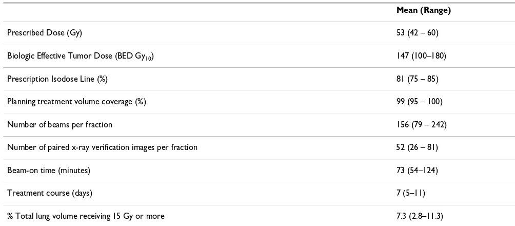
Treatment plans were composed of hundreds of pencil beams shaped using a single 20, 25, 30 or 35-mm diame-ter circular collimator (Table 3). An average of 53 Gy was delivered to the prescription isodose line in three 1–2 hour treatments over a 5 to 11 day period (mean, 7 days). The percentage of the total lung volume receiving 15 Gy or more was low (range, 2.8 – 11.3%) despite the radical treatment intent. On average, 53 paired orthogonal x-ray images of the fiducials were taken during each treatment to confirm the accuracy of the correlation model. Two patients received concurrent systemic therapy as
pre-scribed by their treating oncologists. One of these patients completed treatment flanked by cycles 2 and 3 of full-dose carboplatin and docetaxel. The second patient received concurrent gefitinib.
Complications
Pneumothorax requiring tube thoracostomy developed in 25% of patients following fiducial placement. Subse-quently, all patients completed treatment without inter-ruption or noteworthy side effects. Following treatment, acute toxicity consisting of mild transient fatigue was reported in the majority of patients. Chest wall discom-fort, typically lasting several weeks, developed in 8 of 12 patients with tumors in close proximity to the pleura (5 mm). Classic acute grade III radiation pneumonitis was observed in 1 patient who had received 60 Gy with con-current gefitinib treatment. Despite her relatively good lung function (FEV1 = 1.51 L), small GTV (7.56 cc) and low V15 (9.5% of total lung volume), she developed an infiltrate corresponding with the high dose treatment vol-ume and hypoxia requiring supplemental oxygen 4 weeks following CyberKnife treatment. Her acute symptoms appeared unrelated to her severe underlying heart disease and resolved with steroids. She discontinued gefitinib and is well two years following treatment.
Post-treatment Pulmonary Status
Among the entire group, no statistically significant change was seen in percent predicted FEV1 and TLC at 6, 12, 18 or 24 months. Statistically significant reductions of 9% (from 57% to 48%; p = 0.005) and 11% (from 57% to 46%; p = 0.05) in the mean percent predicted DLCO were seen at 6 and 12 months, respectively. Reductions in DLCO at 18 and 24 months did not reach statistical signif-icance.
Table 2: Patient and Tumor Characteristics
Mean (Range)
Age (years) 74 (64 – 86)
Weight (lbs) 156 (116 – 225)
FEV1 (L) 1.08 (0.53 – 1.71)
% predicted FEV1 52 (21 – 84)
% predicted TLC 103 (69 – 136)
% predicted DLCO 57 (44 – 83)
Maximum Tumor Diameter (cm) 2.2 (1.1 – 3.5)
Gross Tumor Volume (cc) 9.7 (1.3 – 24.4)
Table 3: Treatment Characteristics
Mean (Range)
Prescribed Dose (Gy) 53 (42 – 60)
Biologic Effective Tumor Dose (BED Gy10) 147 (100–180)
Prescription Isodose Line (%) 81 (75 – 85)
Planning treatment volume coverage (%) 99 (95 – 100)
Number of beams per fraction 156 (79 – 242)
Number of paired x-ray verification images per fraction 52 (26 – 81)
Beam-on time (minutes) 73 (54–124)
Treatment course (days) 7 (5–11)
CT Tumor Response
Six-month CT scans were available for all 20 patients. Thirteen lesions responded to treatment as documented by a decrease in tumor volume. Seven lesions were obscured by radiation fibrosis at 6 months. At 12 months, 16 patients' CT scans were available for review. Eight lesions continued to respond to treatment, three of which had resolved completely. Eight lesions were obscured by radiation fibrosis at 12 months. At 18 months, 12 patients' CT scans were available for review. Two lesions responded completely, 2 exhibited a partial response to treatment with only minimal residual soft tissue abnor-mality remaining, and 8 were completely obscured by radiation fibrosis. In each case fibrosis corresponded with the planned high-dose treatment volume and uniformly encompassed the fiducials (Figure 1). There were no major changes in tumor response following the 18 month evaluation (Table 4). Serial imaging characteristics sug-gesting local failure were not observed during early fol-low-up and consequently no confirmatory biopsies have been completed.
Disease Spread and Survival
No regional lymph node failures or distant metastases were observed during early follow-up. However, two oxy-gen-dependent patients with pre-treatment FEV1 values of 0.53 and 0.76 liters died of progressive lung dysfunction at 9 and 18 months, respectively. Therefore, with a median follow-up of 25 months for surviving patients, Kaplan-Meier overall survival at 2 years was 87% (Figure 2).
Discussion
Stage I NSCLC is curable.[22] Peripheral lung tumors are more likely to be cured with radiosurgery than central can-cers because there is less untreated lymphatic spread[23] and a more favorable therapeutic window.[11] However, consistently curing these patients without surgery will require adequate gross tumor doses with finely tailored dose gradients capable of eradicating known relatively radiation-sensitive microscopic tumor extensions, while adequately preserving lung function.
In late 2004, we initiated a radical CyberKnife protocol for medically inoperable patients with small, peripheral, stage I NSCLC. Ultimately, we treated a select group of patients with relatively good performance status and small tumor volumes because we were concerned about CT-guided fiducial placement and treatment-related pul-monary toxicity. Mandatory minimum gross (42 Gy)[24] and microscopic tumor doses (30 Gy) [25-27] were derived from historical clinical data. Continuous tracking of respiratory tumor motion and highly accurate beam alignment throughout treatment with the CyberKnife allowed us to deliver radical dose distributions with
tighter margins on the GTV than historically feasible (5 mm).[14] Numerous pencil beams were used to produce dose gradients that conform closely to the shape of the tar-get, resulting in theoretically adequate microscopic dis-ease doses extending an ample 1 cm or more from the tumor.[28] Twenty patients have been treated in 30 months. With a median follow-up of 25 months for sur-viving patients, the 2-year Kaplan-Meier local control rate was 100%, the 2-year Kaplan-Meier overall survival rate was 87%, and there have been no severe (grade IV) treat-ment-related complications or early mortalities. Further-more, despite the comprehensive nature of the treatment, the decrement in lung function remained at acceptable levels. Thus, we conclude that radical radiosurgery with real-time tumor motion tracking using the CyberKnife is a safe and effective treatment option for small peripheral Stage I NSCLC.
Despite promising preliminary results, critical issues con-cerning the evaluation of treatment efficacy and selection of patients merit additional consideration. Radiosurgery delivered to small peripheral tumors with margins ade-quate to treat radial microscopic extension (> 1 cm) will
Right upper lobe clinical stage IA NSCLC treatment planning CT (A), planned radiation dose distribution (B: the planning treatment volume is shown in red and the 30 Gy isodose line in blue), and CT at 3 and 6 months post-treatment (C and D) show an initial decrease in the tumor volume followed by radiation fibrosis which correlates with the planned dose dis-tribution, engulfs the fiducials and impedes evaluation of tumor response
Figure 1
damage peritumoral lung tissue. The acute lung injury will often result in asymptomatic focal lung parenchyma fibrosis corresponding with the high-dose radiation vol-ume. [29-32] In the present study all tumors initially responded to treatment with a decrease in volume on CT. However, by the six-month mandatory evaluation, all patients had developed CT evidence of peritumoral radia-tion fibrosis. At 18 months, two thirds of the tumors were obscured by such fibrosis, making CT tumor assessment difficult. Preliminary analysis of PET imaging suggests that it too is unreliable following radiosurgery.[33] Although this trial did not require PET/CT imaging, it was routinely completed. Moderate PET activity was often observed following treatment, but could uniformly be attributed to radiation fibrosis rather than tumor progres-sion on serial imaging. Until a dependable noninvasive means to identify early local recurrence in the presence of fibrosis is developed, inoperable patients with fibrosis will require close follow-up which may include biopsy.[31]
In late 2003, Timmerman et al. published preliminary results of the Indiana University inoperable stage I NSCLC
SBRT dose escalation trial.[12] The primary finding of this study was that 60 Gy in 3 fractions could be safely deliv-ered to inoperable stage I NSCLC patients in less than 2 weeks if relatively tight gross tumor margins (1 cm) were used. However, prior to proceeding with phase II studies, maturing data suggested that critical central thoracic struc-tures tolerated high-dose hypofractionated radiation poorly. Accordingly, the Radiation Therapy Oncology Group (RTOG) protocol 0236 was limited to small (< 5 cm) tumors that lay outside the central bronchial tree, a large area that extends 2 cm from the major airways.[11] We chose to deliver a range of radical doses (42–60 Gy) to smaller tumors (< 4 cm) with tighter margins (5 mm) than RTOG 0236. Therefore, we felt confident selecting a more inclusive definition of peripheral tumor as those tumors located a sufficient distance from sensitive critical central thoracic structures so that radical radiation doses could be delivered to the PTV while adhering to maxi-mum point dose limits (Table 1). To date, with sufficient follow-up to detect late radiation toxicity, clinically appar-ent radiation damage to critical cappar-entral structures has not been observed despite our delivery of a mean dose of 53 Gy to the PTV.
Peripheral thoracic structures such as the lung paren-chyma and the chest wall did sustain clinically apparent damage as anticipated. Despite the radical treatment intent, the mean percentage of the total lung volume receiving a minimum of 15 Gy was only 7.3% (range, 2.4% to 11.3%). Nonetheless, acute Grade III radiation pneumonitis occurred in a single patient treated with con-current gefitinib, an alleged potentiator of radiation-induced lung fibrosis.[34] Although it is tempting to fur-ther limit the percentage of lung receiving greater than 15 Gy in an attempt to decrease the risk of radiation pneumo-nitis, this should only be considered if the radical treat-ment intent is maintained.
All evaluated patients were heavy smokers, 80% of whom had stopped smoking in the distant past (> 3 years). Therefore, although the baseline pulmonary function was poor, PFTs just prior to and following the treatment were largely unaffected by recent smoking. The treatment did not adversely affect FEV1 or TLC; however, it did cause sig-nificant early reductions in the mean percent predicted
Table 4: Tumor response per CT imaging
6 months (%) 12 months (%) 18 Months (%)
Complete Response 0 19 17
Partial Response 65 31 17
Obscured by Fibrosis 35 50 66
Kaplan-Meier plot of overall survival
Figure 2
DLCO. Predictably, the magnitude of this decline was small, as it was in a recently reported segmentectomy series.[35] Regrettably, 2 patients with severe COPD, who required continuous supplemental oxygen prior to treat-ment, died 9 and 18 months after radiosurgery secondary to progressive pulmonary dysfunction. It is unknown whether their deaths were premature and attributable to treatment. Nonetheless, it is possible that a population of inoperable stage I NSCLC patients exists, possibly those whom are oxygen dependent, who may not benefit from radical treatment. Such patients may benefit from a more conservative radiosurgery approach with lower doses [36,37] or tighter margins.[38]
Thoracic surgery uniformly results in permanent chest wall scarring and acute pain, which may persist. Nonethe-less, it remains the standard treatment for stage I NSCLC. In contrast, carefully designed early stage lung cancer radi-osurgery may result in only trivial radiation skin reactions and transient, mild to moderate chest wall pain. The use of hundreds of lightly weighted beams in this study rather than a few heavily weighted ones has prevented the infre-quent but potentially severe skin injuries reported in prior thoracic radiosurgery series conducted using other radio-surgical instruments.[12,39] However, mild-to-moderate chest wall pain, typically lasting several weeks, was seen following treatment in the majority of patients with lesions within 5 mm of the pleura. Limiting the dose to the chest wall would likely diminish the severity of this complication; however, this may have led to potentially life threatening local failures and therefore is not recom-mended at this time given the acceptable and transient nature of the toxicity observed to date.
The current study required CT-guided fiducial implanta-tion prior to treatment. This procedure frequently results in pneumothorax in high-risk patients, often necessitating tube thoracostomy and a short hospital stay.[37] Fortu-nately, alternative fiducial placement approaches and a fiducial-free peripheral lung tumor tracking system are now available. [40-42] Ongoing research will determine appropriate patient selection for these new approaches and their efficacy. In the meantime, fiducial tracking and CT-guided fiducial placement will remain our standard approach for small, peripheral lung tumors. The risk of pneumothorax is justified by the optimal intrafraction tar-geting verification made possible by properly placed fidu-cials. We have found that frequent intrafraction targeting verification and continuous tumor tracking with the Syn-chrony system allows the delivery of adequate dose to the gross tumor and its microscopic extension while keeping the volume of healthy lung exposed to radiation at safe levels. Although there are other ways of dealing with the problem of treating tumors that move with respiration, our experience has made us confident of the safety and accuracy of this approach.
Conclusion
Thoracic radiosurgery is a new treatment option for stage I NSCLC.[13,24,39] Optimal clinical outcomes will require proper patient selection and adequate radiation doses. Inoperable oxygen-independent patients with small peripheral lung tumors are ideal candidates.[11] The delivery of hundreds of radiation beams while contin-uously tracking and compensating for respiratory tumor motion will ensure that the gross tumor and radial micro-scopic extension are effectively treated. Our early experi-ence suggests that radical CyberKnife radiosurgery will result in durable local control with acceptable toxicity. However, larger studies with pathologic confirmation of tumor eradication and ample follow-up are needed to confirm our preliminary findings. At this time, surgery remains the standard treatment option for operable patients with stage I NSCLC.
Abbreviations
BED Gy10: biologic effective tumor dose; CT: computed
tomography; DLCO: diffusing capacity of the lung for car-bon monoxide; FEV1: forced expiratory volume in 1 sec; GTV: gross tumor volume; Gy: Gray; NSCLC: non-small cell lung cancer; PET: positron emission tomography; PFT: pulmonary function tests; PTV: planning treatment vol-ume; TLC: total lung capacity; V15: total lung volume receiving 15 Gy or more.
Competing interests
BC is an Accuray clinical consultant. EA is paid by Accuray to give lectures.
Authors' contributions
References
1. Ginsberg RJ, Rubinstein LV: Randomized trial of lobectomy ver-sus limited resection for T1 N0 non-small cell lung cancer. Lung Cancer Study Group. Ann Thorac Surg 1995, 60:615-622. discussion 622–613
2. Swanson SJ, Herndon JE 2nd, D'Amico TA, Demmy TL, McKenna RJ Jr, Green MR, Sugarbaker DJ: Video-assisted thoracic surgery lobectomy: report of CALGB 39802 – a prospective, multi-institution feasibility study. J Clin Oncol 2007, 25:4993-4997. 3. Allen MS, Darling GE, Pechet TT, Mitchell JD, Herndon JE 2nd,
Lan-dreneau RJ, Inculet RI, Jones DR, Meyers BF, Harpole DH, Putnam JB Jr, Rusch VW: Morbidity and mortality of major pulmonary resections in patients with early-stage lung cancer: initial results of the randomized, prospective ACOSOG Z0030 trial. Ann Thorac Surg 2006, 81:1013-1019. discussion 1019–1020 4. Handy JR Jr, Asaph JW, Skokan L, Reed CE, Koh S, Brooks G, Douville
EC, Tsen AC, Ott GY, Silvestri GA: What happens to patients undergoing lung cancer surgery? Outcomes and quality of life before and after surgery. Chest 2002, 122:21-30.
5. El-Sherif A, Gooding WE, Santos R, Pettiford B, Ferson PF, Fernando HC, Urda SJ, Luketich JD, Landreneau RJ: Outcomes of sublobar resection versus lobectomy for stage I non-small cell lung cancer: a 13-year analysis. Ann Thorac Surg 2006, 82:408-415. dis-cussion 415–406
6. Qiao X, Tullgren O, Lax I, Sirzen F, Lewensohn R: The role of radi-otherapy in treatment of stage I non-small cell lung cancer. Lung Cancer 2003, 41:1-11.
7. Saunders M, Dische S, Barrett A, Harvey A, Griffiths G, Palmar M: Continuous, hyperfractionated, accelerated radiotherapy (CHART) versus conventional radiotherapy in non-small cell lung cancer: mature data from the randomised multicentre trial. CHART Steering committee. Radiother Oncol 1999, 52:137-148.
8. Withers HR, Taylor JM, Maciejewski B: The hazard of accelerated tumor clonogen repopulation during radiotherapy. Acta Oncol
1988, 27:131-146.
9. Lax I, Panettieri V, Wennberg B, Amor Duch M, Naslund I, Baumann P, Gagliardi G: Dose distributions in SBRT of lung tumors: Comparison between two different treatment planning algo-rithms and Monte-Carlo simulation including breathing motions. Acta Oncol 2006, 45:978-988.
10. McGarry RC, Papiez L, Williams M, Whitford T, Timmerman RD: Stereotactic body radiation therapy of early-stage non-small-cell lung carcinoma: phase I study. Int J Radiat Oncol Biol Phys 2005, 63:1010-1015.
11. Timmerman R, McGarry R, Yiannoutsos C, Papiez L, Tudor K, DeLuca J, Ewing M, Abdulrahman R, DesRosiers C, Williams M, Fletcher J: Excessive toxicity when treating central tumors in a phase II study of stereotactic body radiation therapy for medically inoperable early-stage lung cancer. J Clin Oncol 2006, 24:4833-4839.
12. Timmerman R, Papiez L, McGarry R, Likes L, DesRosiers C, Frost S, Williams M: Extracranial stereotactic radioablation: results of a phase I study in medically inoperable stage I non-small cell lung cancer. Chest 2003, 124:1946-1955.
13. Timmerman RD, Park C, Kavanagh BD: The North American experience with stereotactic body radiation therapy in non-small cell lung cancer. J Thorac Oncol 2007, 2:S101-112. 14. Collins BT, Erickson K, Reichner CA, Collins SP, Gagnon GJ,
Diet-erich S, McRae DA, Zhang Y, Yousefi S, Levy E, Chang T, Jamis-Dow C, Banovac F, Anderson ED: Radical stereotactic radiosurgery with real-time tumor motion tracking in the treatment of small peripheral lung tumors. Radiat Oncol 2007, 2:39. 15. Wong KH, Dieterich S, Tang J, Cleary K: Quantitative
measure-ment of CyberKnife robotic arm steering. Technol Cancer Res Treat 2007, 6:589-594.
16. Giraud P, Antoine M, Larrouy A, Milleron B, Callard P, De Rycke Y, Carette MF, Rosenwald JC, Cosset JM, Housset M, Touboul E: Eval-uation of microscopic tumor extension in non-small-cell lung cancer for three-dimensional conformal radiotherapy plan-ning. Int J Radiat Oncol Biol Phys 2000, 48:1015-1024.
17. Goldstein NS, Ferkowicz M, Kestin L, Chmielewski GW, Welsh RJ: Wedge resection margin distances and residual adenocarci-noma in lobectomy specimens. Am J Clin Pathol 2003, 120:720-724.
18. Colice GL, Shafazand S, Griffin JP, Keenan R, Bolliger CT: Physio-logic evaluation of the patient with lung cancer being consid-ered for resectional surgery: ACCP evidenced-based clinical practice guidelines. Chest 2nd edition. 2007, 132:161S-177S. 19. Banovac F, McRae D, Dieterich S, Wong K, Dias L, Chang T:
Percu-taneous Placement of Fiducial Markers for Thoracic Malig-nancies. In Robotic Radiosurgery: Treating Tumors that Move with Respiration Edited by: Urschel HC, Kresel JJ, Luketich JD, et al. Berlin: Springer-Verlag; 2007:15-29.
20. Yousefi S, Collins BT, Reichner CA, Anderson ED, Jamis-Dow C, Gag-non G, Malik S, Marshall B, Chang T, Banovac F: Complications of thoracic computed tomography-guided fiducial placement for the purpose of stereotactic body radiation therapy. Clin Lung Cancer 2007, 8:252-256.
21. Program, CTE: Common Terminology Criteria for Adverse Events, Version 3.0. DCTD N, NIH, DHHS; 2006.
22. Wisnivesky JP, Yankelevitz D, Henschke CI: The effect of tumor size on curability of stage I non-small cell lung cancers. Chest
2004, 126:761-765.
23. Ketchedjian A, Daly BD, Fernando HC, Florin L, Hunter CJ, Morelli DM, Shemin RJ: Location as an important predictor of lymph node involvement for pulmonary adenocarcinoma. J Thorac Cardiovasc Surg 2006, 132:544-548.
24. Onishi H, Araki T, Shirato H, Nagata Y, Hiraoka M, Gomi K, Yamas-hita T, Niibe Y, Karasawa K, Hayakawa K, Takai Y, Kimura T, Hirokawa Y, Takeda A, Ouchi A, Hareyama M, Kokubo M, Hara R, Itami J, Yamada K: Stereotactic hypofractionated high-dose irradiation for stage I nonsmall cell lung carcinoma: clinical outcomes in 245 subjects in a Japanese multiinstitutional study. Cancer 2004, 101:1623-1631.
25. Barton M: Tables of equivalent dose in 2 Gy fractions: a simple application of the linear quadratic formula. Int J Radiat Oncol Biol Phys 1995, 31:371-378.
26. Group LCS: Effects of postoperative mediastinal radiation on completely resected stage II and stage III epidermoid cancer of the lung. N Engl J Med 1986, 315:1377-1381.
27. Keller SM, Adak S, Wagner H, Herskovic A, Komaki R, Brooks BJ, Perry MC, Livingston RB, Johnson DH: A randomized trial of postoperative adjuvant therapy in patients with completely resected stage II or IIIA non-small-cell lung cancer. Eastern Cooperative Oncology Group. N Engl J Med 2000, 343:1217-1222.
28. Papiez L, Timmerman R, DesRosiers C, Randall M: Extracranial stereotactic radioablation: physical principles. Acta Oncol
2003, 42:882-894.
29. Aoki T, Nagata Y, Negoro Y, Takayama K, Mizowaki T, Kokubo M, Oya N, Mitsumori M, Hiraoka M: Evaluation of lung injury after three-dimensional conformal stereotactic radiation therapy for solitary lung tumors: CT appearance. Radiology 2004, 230:101-108.
30. Kimura T, Matsuura K, Murakami Y, Hashimoto Y, Kenjo M, Kaneyasu Y, Wadasaki K, Hirokawa Y, Ito K, Okawa M: CT appearance of radiation injury of the lung and clinical symptoms after ster-eotactic body radiation therapy (SBRT) for lung cancers: are patients with pulmonary emphysema also candidates for SBRT for lung cancers? Int J Radiat Oncol Biol Phys 2006, 66:483-491.
31. Takeda A, Kunieda E, Takeda T, Tanaka M, Sanuki N, Fujii H, Shige-matsu N, Kubo A: Possible Misinterpretation of Demarcated Solid Patterns of Radiation Fibrosis on CT Scans as Tumor Recurrence in Patients Receiving Hypofractionated Stereo-tactic Radiotherapy for Lung Cancer. Int J Radiat Oncol Biol Phys
2008, 70:1057-1065.
32. Takeda T, Takeda A, Kunieda E, Ishizaka A, Takemasa K, Shimada K, Yamamoto S, Shigematsu N, Kawaguchi O, Fukada J, Ohashi T, Kuribayashi S, Kubo A: Radiation injury after hypofractionated stereotactic radiotherapy for peripheral small lung tumors: serial changes on CT. AJR Am J Roentgenol 2004, 182:1123-1128. 33. Hoopes DJ, Tann M, Fletcher JW, Forquer JA, Lin PF, Lo SS,
Timmer-man RD, McGarry RC: FDG-PET and stereotactic body radio-therapy (SBRT) for stage I non-small-cell lung cancer. Lung Cancer 2007, 56:229-234.
Publish with BioMed Central and every scientist can read your work free of charge
"BioMed Central will be the most significant development for disseminating the results of biomedical researc h in our lifetime."
Sir Paul Nurse, Cancer Research UK
Your research papers will be:
available free of charge to the entire biomedical community
peer reviewed and published immediately upon acceptance
cited in PubMed and archived on PubMed Central
yours — you keep the copyright
Submit your manuscript here:
http://www.biomedcentral.com/info/publishing_adv.asp
BioMedcentral 35. Wang JS, Abboud RT, Wang LM: Effect of lung resection on
exer-cise capacity and on carbon monoxide diffusing capacity dur-ing exercise. Chest 2006, 129:863-872.
36. Le QT, Loo BW, Ho A, Cotrutz C, Koong AC, Wakelee H, Kee ST, Constantinescu D, Whyte RI, Donington J: Results of a phase I dose-escalation study using single-fraction stereotactic radi-otherapy for lung tumors. Journal of Thoracic Oncology 2006, 1:802-809.
37. Pennathur A, Luketich JD, Burton S, Abbas G, Heron DE, Fernando HC, Gooding WE, Ozhasoglu C, Ireland J, Landreneau RJ, Christie NA: Stereotactic radiosurgery for the treatment of lung neo-plasm: initial experience. Ann Thorac Surg 2007, 83:1820-1824. discussion 1824–1825
38. Brown WT, Wu X, Amendola B, Perman M, Han H, Fayad F, Garcia S, Lewin A, Abitbol A, de la Zerda A, Schwade JG: Treatment of early non-small cell lung cancer, stage IA, by image-guided robotic stereotactic radioablation – CyberKnife. Cancer J
2007, 13:87-94.
39. Nyman J, Johansson KA, Hulten U: Stereotactic hypofractionated radiotherapy for stage I non-small cell lung cancer – mature results for medically inoperable patients. Lung Cancer 2006, 51:97-103.
40. Anantham D, Feller-Kopman D, Shanmugham LN, Berman SM, DeCamp MM, Gangadharan SP, Eberhardt R, Herth F, Ernst A: Elec-tromagnetic navigation bronchoscopy-guided fiducial place-ment for robotic stereotactic radiosurgery of lung tumors: a feasibility study. Chest 2007, 132:930-935.
41. Fu D, Kahn R, Wang B, Wang H, Mu Z, Park J, Kuduvalli G, Maurer CR: Xsight Lung Tracking System: A Fiducial-less Method for Respiratory Motion Tracking. In Robotic Radiosurgery: Treating Tumors that Move with Respiration Edited by: Urschel HC, Kresl JJ, Luketich JD, et al. Berlin: Springer-Verlag; 2007:15-29.