M O L E C U L A R C L O N IN G AND SEQ U EN C E ANALYSIS O F c D N A S E N C O D IN G T H E T R A N S F O R M A T IO N -S E N S IT IV E A C T IN C R O S S -
L IN K IN G PR O T E IN T R A N S G E L IN
by
Rabinder Prinjha
Thesis presented for the degree of
Doctor of Philosophy
University College London
ProQuest Number: 10106590
All rights reserved
INFORMATION TO ALL USERS
The quality of this reproduction is dependent upon the quality of the copy submitted.
In the unlikely event that the author did not send a complete manuscript and there are missing pages, these will be noted. Also, if material had to be removed,
a note will indicate the deletion.
uest.
ProQuest 10106590
Published by ProQuest LLC(2016). Copyright of the Dissertation is held by the Author.
All rights reserved.
This work is protected against unauthorized copying under Title 17, United States Code. Microform Edition © ProQuest LLC.
ProQuest LLC
789 East Eisenhower Parkway P.O. Box 1346
ABSTRACT
The central role of actin in crucial cellular activities including muscle contraction, locomotion, cytokinesis, maintenance of cell shape and movement of cell surface receptors has been widely studied. Controlled modulation of the actin cytoskeleton is mediated by an array of molecularly diverse actin associated proteins that variously regulate its polymerisation state, geometric organisation and interactions with other ligands.
I have cloned cDNAs encoding the transformation-sensitive actin gelating higher molecular weight isoform of a 21kDa polypeptide doublet (protein C4) found uniformly distributed along stress fibres in normal mesenchymal cells. This isoform, designated transgelin, was found to be the product of a single gene, conserved at the nucleotide level in the H sapiens, R norvégiens, D melanogaster, and Aplysia genomes with a single strong band as far back as the fission yeast S pombe .
Acknowledgements
It is a great pleasure to thank Dr Durward Lawson for many years of kindness along with excellent and always rigorous supervision. Many thanks to Claire for helpful advice and for the use of purified transgelin and to Suzane for enlivening the lab. Thanks also to Stefanello for many long evenings of fascinating discussion, to Martin Smith for allowing the inclusion of unpublished C4^ sequence, use of human T cell RNA and obviously for his advice and friendship.
Publication:
Prinjha, R K; Shapland, C E; Hsuan, J J; Totty, N F; Mason, I J; Lawson,
D. (1994) Cloning and sequencing of cDNAs encoding the actin
cross-linking protein transgelin defines a new family o f actin-associated
CONTENTS
Title 1
Abstract 2
Acknowledgements 3
Publication 4
Contents 5
List of Figures 10
List of Tables 11
1. Introduction 12
A The Cytoskeleton 12
B 1 Intermediate Filaments 12
B2 Microtubules 17
B3 Microfilaments 21
1 Actin Supergene Family 24
2 Cell Motility 30
3 Cell Division 31
4 Cell Adhesion 33
5 Stress Fibres 36
6 Cell Transformation 38
C Actin-Associated Proteins 46
1 Thymosins 46
2 Profilins 49
3 Cofilins 52
4 Alpha-Actinin Family 55
5 Myosins 64
6 Caldesmons 68
7 Gelsolin Family 69
8 Tropomyosin Family 73
9 Troponins 76
10 Synapsins 77
11 Dimeric Barbed-End Capping Proteins 78
12 Protein 4.1 -ERM Family 7 8
13 Vincuhn Family 82
14 MARCKS Family 84
15 Tensin Family 85
16 Scruin Family 85
17 Fascin Family 86
18 Titin Family 87
19 Ponticulins 87
20 Unclassified Actin-Binding Proteins 88
2. Methods
1.1 Abbreviations 104
1.2 Buffers & Solutions 106
2 Tissue Culture 112
1 Media & Plastics 112
2 Cell Lines 112
3 Cell Passaging 112
4 Cell storage, freezing & thawing 113
5 REF production 113
3 Anti-C4 Monoclonal Antibody Production 113
4SDS-PAGE 114
1 Sample preparation 114
2 Gel preparation & Electrophoresis 114
5 Immunoblotting 115
1 Electroblotting 115
2 Post-blot procedure 115
3 lodination 116
6 Molecular Genetics 117
1 Bacteriological Procedures 117
2 Plating and titre determination 118
3 Bacteriophage stock preparation & storage 118
7.1 Ohgonucleotide labelling 119
7.2 Oligonucleotide Controls 119
8 Xgtl 1 Library Screening 120
2 Filter lifts 120
3 Filter treatment 120
4 Filter hybridisation 120
5 Plaque purification 121
9 PCR amplification 121
10 Reverse-Transcriptase Reactions 122
11 “Hot-Starf ’ PCR Amphfication 122
12 Cloning of PCR Products into Vectors 122
1 Vector Treatment 122
2 Cohesive-End Cloning 123
3 Blunt-End Cloning 123
4 Ligation Reactions 123
5 Transformation into DH5a Cells 124
6 Plasmid DNA Extraction & Purification 124
13 Restriction digests 126
15 Vacuum blotting 127
16 Glassmilk Purification 128
17 Oligonucleotide Random Priming 129
18 Spun Columns 129
19 Hybridisation of clone R1 to Human gut clones HI 130
20 Autoradiography 130
21 RNA Techniques 131
1 RNA Preparation 132
2 RNA gel electrophoresis 133
3 Hybridisation 133
22 Sequencing 133
1 Reactions 133
2 Wedge gel preparation 134
3 Electrophoresis 135
4 Fixation and autoradiography 135
23 Computer Analysis of sequence data 136
24 Genome Analysis 137
1 DNA preparation 137
2 Digestion 138
3 Electrophoresis 138
4 Blotting 138
5 Hybridisation 138
6 Cross-species Southern Blot Hybridisation 139
3 Results 140
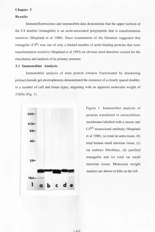
1 Immunoblot Analysis 140
2 Molecular Genetics 141
3 Pre-Screening Negative Controls 142
4 Library Screening 142
5 PCR Analysis of Rat Aorta ^gtl 1 Clones 143
6 Cross-Hybridisation 144
7 Derived Rat Aorta Sequences 144
8 Isolation of Clones Encoding the N-Terminus of Transgelin 145
9 Reverse-T ranscriptase-PCR 148
10 Cloning of PCR Products into Plasmids 150
11 Translation 150
12 Gene-Product Characteristics 153
13 Amino Acid Composition of Transgelin 153
14 Sequence Motifs 154
15 Hydropathy Plot 155
16 Secondary Structure Predictions 156
18 Database Homology Searches 158
19 Dot-Plots 160
20 Homologous Peptides 162
21 Genome Analysis 162
1 Rat Genome 162
2 Cross-Species Hybridisation 164
22 Northern Blot Analysis 165
1 Distribution of Transgelin mRNA 165
2 Transformation Sensitivity of Transgelin mRNA Expression 166
4 Discussion 167
1.1 Selection of Tissue and Library Sources 167
2 Sequence of Purified Transgelin Protein 167
3 Oligonucleotide Selection 168
4 Choice of Vectors 169
5 Oligonucleotide Controls for Xgtl 1 cDNA Library Screening 169 2.1 A,gtl 1 cDNA Library Screening with Oligonucleotide Probe 1 169
2 PCR Analysis of cDNA Inserts 170
3 Sequence of cDNA Inserts 171
3 Isolation of Clones Encoding the 5’ Region of Transgelin 173
4 Reverse-Transcriptase PCR 174
5.1 Initiation of Transgelin Translation 176
2 T ermination of T ransgelin T ranslation 178
3 3’Non-Coding Sequence of cDNA Inserts 178
6.1 Physical Properties of cDNA Encoded Protein 180
A Molecular Weight 180
B Isoelectric Point 180
C Charge Distribution 181
(i) Positive Cluster 181
(ii) Negative Cluster 181
D Cysteine Distribution 182
7.1 Sequence Motifs 183
A Phosphorylation Sites 183
B Calcium Binding EE-Hand 184
8 Hydropathy Plots 185
9 Secondary Structure Predictions 186
10.1 Sequence Conservation 187
2 Sequence Database Searches 187
11 Dot-Plots 193
12 Homologous Peptides 195
13.1 Genome Analysis 197
14.1 Northern Blot Analysis 199
2 Actin mRNA Content 200
3 Transgelin mRNA Tissue Distribution 200
4 Transformation Sensitivity of Transgelin Expression 201
Future Work 203
List of Figures
Figure 1. Immunoblot analysis of proteins transferred to nitrocellulose membranes labelled with a mouse anti C 4 ^ monoclonal antibody.
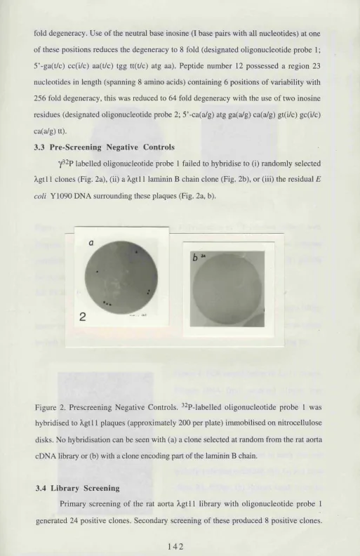
Figure 2. Negative controls.
Figure 3. Tertiary probe 1 positive clones. Figure 4. PCR amplification of Xgtl 1 clones. Figure 5. Cross-hybridisation.
Figure 6. Complete sequences of Xgtl 1 clones.
Figure 7. Schematic representation of the extent of overlapping sequence in clones R l, R2, R3 and H I.
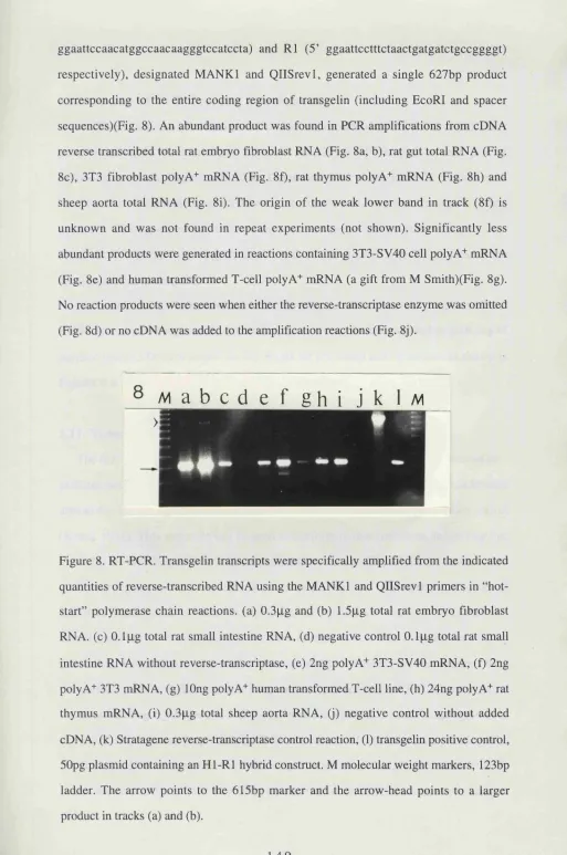
Figure 8. RT-PCR.
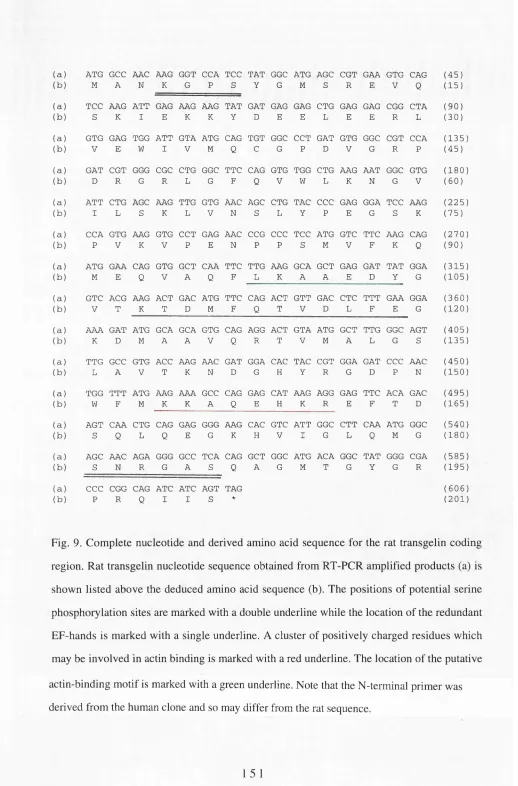
Figure 9. Complete nucleotide and derived amino acid sequence for the rat transgelin coding region.
Figure 10. Complete nucleotide and derived amino acid sequence for the sheep transgelin coding region.
Figure 11. 5’-non-coding region in clone HI. Figure 12. EF-Hand Sequence Alignments. Figure 13. Hydropathy profile.
Figure 14. Secondary Structure.
Figure 15. Alignment of homologous sequences. Figure 16. Alignment of the unc-87 repeat motif. Figure 17. Dot-Plots.
Figure 18. Low stringency genomic Southern blot. Figure 19. High stringency genomic Southern blot. Figure 20. Cross-species Southern blot.
Figure 21. Northern blot analysis-Tissue distribution.
List of Tables.
Table 1. Buffers & Solutions. Table 2. Cell Lines.
Table 3. Hydropathic Index of Amino Acids. Table 4. Amino Acid Dissociation Constants.
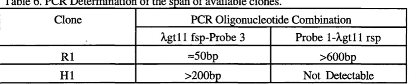
Table 5. Purified Transgelin Sequence and Oligonucleotide Probe Synthesis. Table 6. PCR Determination of the Span of Available Clones.
Chapter 1
I
n t r o d u c t i o nA
The complex, highly dynamic network of protein filaments within the cytoplasm, known as the cytoskeleton, has been intimately implicated in a diverse array of fundamental cellular functions. The most significant of these include: cell movement; cell division; maintenance of cell shape and volume; regulation of organelle localisation and movement; vesicle transport coupled to the regulation of secretion and the generation of intracellular mRNA/ gene product gradients by differential message localisation and translation (for a general review see Alberts et al 1994).
The cytoskeleton is composed of three main groups of filamentous polymers: (a) Intermediate filaments (=8-14nm diameter) (reviewed in Stewart 1993);
(b) Microtubules («22-25nm diameter) (reviewed in Mandelkow & Mandelkow 1989); (c) Microfilaments («7nm diameter) (reviewed in Bretscher 1991).
Each of these networks is regulated by a rapidly growing list of binding proteins that alter the distribution and structure of the filamentous proteins according to the specific requirements of the cell or tissue.
B 1 In t e r m e d ia t e f il a m e n t s
Intermediate filaments are composed of a large family of related proteins that form cell type specific 8-14nm filaments. The intermediate filament protein family can be sub divided into a growing number of main groups (briefly summarised below) based on immunological reactivity, sequence comparisons (50-99% identity within groups and 25- 30% identity between groups) and site of synthesis (for review see Steinert & Roop
1988).
(I) Cytokeratins (40-65kDa)- expressed in epithelial cells;
(H) Vimentins (52-58kDa)- expressed in mesenchyme derived tissues; (HI) Desmins (50-54kDa)- expressed in muscle tissues;
(VI) Lamins (60-75kDa)- expressed in a range of cell types and found in association with the nuclear membrane.
(VII) Nestin (240kDa)- transiently expressed in neuronal precursor cells and myoblasts.
Intermediate filament subunits are composed of a highly conserved alpha-helical rod domain of =310 residues and more divergent N and C terminal domains (reviewed in Skalli et al 1992) it was demonstrated that of these only the N terminus plays an essential role in intermediate filament assembly (Van Den Heuvel et al 1987; Letai et al 1992). The basic protofilament building block of cytoplasmic intermediate filaments involves a tetramer composed of two double-stranded coiled-coils that may possibly have an antiparallel alignment (Geisler et al 1985 and reviewed in Steinert & Roop 1988). Eight protomers (16 subunits) are thought to associate in a staggered manner forming thick rope like structures approximately lOnm in diameter. However, the mechanisms by which these protofilament subunits polymerise into coordinated intermediate filaments remains unclear (for review see Shoeman & Traub 1993).
The low solubility of intermediate filament proteins and the apparent absence of a pool of unpolymerised subunits was originally taken as a reflection of the static nature of intermediate filaments (reviewed in Eriksson et al 1992). However, several recent independent studies using newly synthesised transfected subunits or microinjected fluorescently labelled subunits have indicated that these are readily incorporated into endogenous intermediate filament networks (Ngai et al 1990; Miller et al 1991) suggesting that a continuous turnover of filament subunits occurs in most normal cells.
combination of factors acting on a variety of sequence elements (including a proximal enhancer, a proximal promoter, CAAT, TATA and multiple GC boxes) that were restricted to the 5' end of the genes (Zehner et al 1987). At least two desmin genes were detected and analysis of sequence homology between vimentin and desmin indicated 70% homology (Herrmann et al 1989).
Peripherin was originally isolated as a neurofilament binding protein but has since been sequenced and shown to be an intermediate filament protein more closely related to the desmin/ vimentin groups than to the neurofilament proteins (Leonard et al 1987; Leonard et al 1988; Landon et al 1989). Glial fibrillary acidic protein (GFAP) was found to be encoded by a single gene whose expression was primarily restricted to astrocytes and preceded that of vimentin during astroglial maturation in vivo (reviewed in Zehner 1991). While only three coordinately regulated neurofilament genes were originally described (NF-H; NF-L; and NF-M) (Lees et al 1988) more recent analysis of primary sequence data has suggested that a-internexin, which had previously been characterised as an intermediate filament associated protein, is actually a member of the neurofilament gene family (Fliegner et al 1990). The three lamins A, B and C; found localised at the nuclear membrane, were shown to be encoded by two distinct genes with A and C being generated from one gene (Fisher et al 1986) that was not expressed until late in development (Rober et al 1989). The lamin B gene product was shown to function as an intermediate filament attachment site at the interface with the nuclear envelope (Georgatos & Blobel 1987). A highly divergent 240kDa IF protein, designated nestin, with a very short N-terminal head domain and an extended acidic C terminus was found to be expressed in neuronal cell precursors and myoblasts. Its lack of a head-domain may require it to function as an obligate heterodimer with vimentin, with which it colocalises while its sequence places it in a distinct class (Lendahl et al 1990). Overall analysis of gene structure and sequence for all the intermediate filament proteins including those identified in nematodes suggested that they all arose from a single ancestral nuclear lamin-like gene (Dodemont et al 1990; Doring & Stick 1990) following the loss of the CAAX box and nuclear localisation sequence (Dodemont et al 1994).
(reviewed in Eriksson et al 1992). Several kinases can phosphorylate vimentins, desmin, nuclear lamins and keratins in vitro (Eriksson et al 1992). For example, vimentin residue ser^^ was found to be hyper-phosphorylated by kinase when cells entered mitosis and this was accompanied by disassembly of the vimentin network into its component subunits (Chou et al 1990; Chou et al 1991). Nuclear lamins also act as a target for p34^4c2 (Enoch et al 1991) potentially allowing coordinated disassembly of both cytoplasmic networks and the nuclear matrix immediately prior to mitosis (for review see Nigg 1992).
Despite a vast body of work, the composition, nature and function of intermediate filaments, beyond basic structural roles (Lazarides 1980) remains a matter of considerable debate (Fuchs 1991; Bloemendal & Pieper 1989). Recent studies attempting to elucidate intermediate filament function have used the lamins, these are found forming a thin fibrous network underlying the inner nuclear membrane (reviewed in Nigg 1992). Inactivation of the lamin III gene in Xenopus oocytes allowed the initial stages of nucleus formation (encapsulation of chromatin by fused membrane vesicles and nuclear pore assembly) to proceed normally, however, these nuclei were very fragile and were unable to synthesise DNA (Newport et al 1990). Lamins would therefore appear to be functioning by imparting integrity to the nuclear envelope and also as a potential scaffold for DNA decondensation and replication. Like cytoplasmic intermediate filaments lamin depolymerisation/ disassembly is regulated by phosphorylation by cdc2 kinase at specific points in the cell cycle (Ward & Kirschner 1990; Heald & McKeon 1990; Peter et al 1990). Déphosphorylation and chromosomal contact were found to be sufficient to promote reassembly of the lamin network after mitosis was completed (Glass & Gerace 1990).
that intermediate filaments play a significant role in the organisation of diverse cellular systems. The difficulties experienced with studies on IF deficient cell lines, microinjection (Klymkowski 1981) and gene ‘knock-out’ experiments (Newport et al 1990) in determining the function of intermediate filaments, rather than suggesting an absence of function, may signal (as with actin-binding proteins see Bray & Vasiliev 1989) the existence of parallel networks of subtly different redundant systems that allow their as yet undetermined, essential function to be performed (Goldstein & Vale 1992).
Intermediate Filament Associated Proteins
B2
M i c r o t u b u l e sMicrotubules are considered to be important in mitosis, intracellular vesicle transport, organisation and positioning of membrane organelles, determination of cell shape, directionality and persistence of cell motility (for review see Mandelkow & Mandelkow 1989). Vertebrates possess five a tubulin and six P tubulin genes that display 36-42% amino acid identity with each other (with most of the differences concentrated in the highly acidic carboxy-terminus)(for reviews see Luduena et al 1992; Burns 1991). Tubulin subunits contain two GTP binding sites per a p heterodimer, head-tail polymerisation of these subunits into protofilaments is accompanied by (but not dependent on) the hydrolysis of one of these (p-tubulin bound) to GDP-Pi (Carlier 1988; 1989). Eleven to fifteen of these protofilaments associate side by side to form the hollow (22- 25nm diameter) tubular microtubules seen in electron micrographs. Microtubules, like actin filaments, are polar structures with different equilibrium constants at the two ends representing the faster growing + end and the slower growing - end (in cells the minus end is usually attached to nucleating sites). While these filaments should theoretically undergo “treadmilling” direct observation of microtubules indicates that they often display a process designated as “dynamic-instability”, such that individual microtubules never reach steady- state, switching abruptly from the growth (termed rescue) state to a rapidly shrinking phase (termed the catastrophe state)(reviewed in Mandelkow & Mandelkow 1992). The probabilities of transition from catastrophe to rescue states are such that the formation of fewer, longer microtubules is favoured (for review see Gelfand & Bershadsky 1991). It has been postulated that at high tubulin concentrations the rate of GTP-hydrolysis “lags” behind the filament growth rates such that filaments are likely to possess GTP-tubulin caps at their growing + end that stabilise the filament, while at low tubulin levels the filaments are likely to have GDP-tubulin caps that have a higher probability of switching to the catastrophe state of rapid shrinkage (Carlier 1988).
in mitosis, this property is used in the treatment of some cancers (for general review see Alberts et al 1994).
The stability of p-tubulin mRNA has been found to be regulated by the cellular concentration of free tubulin subunits by a mechanism designated as autoregulated instability. Treatment of cells with agents that raised the unpolymerised tubulin concentration were found to reduce the level of p-tubulin mRNA without altering nuclear transcription or processing rates. Translation of the first four amino acids (MREI) was found to be essential for this mechanism, suggesting that binding of the tubulin dimer to these residues in the nascent polypeptide during translation acivated a specific RNAase that terminated translation (reviewed in Cleveland 1988).
While purified tubulin isotypes appear to be functionally interchangeable in in vitro assay systems and are often found to be coexpressed in different cell types, some recent evidence from the genetically amenable systems of Drosophila and C elegans has indicated that specific isotypes cannot be functionally replaced by others (for review see Moritz 1993). For example, although cytoplasmic microtubules usually consist of thirteen parallel protofilaments those in some C elegans neurons consist of eleven protofilaments while those in a class of touch-sensitive neurons contain fifteen proto-filament microtubules. Analysis of mec -7 mutants (lacking touch sensitivity) found only neurons with eleven protofilament microtubules, subsequent sequencing of the mec -1 gene indicated that it encoded a P-tubulin isotype (90-93% identical to vertebrate p- tubulins)(Savage et al 1989).
The tubulin isotypes are subject to a number of post-translational modifications that function to “mature” microtubules and often increase their stability (for review see Luduena et al 1992):
(1) Acétylation of a-tubulins, at the conserved lysine at residue 40, has been shown to increase microtubule stability (Pipemo et al 1987).
(3) Neuron specific tubulin pm was found to be phosphorylated at tyrosine 437 by pp60^‘src or c-mos (Matten et al 1990; Zhou et al 1991) and serine 444, the functional significance of these modifications remains unknown (Alexander et al 1991).
(4) Glutamic acid residues in both alpha (0445) and beta
in
(G438) tubulins were found to undergo the unusual post-translational modification of glutamylation (addition of up to six gluatmic acid residues to the y-COOH group of the glutamic acid residue), again the functional significance of the resultant highly acidic branched tail is unknown, although it has been proposed to allow the binding of additional microtubule associated proteins (Alexander et al 1991; Edde et al 1990).y-tubulins (454aa, 50,825Da) represent a distinct class of tubulins with -33% amino acid identity with p-tubulins and significantly less acidic C termini (Oakley & Oakley 1989; Burns 1991). They are expressed at levels substantially lower than a/p - tubulins and have been shown to principally localise to microtubule poles and microtubule organising centres, particularly the pericentriolar material of the centrosome (Oakley et al 1990) and have thus been inferred to function as potential nucléation sites (for reviews see Oakley 1992; Joshi 1993).
Organisation of Microtubules into Supramolecular Structures
towards the minus end of the adjacent microtubule. The strength of the cross-links in the axoneme is sufficient to prevent free relative movement of the microtubules, resulting in deformation (bending) that progresses from the base to the tip during the force generating stroke of the cihum or flagellum.
Non-Motor MAPs
These widely studied, numerous and diverse proteins are generally thought to have a structural role in regulating the organisation and stability of cytoplasmic microtubules and are beyond the scope of this discussion (for reviews see Olmstead 1991; Lee & Brandt
1992; Lee 1993; Kreis & Vale 1993; Hirokawa 1994).
Microtubule Motors
This extremely large class of proteins has commonly been divided into the axoneme motor proteins such as flagellar dynein, long known to be responsible for axonemal motility (reviewed in Witman 1992) and the cytoplasmic motors such as the kinesins, cytoplasmic dyneins and dynamins, principally but not exclusively responsible for vesicle movement along microtubules (for reviews see Witman 1992; Vallee & Shpetner 1990; Bloom 1992; Skoufias & Scholey 1993; Sawin & Endow 1993; Hoyt
B3 M
ic r o f il a m e n t sActin is a 42-43kDa abundant globular protein with Mg^^-ATPase activity that is able to associate with itself to form polymeric filaments. These two states are referred to as G- and F-actin respectively (for review see Pollard 1990). Models derived from the atomic resolution X-ray crystallographic structure of G-actin (Kabsch et al 1990) schematically present actin within a 5.5x5.5x3.5nm box (reviewed in Bremer & Aebi 1992) in which the molecule is seen as a bilobed structure with a significant cleft between the major domains. The major domains can be further divided into subdomains 1-4 (with both the N- and C- termini localising within subdomain 1). G-actin is able to bind divalent cations (Ca^'*’, or in physiological conditions Mg^'*') with nM affinity (for review see Estes et al 1992) and ATP or ADP (for review see Carlier 1991b) and these ligands have been shown to locate at adjacent sites in the major cleft between the lobes (with the Mg^'^ binding the p and y phosphates of the AXP), also numerous salt-bridges and hydrogen bonds are thought to associate with these ligands, stabilising the overall conformation of the four subdomains (for review see Reisler 1993; Holmes & Kabsch 1991). Despite a lack of amino acid similarity, the crystallographic structure of actin can be readily aligned with other proteins with an ATPase activity, namely hexokinase and a 44kDa fragment of the heat shock (cognate) protein designated HSC70 (Flaherty et al 1991) suggesting that they share a common ancient ancestor and are under significant structural constraints.
in the process of elongation to yield filaments which are capable of annealing with the ends of other filaments (for reviews see Carlier 1991b; Stossel 1989; Cooper 1991; Pollard
1990). While the hydrolysis of the bound ATP is not essential (since it proceeds in the presence of non-hydrolysable nucleotides (reviewed in Carlier 1989)) its irreversible nature appears to affect the thermodynamics of filament formation, making the two ends unequal and allowing the process known as ‘treadmilling’ (reviewed in Carlier 1991a; Carlier 1991b), ATP hydrolysis may also influence filament structure (Janmey et al 1990) although the latter has been questioned (Pollard et al 1992). This hydrolysis appears to occur vectorially towards the barbed growing end and results in the presence of ATP- and ADP-actin caps at high and slow filament growth rates respectively (Carlier et al 1987).
The growing filaments are composed of two intertwined helical strands (with adjacent actin molecules shifted by approximately half of one subunit). By convention (derived from the results of labelling of actin filaments with myosin heads in which an arrow head pattern is seen in electron-micrographs) the two unequal ends of the filament are referred to as the barbed (faster growing) and pointed ends (rabbit skeletal muscle alpha-actin ends have critical concentrations of 0 . 1 |liM and 1 .5 |liM respectively)(for
reviews see Stossel 1989; Pollard 1990). Alignment of the deduced G-actin structure within a filament is possible with minor conformational adjustments (Bremer & Aebi
1992). In these models the actin molecule is ahgned with the long-axis of the filament with sub-domain 1 oriented towards the barbed-end, other subunits contribute to the stability of the filament through interactions with actin residues above, below and in the adjacent strand (reviewed in Holmes & Kabsch 1991). The observation of ‘locally unravelled’ actin strands in enhanced EM images of filaments (Bgelman & DeRosier 1983) has been taken to suggest that interactions along the filament are stronger than those between strands and various controversial models proposing a physiological role for this in mechanotransduction have been proposed (Bremer et al 1991; Schutt et al 1989; Schutt & Lindberg 1992; reviewed in Reisler 1993).
levels are below the critical concentrations) and capping of filament barbed-ends by specific actin-binding proteins provides independent regulatory mechanisms for controlling polymerisation. Modulation of the affinity of these binding proteins for actin in response to signals such as calcium, ATP/ADP ratios, osmotic stress, activation of members of the ras superfamily, phosphorylation or inositol phosphate pathway components allows the cell to rapidly and accurately produce actin dependent responses to physiological signals (for reviews see Stossel 1993; Carlier 1993; Weeds & Maciver 1993; Hall 1993).
B3.1 Ac t in Su p e r g e n e Fa m i l y
The acting represent a highly conserved family of widely distributed proteins thought to be variously involved in contractile muscle force generation, maintenance of cell shape, control of cell motility and a panoply of other cellular processes (for reviews see Pollard 1990; Cooper 1991; Bretscher 1991; Reisler 1993; Stossel 1993; Herman 1993). While S cerevisiae contains only a single conventional actin gene with 89% amino acid identity with mammalian p-actins (Gallwitz & Sures 1980), Drosophila melanogaster has been shown to have at least six functional actin genes (Tobin et al 1980; Fryberg et al 1980; Fryberg et al 1982) and all vertebrates contain at least six actin genes only differing from each other by between 2 and 7% (for reviews see Rubenstein 1990; Hightower & Meagher 1986; Herman 1993). In humans these six genes encode «skeletal (377aa),
«cardiac (377aa), «sm ooth (377aa), Yenteric (376aa), Ycytoplasmic (375aa) and Pcytoplasmic
(375aa) isoforms. The expression of each of these genes, where examined, is regulated in a precise (stage and tissue specific) manner during development (see references in Herman
1993). The expression of various isoforms during in vitro chick muscle development remains one of the best studied systems displaying initial Ycyt and pcyt coexpression followed by a switch, first to «card and then to « sk el actin isoforms (for review see
Buckingham 1985) with some reports of nonmuscle isoforms being specifically ‘exported’ from the mature myotubes in actin-rich ‘macules’ (reviewed in Rubenstein 1990). The complexity of regulation in other systems is illustrated by Northern blot experiments which have indicated that the «smooth actin gene promoter was affected by TGFpi (Kocher & Madri 1989), cAMP (Nomura et al 1992), hormones, cell proliferation (Reddy et al
1990) and oncogenic transformation (Leavitt et al 1985). In vitro CAT transcription assays characterised a 200bp region 5’ to the « s k e l actin gene containing various regulatory elements (an ATAAAA box at -24 and paired CCAAT box associated repeats at -83 and -127bp) that were shown to be responsible for specifically inducing very high levels of expression only in adult skeletal muscle tissues (Chow & Schwartz 1990).
minor actin isoforms, actin related proteins (see below), processed ‘pseudogenes’ and other currently uncharacterised actin genes (reviewed in Buckingham 1985).
C l a s s I a n d II A c t i n s : The variations in amino acid sequence between the conventional actins are clustered, with few exceptions, near their N termini and these differences have been used to classify them into separate classes (reviewed in Rubenstein 1990; Herman 1993). The Class I actins (pcyt> Ycyt and Ysmooth) are synthesised with three acidic residues at positions 2, 3 and 4 and acetylated-methionine 1 is cleaved by a specific cytoplasmic 77kDa acetyl-aminopeptidase (Sheff & Rubenstein 1992) to expose the acidic residue at position 2 which rapidly becomes acetylated. Class II actins (oCcardiac, (^skeletal,
asmooth) are characterised by a cysteine residue at position 2 and four acidic residues at positions 3-6. The acetylated methionine 1 is removed from the nascent polypeptide during translation, the exposed cysteine residue 2 becomes acetylated and this is cleaved by the same cytoplasmic aminopeptidase as the Class I actins to expose the acidic residue which in turn becomes acetylated (Sheff & Rubenstein 1992). Yeast actins do have an acetylated methionine at position 1 but are not proteolytically processed (Cook et al 1992). The Class m or unconventional actins have highly divergent N-termini and remain poorly studied in regards to processing (Herman 1993), and will be discussed separately.
While the precise physiological functions of actin in general and these distinct isoforms in particular have proven difficult to elucidate, a number of different approaches have begun to characterise their properties and possible roles and are discussed below (also see Hennessey et al 1993 for review).
(1) Class I and II actin isoforms were purified and shown to display near identical polymerisation kinetics and barbed- and pointed-end critical concentrations under physiological conditons. However, when assays using identical salt conditions were performed at reduced temperatures «skeletal actin was found to polymerise ten fold more efficiently than platelet Ycytoplasmic actin (Gordon et al 1977).
(Reuner et al 1987) and this was taken to suggest a specific role for these isoforms in regulating stress-fibre integrity (Rubenstein 1990).
(3) Use of isoform specific antibody probes used to analyse the actin distribution in cultured pericyte cells (vascular cells associated with smooth muscle cells) suggested that while all Class I actins colocalised in stress-fibres, the pcyt and Ycyt isoforms were additionally found in the cell cortex and leading lamellae while asm was restricted to the microfilaments (De Nofrio et al 1989). Observation of askel and pcyt actins present at identical concentrations indicated that the former incorporated into myofibrils three to four fold more efficiently (Peng & Fischman 1991) suggesting in vivo mechanisms for isoform sorting. Isoform specific antibodies were also used to demonstrate the preferential localisation of pcyt actin in submembranous regions, filopodia, pseudopodia, fan lamellae and at the ends of stress-fibres (Hoock et al 1991). During injury induced migration in pericyte monolayers pcyt mRNA synthesis increased 2-3 fold and importantly, was found by in situ hybridisation to colocalise with the protein at the wound edge (Hoock et al
1991). Similarly, serum stimulation of starved cells resulted in the redistribution of p-actin mRNA from a diffuse pattern to concentration in lamellae at the cell periphery within minutes, the process was dependent on the 3’-UTR of the mRNA (Hill et al 1994) and on tyrosine kinase activity (Latham et al 1994). This possibility of actin isoform sorting through specific mRNA localisation with microfilaments may provide a very elegant general method for compartmentalised protein expression (Zambetti et al 1990; Sundell & Singer 1991; Biegel & Pachter 1992; Taneja et al 1992; for reviews see Singer 1992; Kislauskis & Singer 1992; Wilhelm & Vale 1993).
(4) The directed overexpression of pcyt actin in cultured myogenic C2 myoblasts caused a loss of stress-fibres, while overexpression of Ycyt actin resulted in reduced cell adherence and cell rounding (Schevzov et al 1992; Lloyd et al 1992).
with a reduced ability to activate myosin ATPase activity, reduced motility (30-40%) in motor assays and increased filament fragmentation (Aspenstrom et al 1992).
(6) Mutation and réintroduction of a Drosophila actin gene designated Act88F expressed only in indirect flight muscles (in which stretch activation allows very high contraction rates) into flightless null mutants (lacking both endogenous Act88F genes) indicated that E316K and G368E mutants formed near normal myofibrils and yielded flies with «80% of normal indirect flight muscle function but V339I, E364K and G366D mutants failed to assemble myofibrils and along with E316K were flightless (Drummond et al 1990; reviewed in Sparrow et al 1991; Hennessey et al 1993). The residue at position 316 was mapped to a region away from the conserved ATP/Mg^+ binding cleft and was, surprisingly, found to have the greatest effect on actin assembly and myosin binding, probably by disrupting actin folding or actin-actin interactions (Kabsch et al 1990; Drummond et al 1991).
(7) Disruption of the single actin gene in S cerevisiae was found to be lethal (Shortle et al 1982). Subsequent generation and characterisation of temperature sensitive mutants indicated multiple functional roles for actin manifest as disorganised actin cables; non directed/ delocalised deposition of chitin (normally deposited at the site of daughter bud formation); aberrant secretion, with intracellular accumulation of vesicles; increased sensitivity to osmotic stress and lethality in budding cells at the elevated temperature (Novick & Botstein 1985; for review of yeast actin mutations and their suppressors see Drubin 1990). These observations were supported by the finding that while substitution of residues at 191, 336, 356, 373 and 374 were essentially neutral, mutation of D l l or removal of three amino acids (KCF) at the C terminus were both lethal and also removal of both CF or just the F residue produced temperature sensitive mutants with increased cell size, sensitivity to osmotic stress and actin filament disorganisation (Johannes & Gallwitz 1991).
more numerous, shorter but tighter spontaneous bundles than ADSE, possibly by allowing closer association of filaments. It was found that mutant actin copolymerised with wild-type actin (these combined filaments will have a higher negative charge than mutant filaments) was able to activate myosin S 1 ATPase better than mutant actin alone (Cook et al 1992) suggesting that electrostatic interactions are important in bundle formation and myosin interactions.
These investigations, amongst others, would therefore suggest a specific contractile role for the a (skeletal, cardiac and smooth) actin isoforms, either in muscle myofibrils or tension generating stress-fibres; for Pcyt actin in cell motihty at the leading edge and a function for y (cytoplasmic, enteric smooth muscle) in ventral adhesive and cortical cytoskeletal arrays and in areas of moving cytoplasm. The finely controlled expression of each of the actin isoform genes, allied with isoform specific microfilament dependent mRNA sorting and translation combined with preferential incorporation or association with actin-binding proteins provides the several independent control points necessary to exquisitely regulate cell behaviour.
Classin Ac t in s: While class I and II actins are very highly conserved («90% amino
acid identity between yeast and human actins) a variety of proteins displaying 50-60% identity with vertebrate actins have been positioned in a separate class (for reviews see Clark & Meyer 1993; Herman 1993). Phylogenetic analysis of available actin sequences has recently allowed their reclassification into five sub-classes (Fryberg et al 1994). The actin-related proteins in the class designated Arpl represent those most recently diverged from conventional Class I and II actins. Alignment of A rpl sequences onto the alpha- skeletal actin crystallographic structure indicates structural conservation and colinearity while those in Arp2 and Arp3 subclasses contain sequence insertions at various points which have been proposed to occur as surface loops that do not disrupt the core structure or the folding of their highly conserved ATP and divalent cation binding sites (Clark & Meyer 1992).
(Clark & Meyer 1992) and within the multisubunit activator of dynein (a motor protein involved in movement on microtubules) that is known as dynactin (Lees-Miller et al 1992b). The dynactin complex contains multiple copies of the centractin/ actin-RPV protein that have been shown to be able to form short filaments associated with dimeric capping proteins (Schafer et al 1994), bind nucleotides and cosediment with filaments of conventional muscle actin (Melki et al 1993).
Null mutants of the yeast homologue of these proteins, Act5 was found to have a phenotype similar to that of dynein heavy chain mutants (namely abnormal spindle orientation and nuclear migration)(Muhua et al 1994). Consistent with the localisation of dimeric barbed-end actin-capping proteins to the dynactin complex (Schafer et al 1994) Act5", c a p r double mutants displayed poorer nuclear migration and spindle orientation than single Act5‘ mutants, suggesting that the length of the mini-actin filaments within the dynactin complex is critical for normal dynein motor function (Muhua et al 1994).
Arp2 proteins include S cerevisiae Act2 (47% identity with its Actl)(Schwob & Martin 1992) and D melanogaster Arpl4D (Fryberg et al 1994). Yeast Act2 was found to be encoded by a single essential gene with a possible role in cytokinesis (heterozygotes produce large cell-cycle arrested cells with nuclei containing buds) this view was supported by the presence of a cdc28 (p34^^^^) kinase phosphorylation motif that is absent in conventional actins (Schwob & Martin 1992).
Arp3 proteins include bovine Act2 containing a protein kinase C target site (Act2 is 36% identical with vertebrate beta-actin)(Tanaka et al 1992) and S pombe act2 (35-40% identity with vertebrate actins and 60% identity with bovine Act2), gene disruption experiments suggest a role for S pombe act2 in spore germination (Lees-Miller et al
1992a).
B3.2 Cell Motility
The requirement for precisely regulated mechanisms of cell motility in the processes of embryogenesis, immune defence or wound healing and the consequences of a failure to control this in cancer cell metastasis serve to highlight the central role of cell motility in biology. No single model currently exists that satisfactorily explains the mechanics of motility in all cell-types suggesting that, at least to some extent, different aspects of each will prove to be functioning according to specific cellular requirements and organisation (for reviews see Heath & Hollifield 1991; Cypher & Letoumeau 1992; Smith 1988; Cooper 1991; Small 1989; Zigmond 1989; Bray & White 1988; Stossel 1993). However, gathering evidence from transfection studies involving gelsolin (Cunningham et al 1991); ABP-280 (Cunningham et al 1992); a-actinin (Gluck et al 1993); tropomyosin (Prasad et al 1993) and vinculin (Fernandez et al 1992) suggest a critical role for actin and its associated proteins in cell motility.
The results of numerous investigations using cytochalasins on growth cones (Smith 1988), inactivation of myosin Ib or II in Dictyostelium (Knecht & Loomis 1987.; DeLozanne & Spudich 1987; Jung & Hammer 1990), physical separation of lamellae from cell bodies (Malawista et al 1982; Euteneuer & Schliwa 1984), microtubule distribution (Bershadsky et al 1991), fluorescence labelling/ bleaching (Theriot & Mitchison 1991), ‘optical-tweezers’ and/or surface associated particles (Kucik et al 1991) combine to suggest the need for localised, actin polymerisation and myosin I powered protrusive force generation together with distal cortical tension generated at the rear of the cell by myosin II, with microtubule mediated support and regulation of directionality and persistence (for reviews see Bray & White 1988; Stossel 1993), Plasma membrane based events involving receptor activation and intracellular signalling (via calcium, diacylglycerol, IP3, G
fimbrin), stabilised by tropomyosins or caldesmons or cross-linked into highly branched viscoelastic gels (by proteins such as ABP280/ filamin) and subsequent anchorage to the membrane via proteins such as myosin I, annexin II, spectrin or MARCKS or incorporation into transient substrate adhesion sites (focal contacts- involving proteins such as talin, vinculin and integrin beta chains) may provide the rigidity and adherent traction necessary for amoeboid motility. Most actin filaments at the front of the lamella are oriented with their barbed-ends nearest the plasma membrane and the mechanism(s) by which actin polymerisation is able to force the membrane forward are disputed but may involve any or all of the following: (i) localised ‘osmotic-stress’ pushing the membrane away from the growing barbed-ends of filaments, (ii) a myosin I dependent ‘ratchet- mechanism’ pushing the membrane away from the growing barbed-end, (iii) a ‘Brownian- ratchet’ mechanism whereby thermodynamic oscillation of the plasma membrane allows subunit additions at the barbed-end, (iv) integral or membrane associated proteins like insertin/ tensin able to bind the barbed-end but which still allow polymerisation (see Gaertner & Wegner 1991 for mechanics), or (v) actin filaments pushing directly onto a ‘raft’ structure of proteins inserted into the membrane (see Stossel 1993 for a more detailed review and discussion of these mechanisms).
An integrated cycle designated the “Cortical Expansion Model” based on the processes observed in Dictyostelium motility involves three interlinked stages: (I) an agonist induced uncapping of barbed-ends produces rapid polymerisation, (II) cross- linking of these newly formed filaments to yield a gel and (III) an increase in the volume of the gel involving osmotic pressure (water entry into the concentrated environment of the gel coupled with limited severing of filaments and de novo polymerisation) produces pseudopod extension (for review see Condeelis 1992).
While the current understanding of these processes is fragmentary a combination of high resolution observation of moving cells and manipulation of the biochemistry of actin-binding proteins in future experiments may unravel some of the complex interactions known to contribute to cell motility.
B3.3 Cell Division
formation of a contractile ring containing actin and myosin filaments responsible for separating the ‘daughter-cells’ (for reviews see Satterwhite & Pollard 1992; Salmon 1989). It is known that the poleward migration of the chromosomes is accompanied by signals emanating from the mitotic spindles, at opposite ends of the cell (possibly including a calcium wave (Pluck et al 1991)). These signals induce the aggregation of preexistent actin filaments (with some de novo polymerisation)(Cao & Wang 1990) and these processes are accompanied by alterations in the activities of various enzymes including maturation promoting factor (cyclin-p3 4cdc2 kinase), protein phosphatases type 1
and 2A, Ca^^-calmodulin dependent kinase and protein kinase C (PKC). It is also known from other studies that these enzymes can directly affect the actin cytoskeleton (p3 4cdc2
kinase and PKC dependent phosphorylation of caldesmon and regulatory myosin light chains affects their affinity for actin and activity)(for reviews see Sellers 1991; Satterwhite & Pollard 1992). The importance of this specific regulation and correct function was demonstrated in experiments using myosin II null mutants in Dictyosteliun which were found to form normal pseudopods, round-up in mitosis and complete nuclear division but were unable to form a functional contractile ring (Knecht & Loomis 1987; DeLozanne & Spudich 1987).
actin-binding proteins and the signalling events involved in this process remain obscure, but should succumb to the genetic dissection possible with Drosophila , Dictyostelium , and yeast systems.
B3.4 Cell Adhesion
The ability to accurately and rapidly control cell-cell and cell-substrate adhesion is vital in a range of processes including embryogenesis and cell motility. At least four distinct macromolecular structures have been characterised in studies on cell adhesion.
Desmosomes (or macula adherens) and hemidesmosomes are ‘spot-like’ cell-cell and cell-basal lamina contact sites respectively, found in cells subject to large shear forces and they are principally anchored to intermediate filaments with little or no interaction with actin filaments and so are not discussed in detail here (for reviews see Garrod 1993; Luna & Hitt 1992). The other two structures adherens junctions (belt-desmosomes/ zonulae adherens) and focal contacts (adhesions) are partially analagous systems mediating cell cell and cell-substrate adhesion respectively and are often found associated with actin filamentous structures (for reviews see Tsukita et al 1992; Turner & Burridge 1991; Luna & Hitt 1992).
Cell adhesion is currently considered to be mediated by four types of transmembrane receptor: (1) Selectins- carbohydrate binding proteins with lectin domains, involved principally in lymphocyte/ endothelial cell interactions with little or no demonstrated intracellular association with the actin cytoskeleton (for reviews see Lasky 1992; McEver 1992; Rosen & Bertozzi 1994). (2) ICAM-Iike cell adhesion molecules (members of the immunoglobulin superfamily) (for review see Hynes 1992). (3)
Cadherins- members of a superfamily of tissue-specific transmembrane glycoproteins mediating calcium dependent homotypic cell-cell adhesion, they are found associated with actin filaments at adherens junctions (for reviews see Takeichi 1991; Grunwald 1993). (4)
Integrins- members of a superfamily of transmembrane heterodimers with variable cytoplasmic domains, of which specific beta chains permit association with actin filaments (for review see Hynes 1992).
marked in epithelial cells which adhere to form continuous sheets of cells, each containing a ring of actin filaments located apically in a structure also known as ‘belt-desmosomes’ (or zonulae adherens ). Examination of these actin filaments indicates that their barbed- ends are invariably proximal to the membrane (reviewed in Tsukita et al 1992). The cytoplasmic domain of all members of the cadherin family display between 60 and 80% amino acid identity and have been shown to coprecipitate with three components designated a-(102kDa), p-(8 8kDa) and y-(80kDa) catenin. p- and y-catenins are members
of the plakoglobin family and also mediate an association with intermediate filaments (Garrod 1993). a-catenin was shown to be a member of the vinculin gene family with a conserved domain structure, proposed to permit self-association and linkage with actin and also other actin-binding proteins (see Section 1.C.13). Binding of a-catenin/vinculin to a- actinin, a/p-spectrin, zyxin and members of the ERM family of barbed-end capping proteins including radixin in a high affinity macromolecular complex may anchor filament filaments to the membrane cadherin receptor (reviewed in Tsukita et al 1992). The affinity of these components for each other are proposed to be modulated by phosphorylation and this is consistent with the localisation of various kinases (PKC, c- or v-src and c-yes) to adherens junctions (Matsuyoshi et al 1992; Tsukita et al 1991a). While experimental depletion of extracellular calcium normally results in loss of adhesion and is followed by endocytosis of the cadherin receptors and the plasmalemmal undercoat proteins (Kartenbeck et al 1991) these events were substantially reduced by prior inhibition of serine/threonine kinases (Citi 1992). Furthermore, the identification of members of the 4.1/ERM family with both radixin-like and tyrosine phosphatase domains may indicate an elegant mechanism for targeting regulatory enzymes to the adherens junctions (Yang & Tonks 1991; Gu et al 1991; reviewed in Arpin et al 1994).
The association of components of adherens junctions with the signal transduction machinery, mentioned above, together with their structural role in embryonic morphogenesis (eg. formation of the neural tube by coordinated contraction of belt- desmosomes) suggests a number of roles in regulating and being regulated by the cell nucleus and extracellular cues (for review see Tsukita et al 1992).
reflection microscopy, with a general inverse correlation between rates of cell motility and number of focal adhesions (for reviews see Turner & Burridge 1991; Luna & Hitt 1992). While originally characterised as purely structural components anchoring the termini of actin stress-fibres to membranes, emerging data suggests that, like adherens junctions, they may have a role in signal transduction (reviewed in Burridge et al 1992a; Damsky & Werb 1992; Sastry & Horwitz 1993; Ruoslahti & Reed 1994). Focal adhesions in cultured fibroblasts were found to consist of transmembrane as pi integrin heterodimers associated extra-cellularly with fibronectin cables and cytoplasmically with a dense mixture of proteins within the cell (for review see Hynes 1992). A cascade of proteins spanning from the pi subunit of the integrin (cytoskeletal association is regulated by a conformational change transmitted from the alpha subunit) to actin filaments, including talin, vinculin, a - actinin, paxillin, zyxin, VASP, tenuin, tensin and members of the ERM family interact to stabilise the focal adhesion (please see Section C for discussion of these proteins). In addition to these structural components a number of tyrosine or serine/ threonine protein kinases (including PKC, pp60^^^ and ppl25^"^^) are localised to focal adhesions and are found to be variously activated during their formation/ disruption (reviewed in Luna & Hitt
1992). ppl25^^^ is a novel tyrosine kinase in that it has no transmembrane or membrane association domains and lacks a SH2 motif but does contain proline rich regions. Similar regions found in vinculin and zyxin are proposed to function in protein-protein interactions (Schaller et al 1992; reviewed in Schaller & Parsons 1993) and may also allow binding to profilin (Tanaka & Shibata 1985; also see Section C.2).
The importance of phosphorylation during cell adhesion was highlighted by the ability of herbimycin A (blocks tyrosine kinases) to significantly inhibit new focal contact formation (Burridge et al 1992b). Studies of focal adhesion like structures in activated platelets using agpi integrins to spread on a fibronectin substrate indicate that assembly is accompanied by phosphorylation of ppl25^"^^, paxillin, talin, vinculin and pi-integrin (reviewed in Damsky & Werb 1992). The role of signalling proteins was extended further by the observation that in serum-starved fibroblasts microinjection of the GTP-binding protein, rho, stimulated the reassembly of stress-fibres and focal adhesions (Ridley & Hall
the formation of focal adhesions able to resist the applied force, but surprisingly in addition to microfilament recruitment this required intact intermediate filaments and microtubules (Heidemann 1993; Wang et al 1993b).
The absence of significant sequence homology between the cytoplasmic, cytoskeleton interacting, domains of integrins and cadherins suggests that mechanisms for membrane-cytoskeletal associations have developed independently many times during evolution. This, together with the emerging role of these structures in signal transduction highlights the central importance of actin mediated adhesion in biological systems (for reviews see Luna & Hitt 1992; Turner & Burridge 1991; Damsky & Werb 1992; Hynes
1992).
B3.5 Stress Fibres are large linear arrays of anti-parallel actin bundles. They can be seen in vivo in foetal tissue, in scleroblasts of fish scales and in the endothelial cells of the mammalian vena cava and aorta (reviewed in Byers et al 1984) where they are often seen aligned with the direction of blood flow (haemodynamic stress/ shear forces) (Wong et al 1983). Also, in developing chick embryos, removal of the single layer of epithelial cells resulted in the appearance of a linear actin bundle displaying continuous alignment through adjacent cells during subsequent wound healing (Martin & Lewis 1992). In vitro stress fibres are particularly prominent in cultured fibroblastic cells, examined by immuno fluorescence, present as thick (2|xm) bundles spanning the length of the cell (Culp et al
morphology, dissolution of stress fibres and enhanced motility (Honer et al 1988). Microinjection (Cao et al 1992) or stable overexpression of profilin (Finkel et al 1994) resulted in the reduction of cellular stress fibre content and an increase in cortical F-actin structures.
Serum starvation of cultured fibroblasts results in reduced stress fibre content, subsequent réintroduction of serum, individual growth factors (eg PDGF) or the phospholipid lysophosphatidic acid (LPA) rapidly stimulates the formation of focal adhesions and stress fibres along with membrane ruffles and these processes have been shown to involve the ras related GTPases rho and rac respectively (Ridley & Hall 1992; Ridley et al 1992; for review see Ridley 1994). Analysis of the signals resulting from LPA stimulation suggests that the activation of cellular tyrosine kinases is an obligatory step in stress fibre formation and could not be by-passed even by the microinjection of constitutively active rho protein (Ridley & Hall 1994).
Ventral stress fibres are generally thicker and found in close association with the basement membrane, often ending in adhesive regions known as focal contacts and can be seen to run continuously with surface fibronectin fibrils (Hynes & Destree 1978). Dorsal stress fibres are often aligned in near parallel arrays (with their main axis shifted slightly relative to ventral fibres) and can be seen to arch over the nucleus where they terminate probably in cortical receptor-rich regions of the membrane (reviewed in Byers et al 1984). Surprisingly, daughter cells have been observed to display ‘cytoskeletal memory’ adapting similar arrangements of stress-fibres and cell extensions, this may be important in pattern generation during embryogenesis or wound-healing (Hagmann 1993).
direction of the applied force. This reduced motility and cell protrusion was accompanied by the appearance of stress fibres (Kolega 1986). In some cells disruption of stress fibres with cytochalasin B was found to collapse the intermediate filament network while in other cells they were found to be more closely allied with the microtubule network (Roy 1993).
These data, along with the immunofluorescent codistribution of fibronectin cables with stress fibres and their termination at focal contacts (Hynes & Destree 1978; Heath & Dunn 1978) suggest that one of their functions is the generation of tension and maintenance of cell shape. In line with this, disruption of actin microfilaments with cytochalasin rapidly and reversibly disrupts cell shape (reviewed in Cooper 1987) and enforced culture of cells on non-adherent substrates causes the loss of stress-fibres (Shapland et al 1988; for review see Ben-Ze’ev 1991). Physical disruption of tension generating fibroblasts in collagen matrices resulted in the rapid loss of stress fibres and the expulsion of vesicles (ectocytosis) containing actin, annexins and integrins (Lee et al
1993). Similarly, transformation of fibroblastic cells with oncogenic viruses results in a significant reduction in stress fibre content and is accompanied by marked changes in cell shape and behaviour (reviewed in Ben-Ze’ev 1985; Vasiliev 1985). Many of the early changes in stress fibre organisation in RS V transformed cells have been correlated with the activity of specific tyrosine kinases such as pp60^^(^ (Kellie et al 1991) and inhibition of this kinase by the fungal metabolite radicicol (without affecting protein kinases A or C) induced the reappearance of stress fibres (Kwon et al 1992).
B3.6 Cell Transformation
proceed in an alternative direction (termed contact inhibition), the transformed cells will continue to migrate over the adjacent cell and so possess the ability to form multilayered foci in culture (Abercrombie & Heaysman 1976).
Transformed cells able to detach from the primary tumour, disrupt and traverse the underlying basal lamina (laminin rich matrix) before entering the circulatory networks (blood or lymphatic), evade destruction by the immune system and travel to non-hostile environments in other organs can establish secondary tumours in a process known as metastasis (for reviews see Van Roy & Mareel 1992; Birchmeier et al 1991). Considerable evidence from both experimental and epidemiological studies into the incidence of cancers has confirmed the requirement for multiple (three to seven), independent, random events, affecting both dominant (oncogenes) and recessive (tumour suppressors) loci (for review see Kinzler & Vogelstein 1993).
The dominant effector genes known as oncogenes generally produce proteins normally involved in generating or responding to positive growth signals (including constitutively active growth factors (eg PDGF/v-5i5' ) or their receptors (eg EGF-R/v-
erbB), submembranous non-receptor tyrosine kinases (eg c-src ), cytoplasmic kinases (eg ra f ), GTP-binding signaling molecules (eg ras ) or aberrantly expressed transcription factors (c g fo s ))(for reviews see Cantley et al 1991; Hunter 1987; Cooper 1990). Recessive tumour suppressor genes normally function to restrict or limit positive growth signals (eg cyclin D l, Retinoblastoma, Rb; Krev; Adenomatous polyposis carcinoma, APC and p53) or as regulators of morphology and adhesion (eg integrins, cadherins, Schwannomin/ merlin, nm23)(for reviews see Birchmeier et al 1991; Tsukita et al 1993; Kinzler & Vogelstein 1993).
al 1992) and restored their ability to regulate cell volume following transfer into hypotonic medium (Cantiello et al 1993).
The characteristic rounded morphology of transformed cells reflects their reduced adhesion to the underlying matrix and may, at least in part, result from the altered expression of extracellular matrix proteins such as fibronectin (for review see Schwartzbauer 1991) since the addition of exogenous protein to transformed cells in culture was found to increase stress fibre numbers and restore their coalignment with fibronectin cables (Willingham et al 1977).
Degradation and remodelling of the extracellular matrix, by a broad range of secreted and membrane bound proteases and their activators (for reviews see Mignatti & Rifkin 1993; Chen 1992) also contributes to the reduced adhesion and invasive behaviour of transformed cells. Localisation of extracellular matrix degrading proteases in cell protrusions designated as ‘invadopodia’ correlated with activation of cellular kinases and invasion of adjacent regions (Mueller et al 1992) and similarly, inhibition of plasminogen activated proteases with specific peptides or anticatalytic antibodies resulted in enhanced adhesion and conversion from focal cell growth to monolayer formation (Melchiori et al 1992; Sullivan & Quigley 1986; Hebert & Baker 1988).
in Birchmeier et al 1991) and may correlate with the ability of CD44 to interact with the actin cytoskeleton via members of the radixin (ERM) family (Tsukita et al 1994).
Submembranous Protein Complexes
Reduced adhesion following transformation was found to be not limited to changes in receptors since highly metastatic cell lines with apparently normal transmembrane receptor expression patterns could be characterised (reviewed in Takeichi 1993). These cells were found to contain deficiencies in the protein complexes that had been implicated in anchoring ligand-bound receptors to the underlying cytoskeletal meshwork (for reviews see Takeichi 1993; Tsukita et al 1993). The cytoplasmic domain of cadherin molecules clustered in adherens junctions are normally associated with a tripartite complex of catenins (a , p and y) (Section 1.B3.4) but poorly adherent, metastatic PC9 lung carcinoma cells were found to lack a-catenin (Morton et al 1993). They were induced to reaggregate into epithelial structures, with a concomitant reduction in tumourigenicity following transfection with the a-N-catenin isoform (reviewed in Takeichi 1993). Consistent with this was the finding that elevated tyrosine-phosphorylation of a - and p- catenins in src -transformed (RSV) chick fibroblasts correlated with reduced cadherin dependent adhesion and elevated invasive behaviour (Behrens et al 1993; Matsuyoshi et al
1992; Hamaguchi et al 1993).
M icrofilament Alterations
Cellular transformation results in the loss of large microfilament bundles and a reduction in total stress fibre content (Pollack et al 1975) with residual stress fibres failing to pass through peripheral regions of the cell (Goldman et al 1976). These changes were shown to occur without an alteration in the overall concentration of actin by means of a rearrangement of stress fibres into shorter more complex polymeric forms (Heacock et al 1984; Felice et al 1990; Goldman et al 1976; for reviews see Vasiliev 1985; Ben-Ze'ev 1985).
In some cases transformation was found to be accompanied by point mutations in genes encoding actin isoforms (Kakanuga et al 1984; Sadano et al 1988). Mouse fibroblasts transformed with the ras oncogene were found to express significantly lower levels of ttsm-actin and apparently normal levels of other isoforms (Leavitt et al 1985). The preferential localisation of the asm isoform in contractile structures, together with cell-shape alterations following transformation may provide an indication of its role in normal cells in generating the force necessary to maintain an adherent, well spread morphology (for reviews see Herman 1993; Vasiliev 1985).
Actin-binding Protein Alterations
results in elevated levels of a protein possessing F-actin-binding, SH2 and SH3 domains and tyrosine-kinase activities (Section 1.C.20). The extent of colocalisation of the bcr/abl fusion protein with actin was found to correlate directly with stress fibre content and transforming potential (McWhirter & Wang 1991; McWhirter & Wang 1993). Analysis of the promoter elements in the t- and 1-plastin genes (Section 1.C.4) identified regions responsible for the elevated expression of these actin bundling proteins following neoplastic transformation in cells derived from solid tumours (Lin et al 1993a, b).
Many components involved in anchoring actin filaments to the membrane or to focal adhesion sites are found at reduced levels in transformed cells (for example see the cadherin-actin linking protein a-catenin above). Also, as mentioned earlier, ABP280 (the cortical actin cross-linking protein (Section 1.C.4)) was found to be down-regulated following transformation (Cunningham et al 1992). The results of transfection experiments have confirmed the importance of its ability to link widely separated actin fliaments to the integral membrane glycoprotein Gplb/Ix in regulating cytoskeletal stability and persistence of directionality during chemotactic movement, while overexpression actually resulted in cells with impaired motility (Cunningham et al 1992).
Although ezrin (aka cytovillin) expression was increased in carcinoma cells (Suni et al 1984) the mRNA for a highly related member of this family, variously designated merlin (Trofatter et al 1993 ) and schwannomin (Rouleau et al 1993) (Section 1.C.12), was found to be absent in 7 out of 9 examined human tumours (reviewed in Kinzler & Vogelstein 1993). Since sequence homology was used to propose a membrane- cytoskeleton targetting role for this protein its absence in transformed cells together with its reported ability to suppress the morphological effects of ras transformation (Tikoo et al 1994) may also contibute to abnormal signal transmission from the cortical actin network (reviewed in Arpin et al 1994) and as such would represent the first description of the occurrence of a tumour suppressor gene to the actin cortex.