Research and Reports in Tropical Medicine 2018:9 49–62
Research and Reports in Tropical Medicine
Dove
press
submit your manuscript | www.dovepress.com 49
O R I G I N A L R E S E A R C H open access to scientific and medical research
Open Access Full Text Article
Molecular diagnosis of microbial copathogens
with influenza A(H1N1)pdm09 in Oaxaca, Mexico
Luis Román
Ramírez-Palacios1
Diana Reséndez-Pérez2
Maria Cristina
Rodríguez-Padilla2
Santiago Saavedra-Alonso2
Olga Real-Najarro3
Nadia A Fernández-Santos4
Mario A Rodriguez Perez4
1Laboratorio Estatal de Salud Pública
de Oaxaca, Oaxaca, 2Departamento
de Inmunología y Virología, Facultad de Ciencias Biológicas, Universidad Autónoma de Nuevo León, San Nicolás de los Garza, Mexico;
3Consejería de Educación, Madrid,
Spain; 4Instituto Politécnico Nacional
(IPN), Centro de Biotecnología Genómica, Reynosa, Mexico
Background: Multiple factors have been associated with the severity of infection by influenza A(H1N1)pdm09. These include H1N1 cases with proven coinfections showing clinical associa-tion with bacterial contagions.
Purpose: The objective was to identify H1N1 and copathogens in the Oaxaca (Mexico) popu-lation. A cross-sectional survey was conducted from 2009 to 2012. A total of 88 study patients with confirmed H1N1 by quantitative RT-PCR were recruited.
Methods: Total nucleic acid from clinical samples of study patients was analyzed using a Tes-sArray RPM-Flu microarray assay to identify other respiratory pathogens.
Results: High prevalence of copathogens (77.3%; 68 patients harbored one to three pathogens), predominantly from Streptococcus, Haemophilus, Neisseria, and Pseudomonas, were detected. Three patients (3.4%) had four or five respiratory copathogens, whereas others (19.3%) had no copathogens. Copathogenic occurrence with Staphylococcus aureus was 5.7%, Coxsackie virus 2.3%, Moraxella catarrhalis 1.1%, Klebsiella pneumoniae 1.1%, and parainfluenza virus 3 1.1%. The number of patients with copathogens was four times higher to those with H1N1 alone (80.68% and 19.32%, respectively). Four individuals (4.5%; two males, one female, and one infant) who died due to H1N1 were observed to have harbored such copathogens as Strep-tococcus, Staphylococcus, Haemophilus, and Neisseria.
Conclusion: In summary, copathogens were found in a significant number (>50%) of cases of influenza in Oaxaca. Timely detection of coinfections producing increased acuity or severity of disease and treatment of affected patients is urgently needed.
Keywords: bacteria, copathogens, microarray assay, H1N1
Introduction
Influenza viruses A and B are the main pathogens responsible for the onset of epidemics because of their evolving nature. They are RNA viruses that have a high mutation rate and ability to make “drift” changes; however, only influenza A viruses are responsible for pandemics. Worldwide, influenza A viruses are the cause of severe infections in 3–5 million people annually, and these viral infections kills 0.25–0.5 million people annually.44 As such, influenza outbreaks produce high morbidity and mortality rates
with great economic and social impact.44
Early findings in relation to the most recent influenza pandemic occurred in April 2009 in Mexico and soon spread to other countries. The pandemic was caused by an H1N1 variant, which came from two genetic recombination events. The first occurred in 1998, when an avian virus, an American pig virus, and virus fragments of humans had exchanged genetic materials. The following recombination with a European swine
Correspondence: Mario A Rodriguez Perez
Instituto Politécnico Nacional (IPN), Centro de Biotecnología Genómica, Boulevard del Maestro Esquina Elías Piña, Colonia Narciso Mendoza, Reynosa, Tamaulipas 88710, Mexico
Email drmarodriguez@hotmail.com
Journal name: Research and Reports in Tropical Medicine Article Designation: ORIGINAL RESEARCH
Year: 2018 Volume: 9
Running head verso: Ramírez-Palacios et al
Running head recto: Copathogens with H1N1 in Oaxaca, Mexico DOI: http://dx.doi.org/10.2147/RRTM.S144075
Research and Reports in Tropical Medicine downloaded from https://www.dovepress.com/ by 118.70.13.36 on 27-Aug-2020
For personal use only.
Dovepress Ramírez-Palacios et al
virus strain resulted in the pandemic swine origin influenza virus.2,30 In Mexico in recent years, this has caused at least
four outbreaks with high mortality rates compared with that presenting in other countries.6 During the winter of
2017-2018, influenza activity increased in Mexico, and 2,855 cases of influenza and 73 deaths were confirmed by March 02, 2018, of which 46 cases were A(H3N2), 11 cases A(H1N1) pdm09, 10 cases B, and the remaining six cases were not subtyped.39
Results emanating from different studies have shown that influenza outbreaks are characterized by high severity of symptoms with increased mortality,6,8,13,25,32 Several
fac-tors have been associated with H1N1 disease severity, such as factors due to the virus (ie, viral pathogenic mutations, resistance to antivirals), factors inherent to host susceptibility (eg, age, sex, race), including physiological immunosuppres-sion or acquired diseases (ie, diabetes, hypertenimmunosuppres-sion, obesity, asthma), factors associated with available medical services and public health facilities, and factors arising from the pres-ence of bacterial coinfections.1,3,6,8,9,16,18,25,32,34,35,41
Seasonal and pandemic influenza often have complica-tions arising from bacterial coinfeccomplica-tions. Cillóniz et al12
documented that in H1N1 patients with community-acquired pneumonia, the most frequently isolated bacterial pathogens were Streptococcus pneumoniae (26, 62%) and Pseudomo-nas aeruginosa (6, 14%). Staphylococcus aureus was rarely found, and Haemophilus influenzae was not found.12 During
the 1918 pandemic, most deaths had bacterial coinfections. Globally, more than 34% of influenza virus infections needed intensive care among hospitalized patients, from which 0.5% of all cases of influenza corresponded to healthy young indi-viduals and at least 2.5% of total cases the elderly group and those with coinfections harbored the bacteria.27 Symptoms of
influenza cases with bacterial coinfections are similar to those with severe influenza, but the former may have a higher risk of death. Identification of coinfections should be considered in patients with influenza-like illness (ILI) presenting symptoms suggestive of pneumonia, such as dyspnea, tachypnea, and hypoxia, or with evidence of septicemia.27 Many copathogens
are known to be colonizers of the respiratory mucosa, ie,
S. pneumoniae, H. influenzae, and Neisseria meningitidis, including the upper and lower respiratory tracts. Distinc-tion between copathogenic colonizaDistinc-tion and coinfecDistinc-tion is critical, because a proven coinfection is clinically correlated with signs of pneumonia and bacterial contagion producing increased acuity or severity of disease.27
Empirical antiviral treatment should be considered and managed in such critically ill patients. The most commonly
isolated bacterial pathogens are those that colonize the naso-pharynx, and this complex of virus–bacteria contributes sig-nificantly to the pathogenesis of the disease, mainly in periods of endemic influenza.26,34 There have been studies reporting
copathogens between influenza and other viruses, but few cases have observed that this produced severe complications because of coinfection.15,33,42 Two studies have hypothesized
a “viral interference”, suggesting that a rhinovirus infection may interfere with the A(H1N1pdm09) influenza, but this is still not fully understood.24,33
The precise identification of infectious pathogens respon-sible for acute respiratory infections, primarily influenza, is a critical factor for proper treatment of the disease and control during outbreaks and for the appropriate use of antibiotics and antivirals. For these reasons, continuation of investiga-tions into the pathogens commonly associated with influenza cases is urgently needed. Here, the presence of bacterial and viral copathogens are identified using clinical samples for the molecular diagnosis by resequencing microarray in study patients with confirmed influenza A(H1N1)pdm09 in Oaxaca, Mexico. We also document an association between influenza A(H1N1pdm 09) and symptoms of disease severity in dead patients with multiple-microbial infection.
Materials and methods
Ethics statement
The present study involved the collaboration of one gov-ernment health institution in Mexico that performed the sample collection. Before each examination, each adult who had voluntarily come to the examination point and agreed to participate was informed about the microbiological process of his/her sample, and oral consent was obtained. Parents or guardians provided oral consent on behalf of all under age child participants. The ethical committee of the health secretariat of Mexico approved the use of oral consent, given that the studies were conducted as part of the national H1N1-surveillance program and thus part of a routine public health-monitoring program conducted by the Mexican government.
Cross-sectional survey
A total of 88 study patients with confirmed H1N1 by quantitative qRT-PCR from six health districts of Oaxaca in Mexico were examined for other microbial infections. These patients were recruited from April 2009 throughout December 2012. The present study meets the operational definition criteria for ILI cases recommended by the World Health Organization.45
Research and Reports in Tropical Medicine downloaded from https://www.dovepress.com/ by 118.70.13.36 on 27-Aug-2020
Dovepress Copathogens with H1N1 in Oaxaca, Mexico
Inclusion
Only participants with confirmed H1N1 by qRT-PCR and ILI of any age who had a fever ≥38°C, cough, and headache accompanied by one or more of rhinorrhea, rhinitis, arthral-gia, myalarthral-gia, prostration, sore throat, chest pain, abdominal pain, or nasal congestion were included in the study. For patients <5 years of age, irritability was substituted for headache. In those >65 years of age, fever was not required as a cardinal symptom.
Clinical specimens from ILI patients with
H1N1
Individual respiratory clinical samples were collected. These included throat swab, nasopharyngeal swab, bronchoalveolar lavage, and lung biopsy according to each patient’s condition. Throat and nasopharyngeal swabs were collected using a rayon or Dacron hyssop in a plastic tube containing 2–3 mL viral transport medium. Bronchoalveolar wash and lung biopsy samples were collected by trained medical staff and placed in plastic bottles containing 15–20 mL viral transport medium. All samples were kept at 2°C–8°C after sampling and during transport to a local molecular biology laboratory and stored at –80°C until testing.17
Detection of influenza A(H1N1)pdm09
using qRT-PCR
RNA extraction
Viral RNA was extracted using 140 μL of each clinical sample and of a positive control of influenza A(H1N1) pdm09 (donated by the Laboratory of Molecular Validation and Testing of the Institute for Epidemiological Diagno-sis and Reference, Mexico) following the manufacturer’s instructions from the QIAamp viral RNA minikit (Qiagen,
Venlo, Netherlands). RNA extraction was completed using an automated protocol (QIAcube; Qiagen) and an elution volume of 60 μL. The RNA to be used as template was stored at –80°C until testing.
Oligonucleotides (probes and primers)
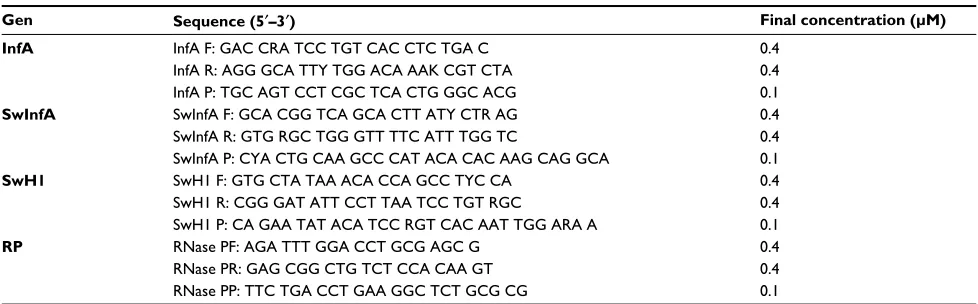
The protocol included four sets of primers and probes (uni-versal influenza A [InfA], swine flu [swInfA], swine H1 [swH1], and RNase P [RP] primers). TaqMan primers and probes were synthesized by Biosearch Technologies (Novato, CA, USA) (Table 1).
qRT-PCR assays
We followed a US Centers for Disease Control and Pre-vention protocol (http://www.who.int/csr/resources/ publications/swineflu/CDCRealtimeRTPCR_SwineH1As-say-2009_20090430.pdf) and used a Fast ABI 7500 thermo-cycler (Thermo Fisher Scientific, Waltham, MA, USA). The total reaction volume was 25 μL, including 5 μL template DNA (4–140 ng/μL), 0.5 μL primer forward (0.4 μM), 0.5 μL reverse primer (0.4 μM), 0.5 μL probe (0.1 μM), 12.5 μL 2× PCR Master Mix, 0.5 μL SuperScript III RT/Platinum Taq Mix, and 5.5 μL sterile molecular biology grade water. The PCR procedure started with one cycle of 50°C for 30 minutes (RT), one cycle of 95°C for 2 minutes (Taq inhibitor activation), followed by 45 cycles of 95°C for 15 seconds and 55°C for 30 seconds, as fluorescence data needed to be collected during the 55°C incubation step. A positive control (10 ng/μL) and a negative control of molecular biology grade water were run in parallel during all experiments. Background fluorescence was considered the correct value of the cycle threshold or cut (Ct). It was thus considered the threshold that went over the background fluorescence for each run in the
Table 1 Primers and probes used to detect influenza A(H1N1)pdm09 virus in study patients of Oaxaca, Mexico
Gen Sequence (5¢–3¢) Final concentration (μM)
InfA InfA F: GAC CRA TCC TGT CAC CTC TGA C 0.4
InfA R: AGG GCA TTY TGG ACA AAK CGT CTA 0.4
InfA P: TGC AGT CCT CGC TCA CTG GGC ACG 0.1
SwInfA SwInfA F: GCA CGG TCA GCA CTT ATY CTR AG 0.4
SwInfA R: GTG RGC TGG GTT TTC ATT TGG TC 0.4
SwInfA P: CYA CTG CAA GCC CAT ACA CAC AAG CAG GCA 0.1
SwH1 SwH1 F: GTG CTA TAA ACA CCA GCC TYC CA 0.4
SwH1 R: CGG GAT ATT CCT TAA TCC TGT RGC 0.4
SwH1 P: CA GAA TAT ACA TCC RGT CAC AAT TGG ARA A 0.1
RP RNase PF: AGA TTT GGA CCT GCG AGC G 0.4
RNase PR: GAG CGG CTG TCT CCA CAA GT 0.4
RNase PP: TTC TGA CCT GAA GGC TCT GCG CG 0.1
Notes: Primers and probes synthesized by Biosearch Technologies (Novato, CA, USA). The probe was labeled with FAM at 5’ and BHQ1 at 3’. A US Centers for Disease Control and Prevention protocol was used. Reprinted from World Health Organization, The WHO Collaborating Centre for influenza at CDC, DC protocol of realtime RTPCR for influenza
A(H1N1), 2009. Available from: http://www.who.int/csr/resources/publications/swineflu/CDCRealtimeRTPCR_SwineH1Assay-2009_20090430.pdf. Accessed March 8, 2018.46 Abbreviations: InfA, universal influenza A; swInfA, swine flu; swH1, swine H1; RP, RNase P.
Research and Reports in Tropical Medicine downloaded from https://www.dovepress.com/ by 118.70.13.36 on 27-Aug-2020
Dovepress Ramírez-Palacios et al
exponential phase of the amplification curve. A cutoff value of Ct=37 was corrected accordingly.
Identification of coinfections using
microarrays (TessArray RPM-Flu 3.1)
Total nucleic acid isolationWith 450 μL of the primary sample, total nucleic acids were recovered following the manufacturer’s instructions from the MasterPure DNA- and RNA-isolation kit (Epicentre Biotechnologies, Madison, WI, USA). A final elution volume of 35 μL was stored at –20°C until testing.
Quantification of total nucleic acids
Amounts of total nucleic acids were estimated according to the ratio A 260:280 nm using a NanoDrop 2000c (Thermo Fisher Scientific).
Synthesis of complementary DNA
With an RPM-Flu 3.1 RT tube containing 4 μL total nucleic acids, RT was performed in order to obtain cDNA. The RT master mix comprised 4 μL 5× buffer for a single chain, 2 μL 0.1 M DTT, 1 μL 40 U/μL RNaseOut, and 1 μL 200 U/μL SuperScript III RT/Platinum Taq Mix. RT-PCR cycling conditions were 25°C for 10 minutes, 50°C for 50 minutes, and 85°C for 5 minutes. The resulting product was stored at –20°C.
Multiplex PCR amplification
With four 3.1 RPM-Flu 3.1 multiplex PCR tubes (A, B, C, and D), multiplex PCR amplification was conducted. The reaction mixture contained 5 μL cDNA, 11 μL Flexi buffer 5× GoTaq, 8.8 μL 25 mM MgCl2, 0.8 μL DNA polymerase GoTaq 5 UI/μL, and 0.4 μL UDG 0.22 KU/220 μL. PCR cycling conditions were initiated with two cycles of 24°C for 10 minutes and 94°C for 2 minutes, followed by 16 cycles of 94°C for 30 seconds, 45°C for 30 seconds (increasing 1°C every cycle until 60°C), and 72°C for 90 seconds. The reaction was ended using 24 cycles of 94°C for 30 seconds and 60°C for 2 minutes. The PCR product was stored at 4°C.
Purification and elution of PCR products
Purification of PCR products was performed following the instructions of the QIAquick PCR purification kit (Qiagen). PCR products of tubes A–D were mixed and purified accord-ing to the manufacturer’s instructions. A final elution volume of 25 μL was stored at 4°C.
Fragmentation and labeling
Reagents were used for the fragmentation and labeling of the PCR products for the resequencing GeneChip assay (Affymetrix; Thermo Fisher Scientific). Briefly, in a PCR tube, fragmentation was conducted using 23 μL purified PCR products and 2.6 μL master mix (2.5 μL fragmentation buffer 10× and 0.1 μL fragmentation reagent). PCR cycling condi-tions were 37°C for 5 minutes and 95°C for 10 minutes. The PCR product was stored at 0°C. Subsequently, a new PCR tube was used to perform the DNA labeling, which contained 25.6 μL fragmented PCR products and 10.4 μL master mix (7.2 μL of TdT buffer 5×, 1.2 μL labeling reagent, and 2 μL TdT 30 UI/μL). PCR cycling conditions were 37°C for 30 minutes and 95°C for 5 minutes. Then, the labeled PCR prod-uct was placed for at least 5 minutes in 0°C for hybridization.
Hybridization, washing, and sample scanning
The hybridization procedure was carried out for 16 hours at 56°C using a hybridization oven (model 640, Affymetrix). Microarrays were then washed and stained in an Affymetrix Fluidics Station. A computer CEL file containing microarray images was produced by an Affymetrix G7 scanner.
Analysis, interpretation, and reanalysis
The scanned image of the microarray was analyzed using GSEQ 4.0 software, which produced a CHP file contain-ing the names and the sequences identified in each sample. Sequences were stored in a FASTA file, which was submitted online to retest the sequences on the manufacturer’s website (http://www2.gsu.edu/~psyrab/gseq/index.html). Sequences of the pathogens identified in the FASTA file contained the results of the positive and negative controls and an overview of the sequences, including their name, C3 score, homology percentage, and length of the longest continuous sequence.
Statistical analysis
Patient data – age, sex, date, flu symptoms, clinical sam-pling date, and place of residence – were captured on an Excel spreadsheet. The proportion of H1N1 patients with microbial infection was calculated as the number of positive patients divided by the total (n=88) number examined and expressed as a percentage (prevalence). The associated 95% CIs of the proportion of patients harboring the pathogens were also determined. Prevalence of patients with influenza A(H1N1)pdm09 and copathogens (additional one to five pathogens) were examined for significant differences at α=0.05, indicated by no overlapping of CIs. The prevalence
Research and Reports in Tropical Medicine downloaded from https://www.dovepress.com/ by 118.70.13.36 on 27-Aug-2020
Dovepress Copathogens with H1N1 in Oaxaca, Mexico
of each pathogen and symptoms in the 88 patients were also examined per age-group.
Results
The influenza A(H1N1)pdm09 virus was detected and con-firmed by qRT-PCR in a total of 88 clinical samples from study patients showing ILI in Oaxaca, Mexico (Table S1). To identify associated pathogens in the 88 samples, we used the microarray RPM-Flu 3.1.TessArray. A total of 71 patients presented copathogens with bacteria and/or viruses other than influenza A(H1N1)pdm09 (Table S1). Of the 71 patients, 27 (30.7%) patients harbored a single pathogen (other than influenza A[H1N1]pdm09), 28 (31.8%) harbored two pathogens, 13 (14.8%) harbored three pathogens, two (2.3%) harbored four pathogens, and one (1.1%) harbored five pathogens. The prevalence of copathogens did not vary among the patients coinfected with one to three pathogens; however, a significantly (P<0.05) low number of patients coharbored four to five pathogens (Table 2).
Genera of associated bacterial pathogens identified were
Streptococcus, Haemophilus, Neisseria, Pseudomonas,
Staphylococcus, Klebsiella, and Moraxella, as well as parain-fluenza coinfections and Coxsackie viruses. The most preva-lent species of bacteria found (P<0.05) were S. pneumoniae
(39.8%), H. influenzae (32.9%), N. meningitidis (25%), and
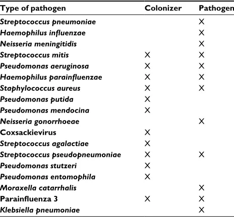
S. mitis (23.9%) (Figure 1). A medium number (14.7%) of prevalent bacteria were P. aeruginosa. A few patients (range of prevalence 2.2%–7.9%) harbored other species of bacteria, such as H. parainfluenzae, S. aureus, P. putida, P. mendocina, and N. gonorrhoeae. Less prevalent species of bacteria and viruses (1.1%) found are also summarized in Figure 1. Table 3 highlights the copathogens identified as colonizers of the respiratory tract and coinfection with pathogens found in the 88 H1N1 patients of Oaxaca.
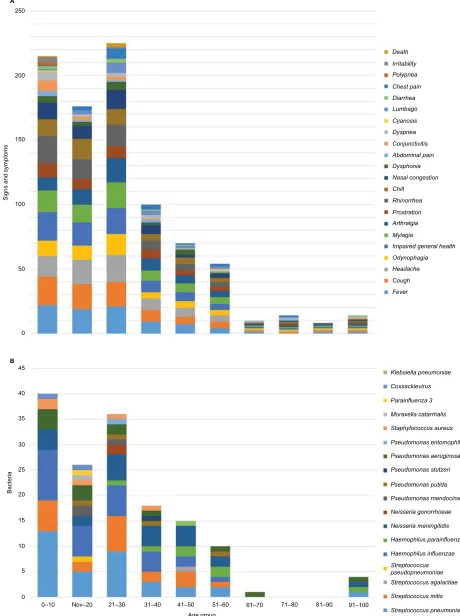
Of the 22 symptoms identified, the most common were fever in 85 patients (96%), impaired general health in 85 (96%), cough in 84 (95%), headache and myalgia in 75 (85%), rhinorrhea in 72 (82%), odynophagia in 57 (65%), cold in 56 (64%), and nasal congestion in 52 (59%). Most signs and/or symptoms were found among study participants 0.4–30 years old infected with influenza A(H1N1)pdm09 and microbial infection (Figure 2). Six of the 88 patients expe-rienced disease severity and were admitted to the intensive care unit (Table S1).
Two of four dead patients harbored three bacteria. One male aged 30 years had S. pneumoniae, H. influenzae, and
N. meningitidis. He was treated on an outpatient basis, and
Table 2 Pathogens associated with influenza A(H1N1)pdm09 virus and microbial prevalence in 88 patients of Oaxaca, Mexico
Associated pathogens, n Samples, n Prevalence* (95% CI)
0 17 19% (12%–28%)
1 27 31% (22%–41%)
2 28 32% (23%–42%)
3 13 15% (8%–22%)
4 2 3% (0.1%–6%)
5 1 1% (0.002%–4%)
Note: *Point estimates.
Figure 1 Number of copathogens positively associated with influenza A(H1N1)pdm09 virus and percentage of prevalence for each pathogen in patients of Oaxaca, Mexico.
Streptococcus pneumoniaeHaemophilus parainfluenzae
Neisseria meningitidisStreptococcus miti s
Pseudomonas aeruginosaHaemophilus parainfluenzaStaphylococcus aureu s
Pseudomonas putid a
Pseudomonas mendocinaNeisseria gonorrhoea e
Streptococcus agalactiae
Streptococcus pseudopneumoniae Pseudomonas stutzeri
Pseudomonas entomophila Moraxella catarrhali
s
Klebsiella pneumoniae Parainfluenza
3
Coxsackievirus B368Coxsackievirus B380
% Prevalence (95% CI
)
0 10 20 30 40 50 60 70 80 90 100
n=35 n=29
n=22 n=21 n=13
n=7
n=5 n=4
Pathogens
n=3 n=2 n=1 n=1 n=1 n=1 n=1 n=1 n=1 n=1 n=1
Research and Reports in Tropical Medicine downloaded from https://www.dovepress.com/ by 118.70.13.36 on 27-Aug-2020
Dovepress Ramírez-Palacios et al
presented such symptoms as fever, impaired general health, odynophagia, cough, and headache. A 29-year-old female had S. aureus, S. mitis, and S. pneumoniae. She had multiple symptoms (Table S1) and died during the period of confine-ment (puerperium) just after childbirth. The remaining two dead patients harbored two bacteria. One male aged 22 years had S. pneumoniae and N. gonorrhoeae. He was treated on an outpatient basis, presented multiple symptoms, and was asthmatic. One 8-month-old male had H. influenza and
S. mitis. The baby presented multiple symptoms, and was also asthmatic.
Discussion
Mexico has suffered at least four outbreaks produced by the influenza A(H1N1)pdm09 virus, with higher mortality rates than those reported by other countries.4,10 As mentioned, there
are viral and host factors,18,7,22 timeliness of medical treatment
of the disease,11,20,37 and presence of copathogens8,25,34,40
asso-ciated with increased severity of disease caused by this virus, among other factors, which may explain the epidemiological scenario in Mexico. During the first outbreaks of the virus in Mexico, increased mortality was noted because of the pres-ence of coinfections with other pathogens.20,31 Therefore, it
is of paramount importance to investigate which organisms are causing coinfections to implement a monitoring and surveillance system for the disease in Mexico.
Here, we used the standard qRT-PCR protocol to identify the influenza A(H1N1)pdm09 virus in conjunction with a microarray assay for detecting other associated pathogens in positive patients to influenza A(H1N1)pdm09. The microar-ray assay has been shown to be capable of detecting in a single sample up to 30 viruses and bacteria that may produce ILI.28
The identification of such pathogens is based on sequence analysis and subsequent search in the GenBank database, which helps to identify specific viral types and species of bacteria present in the sample.28,43
There was a significantly high prevalence rate of influenza patients with one and three associated pathogens (Table 2). The bacteria species most frequently identified was S. pneu-moniae (39.8% of total individuals with copathogens), which differs somewhat with percentages (7.5%–18.6%) reported elsewhere.3,8,13,29H. influenzae (32.9%) and N. meningitidis
(25%) also showed high prevalence rates. N. meningitidis
is associated with greater severity of illness but requires a process of immunosuppression by patients.5,14,23 Other
associ-ated pathogens that presented elevassoci-ated prevalence rates were
S. mitis (23.9%) and P. aeruginosa (14.7%), which coincide with other studies that reported (1%–13.9%) coinfections with those pathogens.3,29 However, when comparing the
results of this study with previous studies, prudence should be exercised, given that anatomic sites sampled and tech-niques of detection (and sensitivity and specificity) were somewhat different.
It is noteworthy that the presence of a high number of patients harboring several pathogens producing respi-ratory infections can be explained by considering the sanitary conditions of a particular country. For example, the prevalence of meningococcal disease varies among countries: 0.3–4 cases per 100,000 people.36 In addition,
there were only three cases where the pandemic virus was found in coinfection with parainfluenza 3 and Coxsackie virus. The results of our study are consistent with other studies that have reported lower rates of viral coinfections with respiratory syncytial virus, coronavirus, influenza B virus, adenovirus, and parainfluenza virus without sig-nificant complications or increase in the severity of the disease.3,15,40 Similarly, some associations were found in
most patients with certain signs and/or symptoms with influenza A(H1N1)pdm09 alone and with other pathogens (Table 3). Although this table does not address severity of illness or indicate that treatment of the bacterial infection was necessary, the evolution in the clinical status of the patients was more severe in some patients who interestingly
Table 3 The 18 bacteria found in the 88 H1N1 patients of
Oaxaca, Mexico: differences between pathogen cocolonizer and
coinfection
Type of pathogen Colonizer Pathogen
Streptococcus pneumoniae X
Haemophilus influenzae X
Neisseria meningitidis X
Streptococcus mitis X X
Pseudomonas aeruginosa X X
Haemophilus parainfluenzae X X
Staphylococcus aureus X X
Pseudomonas putida X
Pseudomonas mendocina X
Neisseria gonorrhoeae X
Coxsackievirus X
Streptococcus agalactiae X
Streptococcus pseudopneumoniae X X
Pseudomonas stutzeri X
Pseudomonas entomophila X
Moraxella catarrhalis X
Parainfluenza 3 X X
Klebsiella pneumoniae X
Note: Bacteria marked in yellow can cause sepsis. N. meningitides is highly pathogenic.
Research and Reports in Tropical Medicine downloaded from https://www.dovepress.com/ by 118.70.13.36 on 27-Aug-2020
Dovepress Copathogens with H1N1 in Oaxaca, Mexico
Figure 2 Signs and/or symptoms of study participants per age group infected with influenza A(H1N1)pdm09 and microbial infection. Notes: (A) Signs/symptoms; (B) bacteria harbored by patients.
0
0
0–10 Nov–20 21–30 31–40 41–50 51–60 Age group
61–70 71–80 81–90 91–100 5
10 15 20 25
Bacteria
30 35 40 45
Death Irritability Polypnea Chest pain Diarrhea Lumbago Cyanosis Dyspnea Conjunctivitis Abdominal pain Dysphonia Nasal congestion Chill
Rhinorrhea Prostration Arthralgia Mylagia
Impaired general health Odynophagia Headache Cough Fever
Klebsiella pneumoniae Coxsackievirus
Parainfluenza 3 Moraxella catarrhalis
Staphylococcus aureus
Pseudomonas entomophila Pseudomonas aeruginosa Pseudomonas stutzeri
Pseudomonas putida Pseudomonas mendocina
Neisseria gonorrhoeae
Neisseria meningitidis Haemophilus parainfluenzae
Haemophilus influenzae Streptococcus pseudopneumoniae Streptococcus agalactiae Streptococcus mitis
Streptococcus pneumoniae
50 100 150
Signs and symptoms
200 250
A
B
Research and Reports in Tropical Medicine downloaded from https://www.dovepress.com/ by 118.70.13.36 on 27-Aug-2020
Dovepress Ramírez-Palacios et al
harbored copathogens: six patients of this group (6.8%) were admitted to the intensive care unit, and four (4.5%) died as a consequence of the coinfections.3,15,40
The 30-year-old male could have died as a consequence of infection with N. meningitidis, as influenza could have facilitated meningococcal colonization.5,19 The 29-year-old
female was in puerperium and presented 11 symptoms, H1N1, and three pathogenic bacteria, such as S. aureus. One study found that the majority of S. aureus isolated in both children and adults were methicillin resistant.27 As a
consequence, she was treated as inpatient in an intensive care unit.
The other two dead patients were asthmatic. They pre-sumably died because adults and children with asthma are more likely to develop pneumonia after getting sick with the flu than people who do not have asthma (https://www.cdc. gov/flu/pdf/freeresources/updated/treating-influenza-2017. pdf). The 22-year-old male harbored S. pneumoniae, which appears to have a synergistic relationship with influenza.38
This patient should have been treated on an inpatient basis and sent to the intensive care unit, as he was asthmatic and presented 10 different symptoms. However, he was treated on an outpatient basis. The 8-month-old male had eight symptoms and harbored S. mitis, which could have elevated the risk of and exacerbated the influenza infection.21
A total of 21 of 88 H1N1 patients harbored S. mitis. All H1N1 patients who had complications and those that were submitted to intensive care units were treated with the neur-aminidase inhibitor oseltamivir (Tamiflu) within 48 hours of first symptoms of infection. An increase in the number of S. mitis cases could also have hampered the efficacy of the viral neuraminidase inhibitor drug.21 Of the four H1N1 patients
who died, S. mitis was present in three.
Copathogens were found in a significant number (>50%) of influenza cases in Oaxaca. Although some copathogens may be simply asymptomatic colonizing bacteria, other manifested proven coinfection and disease severity in some patients. Given that copathogens were found commonly, this could have an impact on the disease, but more studies need to be conducted, eg, investigating if coinfections between the pandemic virus and other pathogens can be the source of secondary infections when patients are in a status of immu-nosuppression. Moreover, other factors explain the increase in the severity of symptoms and mortality rate caused by H1N1 in Oaxaca. It has been noted that in areas of Oaxaca, sanitary conditions are not adequate, and this may allow proliferation of the pathogenic agents detected. A monitor-ing and surveillance system based on molecular diagnostics
for all respiratory pathogens should be implemented, as well as strengthening of sanitary measures in the local Oaxaca population, both of which are urgently needed.
Acknowledgments
MARP holds a scholarship from Comisión de Operación y Fomento de Actividades Académicas COFAA/IPN. The pres-ent work was sponsored by the National Council of Science and Technology (CONACYT, Mexico) and the government of the state of Oaxaca (FOMIX) (grant 122497).
Disclosure
The authors report no conflicts of interest in this work.
References
1. Aktürk H, Sütçü M, Badur S, et al. Evaluation of epidemiological and clinical features of influenza and other respiratory viruses. Turk Pediatri Ars. 2015;50(4):217–225.
2. Mossad SB. The resurgence of swine-origin influenza A (H1N1). Cleve Clin J Med. 2009;76(6):337–343.
3. Blyth CC, Webb SA, Kok J, et al. The impact of bacterial and viral co-infection in severe influenza. Influenza Other Respir Viruses. 2013;7(2):168–176.
4. Borja-Aburto VH, Chowell G, Viboud C, et al. Epidemiological character-ization of a fourth wave of pandemic A/H1N1 influenza in Mexico, winter 2011–2012: age shift and severity. Arch Med Res. 2012;43(7):563–570. 5. Cartwright KA, Jones DM, Smith AJ, Stuart JM, Kaczmarski
EB, Palmer SR. Influenza A and meningococcal disease. Lancet. 1991;338(8766):554–557.
6. Castillo-Palencia JP, Laflamme L, Monárrez-Espino J. Occurrence of AH1N1 viral infection and clinical features in symptomatic patients who received medical care during the 2009 influenza pandemic in central Mexico. BMC Infect Dis. 2012;12:363.
7. US Centers for Disease Control and Prevention. Intensive-care patients with severe novel influenza A (H1N1) virus infection: Michigan, June 2009. MMWR Morb Mortal Wkly Rep. 2009;58(27):749–752. 8. Chertow DS, Memoli MJ. Bacterial coinfection in influenza: a grand
rounds review. JAMA. 2013;309(3):275–282.
9. Chertow DS. Contribution of bacterial coinfection to severe influenza infection. Crit Care Med. 2012;40(5):1664–1665.
10. Chowell G, Echevarría-Zuno S, Viboud C, et al. Recrudescent wave of pandemic A/H1N1 influenza in Mexico, winter 2011–2012: age shift and severity. PLoS Curr. 2012;4:RRN1306.
11. Chowell G, Viboud C Simonsen L, et al. Impact of antiviral treatment and hospital admission delay on risk of death associated with 2009 A/ H1N1 pandemic influenza in Mexico. BMC Infect Dis. 2012;12:97. 12. Cillóniz C, Ewig S, Menéndez R, et al. Bacterial coinfection with H1N1
infection in patients admitted with community acquired pneumonia. J Infect. 2012;65(3):223–230.
13. Domínguez-Cherit G, Lapinsky SE, Macias AE, et al. Critically Ill patients with 2009 influenza A (H1N1) in México. JAMA. 2009;302(17): 1880–1887.
14. Feiterna-Sperling C, Edelmann A, Nickel R, et al. Pandemic influenza A (H1N1) outbreak among 15 school-aged HIV-1-infected children. Clin Infect Dis. 2010;51(11):e90–e94.
15. Goka E, Vallely P, Mutton K, Klapper P. Influenza A viruses dual and multiple infections with other respiratory viruses and risk of hospitalisa-tion and mortality. Influenza Other Respir Viruses. 2013;7(6):1079–1087. 16. Hersh AL, Maselli JH, Cabana MD. Changes in prescribing of antiviral medications for influenza associated with new treatment guidelines. Am J Public Health. 2009;99(Suppl 2):S362–S364.
Research and Reports in Tropical Medicine downloaded from https://www.dovepress.com/ by 118.70.13.36 on 27-Aug-2020
Dovepress Copathogens with H1N1 in Oaxaca, Mexico
17. Hubbard K, Pellar G, Emanuel P. Suitability of commercial transport media for biological pathogens under nonideal conditions. Int J Micro-biol. 2011;2011:463096.
18. Hurt AC, Holien JK, Parker MW, Barr IG. Oseltamivir resistance and the H274Y neuraminidase mutation in seasonal, pandemic and highly pathogenic influenza viruses. Drugs. 2009;69(18):2523–2531. 19. Jacobs JH, Viboud C, Tchetgen ET, et al. The association of
meningo-coccal disease with influenza in the United States, 1989–2009. PLoS One. 2014;9(9):e107486.
20. Jain S, Benoit SR, Skarbinski J, Bramley AM, Finelli L. Influenza-associated pneumonia among hospitalized patients with 2009 pan-demic influenza A (H1N1) virus: United States, 2009. Clin Infect Dis. 2009;54(9):1221–1229.
21. Kamio N, Imai K, Shimizu K, et al. Neuraminidase-producing oral mitis group streptococci potentially contribute to influenza viral infec-tion and reducinfec-tion in antiviral efficacy of zanamivir. Cell Mol Life Sci. 2015;72(2):357–366.
22. Kumar A, Zarychanski R, Pinto R, et al. Critically ill patients with 2009 influenza A (H1N1) infection in Canada. JAMA. 2009;302(17):1872–1879. 23. Legriel S, Merceron S, Tattevin P, et al. Favorable outcome after life-threatening meningococcal disease complicating influenza A (H1N1) infection. Infection. 2011;39(5):477–480.
24. Linde A, Rotzén-Ostlund M, Zweygberg-Wirgart B, Rubinova S, Bryt-ting M. Does viral interference affect spread of influenza? Euro Surveill. 2009;14(40):19354.
25. López-Cervantes M, García J, Pacheco RL, Escamilla RA, Villanueva M. La influenza A/H1N1 2009: una crónica de la primera pandemia del siglo XXl. Rev Digit Univ. 2010;11(4):1–19.
26. Martín-Loeches I, Sanchez-Corral A, Diaz E, et al. Community-acquired respiratory coinfection in critically ill patients with pandemic 2009 influenza A (H1N1) virus. Chest. 2011;139(3):555–562.
27. Metersky ML, Masterton RG, Lode H, File TM Jr, Babinchak T. Epide-miology, microbiology, and treatment considerations for bacterial pneu-monia complicating influenza. Int J Infect Dis. 2012;16(5):e321–e331. 28. Metzgar D, Myers CA, Russell KL, et al. Single assay for simultaneous
detection and differential identification of human and avian influenza virus types, subtypes, and emergent variants. PLoS One. 2010;5(2):e8995. 29. Muscedere J, Ofner M, Kumar A, et al. The occurrence and impact of
bacterial organisms complicating critical care illness associated with influenza A (H1N1) infection. Chest. 2013;144(1):39–47.
30. Neumann G, Brownlee GG, Fodor E, Kawaoka Y. Orthomyxovirus replication, transcription, and polyadenylation. Curr Top Microbiol Immunol. 2004;283:121–143.
31. Palacios G, Hornig M, Cisterna D, et al. Streptococcus pneumoniae coinfection is correlated with the severity of H1N1 pandemic influenza. PLoS One. 2009;4(12):e8540.
32. Randolph AG, Vaughn F, Sullivan R, et al. Critically ill children during the 2009–2010 influenza pandemic in the United States. Pediatrics. 2011;128(6):e1450–e1458.
33. Rhedin S, Hamrin J, Naucler P, et al. Respiratory viruses in hospitalized children with influenza-like Illness during the H1N1 2009 pandemic in Sweden. PLoS One. 2012;7(12):e51491.
34. Rice TW, Rubinson L, Uyeki TM, et al. Critical illness from 2009 pan-demic influenza A virus and bacterial coinfection in the United States. Crit Care Med. 2012;40(5):1487–1498.
35. Rodríguez-Valero M, Calleros HM, Escobar GA, et al. Difference between early clinical features of swine origin A H1N1 influenza confirmed and not confirmed infection in Mexico. J Infect Dev Ctries. 2012;6(4):302–310.
36. Sáfadi MA, Cintra OA. Epidemiology of meningococcal disease in Latin America: current situation and opportunities for prevention. Neurol Res. 2010;32(3):263–271.
37. Serna-Ojeda JC, Castañón-González JA, Macías AE, Mansilla-Olivares A, Domínguez-Cherit G, Polanco-González C. [Survey about responsiveness of third-level hospitals to a medical disaster: after the pandemic influenza in Mexico]. Gac Med Mex. 2012;148(3):227–235. Spanish.
38. Short KR, Habets MN, Hermans PW, Diavatopoulos DA. Interactions between Streptococcus pneumoniae and influenza virus: a mutually beneficial relationship? Future Microbiol. 2012;7(5):609–624. 39. Sistema Nacional de Vigilancia. Dirección General de Epidemiología.
Secretaría de Salud/Sistema de Notificación Semanal de Casos Nuevos, corte al 2 de marzo del 2018. Secretaría de Salud: México; 2018. Avail-able from: https://www.gob.mx/cms/uploads/attachment/file/304904/ INFLUENZA_2018_SE09.pdf. Accessed March 8, 2018.
40. Stefanska I, Romanowska M, Donevski S, Gawryluk D, Brydak LB. Co-infections with influenza and other respiratory viruses. Adv Exp Med Biol. 2013;756:291–301.
41. Volkow P, y Pérez-Padilla JR. Respuesta hospitalaria a la epidemia de influenza. In: Córdova-Villalobos JA, Valdespino-Gómezc JL, de León-Rosales S, editors. La Epidemia de Influenza A/H1N1 en México. Madrid: Editorial Médica Panamericana; 2010:34–44.
42. Wang D, Chen LL, Ding YF, et al. Viral etiology of medically attended influenza-like illnesses in children less than five years old in Suzhou, China, 2011 to 2014. J Med Virol. 2016;88(8):1334–1340.
43. Wang Z, Malanoski AP, Lin B, et al. Resequencing microarray probe design for typing genetically diverse viruses: human rhinoviruses and enteroviruses. BMC Genomics. 2008;9:577.
44. World Health Organization. FluNet. World Health Organization: Geneva; 2018. Available from: http://www.who.int/influenza/gisrs_labo-ratory/flunet/en. Accessed March 8, 2018.
45. World Health Organization. WHO Guidelines for Pharmacological Management of Pandemic Influenza A (H1N1) 2009 and Other Influ-enza Viruses: Part 1 – Recommendations. World Health Organization: Geneva; 2010. Available from: http://www.who.int/csr/resources/ publications/swineflu/h1n1_guidelines_pharmaceutical_mngt.pdf. Accessed March 8, 2018.
46. World Health Organization. CDC protocol of realtime RTPCR for influ-enza A(H1N1). The WHO Collaborating Centre for influenza at CDC Atlanta, United States of America: World Health Organization, Geneva; 2009. Available from:http://www.who.int/csr/resources/publications/ swineflu/CDCRealtimeRTPCR_SwineH1Assay-2009_20090430.pdf. Accessed March 8, 2018.
Research and Reports in Tropical Medicine downloaded from https://www.dovepress.com/ by 118.70.13.36 on 27-Aug-2020
Dovepress Ramírez-Palacios et al
Supplementar
y material
Table S1
Symptoms, clinical evolution, and positivity to bacteria of 88 influenza A(H1N1)pdm09 patients in Oaxaca, Mexico
Age Sex Evolution of patient clinical status
Symptoms Bacteria Fever Cough Headache Odynophagia Impaired general health
Myalgia Arthralgia Prostration Rhinorrhea Chill Nasal congestion Dysphonia Abdominal pain Conjunctivitis Dyspnea Cyanosis Lumbago Diarrhea Chest pain Polypnea Irritability (headache if
<
5 years old)
Death Clinical sample Streptococcus pneumoniae Streptococcus mitis Streptococcus agalactiae Streptococcus pseudopneumoniae Haemophilus influenzae Haemophilus parainfluenzae Neisseria meningitidis Neisseria gonorrhoeae Pseudomonas mendocina Pseudomonas putida Pseudomonas stutzeri Pseudomonas aeruginosa Pseudomonas entomophila Staphylococcus aureus Moraxella catarrhalis Parainfluenza 3 Coxsackievirus Klebsiella pneumoniae 0.4 M ICU + + – + + – – + + – + – – – + – – – – – – – NS + – – – – – – – – – – – – – – – – – 0.5 M I – + – – + – – + + – + + – – + – – – – + + – NS + – – – – – – – – – – – – – – – – – 0.6 F O + + – – – + – – – – – – – – – – – – – – + – TS + + – – + – + – – – – – – – – – – – 0.8 M ICU + + – – + – – + + – + – – + + + – – – – – + LB – + – – + ND – – – – – – – – – – – – 0.8 F O + + + + + + + – + + + – – + + – – – – – – – NS + – – – + – – – – – – – – – – – – – 1 F O + + + + + + – – + + + – – + + – – – – – – – NS + + – – – – + – – – – – – – – – – – 1 F I + + + + + + – – + + – – – + – – + + – – – – NS + – – – + – – – – – – – – – – – – – 1 M O + + – – + – – – + – – – – – + – – – – – + – NS – – – – – – – – – – – – – – – – – – 2 M I + + – – + – – – + + – + + – – – – + – – – – TS + – – – – – – – – – – – – – – – – – 3 F O + + + – + + – + + + + – – – – – – – – – – – TS + + – – + – – – – – – + – – – – – – 4 F O + + + – + + + – + – – – + – – – – – – – – – TS + + – – + – – – – – – + – – – – + – 5 M O + + + + + + + – + – – – + – – – – – – – – – TS + – – – – – – – – – – – – – – – – – 5 M O + + + + + – – + + + + + – + – – – – – – – – TS – – – – – – – – – – – – – + – – – – 6 M O + – + – + + + + + + – – – – – – – – – – – – TS – – – – – – – – – – – + – – – – – – 7 M O + + + + + + + – + + + + – + – – – – – – – – TS – – – – – – – – – – – – – – – – – – 8 M O + + + + + + + + + + + – – – – – – – – – – – TS – – – – – – – – – – – – – – – – – – 8 M I + + + + + + + + + + + + – + – – – – + – – – TS – – – – – – – – – – – + – + – – – – 8 M I + + + + + + + + + + – – – – – – – – – – – – TS + – – – + – – – – – – – – – – – – – 9 M O + + + – + + – – – – – – – – – – – – – – – – TS + – – – + – – – – – – – – – – – – – 9 F O + + + + + + – + + – – – + + – – – – – – – – TS – – – – + – + – – – – – – – – – – – 9 F ICU + + + + + + + + + + + – – – – – – – – – – – BAL – + – – + – + – – – – – – – – – – – 9 M I + + – – + + + – + – + – – – + – – – – + + – TS + – – – – – – – – – – – – – – – – – 10 M I + + + – + + – – + + + – – – – – – – – – – – TS – – – – – – – – – – – – – – – – – – 11 M O + + + + + + + – – + – – – – – – – – – – – – TS + – – – + – + – – – – – – – – – – – ( Continued )
Research and Reports in Tropical Medicine downloaded from https://www.dovepress.com/ by 118.70.13.36 on 27-Aug-2020
Dovepress Copathogens with H1N1 in Oaxaca, Mexico
Age Sex Evolution of patient clinical status
Symptoms Bacteria Fever Cough Headache Odynophagia Impaired general health
Myalgia Arthralgia Prostration Rhinorrhea Chill Nasal congestion Dysphonia Abdominal pain Conjunctivitis Dyspnea Cyanosis Lumbago Diarrhea Chest pain Polypnea Irritability (headache if
<
5 years old)
Death Clinical sample Streptococcus pneumoniae Streptococcus mitis Streptococcus agalactiae Streptococcus pseudopneumoniae Haemophilus influenzae Haemophilus parainfluenzae Neisseria meningitidis Neisseria gonorrhoeae Pseudomonas mendocina Pseudomonas putida Pseudomonas stutzeri Pseudomonas aeruginosa Pseudomonas entomophila Staphylococcus aureus Moraxella catarrhalis Parainfluenza 3 Coxsackievirus Klebsiella pneumoniae 11 M I + + + + + – – – + + + + – – – – – – – – – – TS – – – – – – + – – – – – – – – – – – 12 M I + + + – + – – – + – – – – – – – – – – – – – TS – – – – – – – – – + – – – – – – – – 12 M I + + + – + + – – + – + – – + – – – – – – – – TS – – – – – – – – – – – – – – – – – – 13 F O + + + + + + + – – + – – – – – – – – – – – – TS – – – – – – – – – – – + – – – – + – 13 F O + + + – + + – + + + + – – + – – – – – – – – TS – – – – – – – – – – – – – – – – – – 13 F O + + + + + + + + + + – – – – – – – – – – – – TS + – – – + – – – – – – – – – – – – – 14 F O + + + + – + + – + – – – – – – – – – – – – – TS – – – – + – – – + – – – – + – – – – 14 M O + + + – + – – – – + – – – – – – – – – – – – TS – + – – – – – – – – – – – – – – – – 14 F O + + + – + + + + + + + – – – + – – – + – – – TS – – – + – – – – – – – – – – – – – – 14 M O + + + – + + + + + + + – – – – – + – – – – – TS – – – – – – – – – – – – – – – – – – 15 F O + + + + + + – + + + + – – – – – – – – – – – TS – – – – – – – – + – – – – – – – – – 16 F O + + + + + + + + + + + – – – – – + – + – – – TS – – – – – – – – – – – – – – – + – – 16 M O + + + – + – + – + + – – – – + – – – + – – – TS – – – – – – – – – – – – – – + – – – 17 M O + + + + + + + – – + – – – – – – – – – – – – TS – – – – – – – – – – – + – – – – – – 17 M O + + + + + + + – + + + – – – – – – – – – – – TS – – – – + – – – – – – + – – – – – – 17 M O + + + – + – – + + + – – – – – – – – – – – – TS + + – – + – – – – – – – – – – – – – 17 M O + + + + + + + – + + + + + + – – + – – – – – TS + – – – – – – – – – – – – – – – – – 19 M O + + + + + + + + + + + + – – – – – – – – – – TS + – – – + – – – – – – – – – – – – – 21 M O + + + + + + – – + – + – – – – – + – + – – – TS + – – – + – – – – – – + – – – – – – 21 F O + + + + + + + + + + + – – – – – + – – – – – TS – – – – – – – – – – – – + – – – – – 21 F O + + + – + + + + + + + – – – – – – – + – – – TS + – – – + – + – – – – – – – – – – – 22 M O + + + + + + + + + + + + – + – – + – + – – – TS + – – – – – + – – – – – – – – – – – 22 M O + + + – + + + + + – + – – – + – – – – – – + LB + – – – – – – + – – – – – – – – – – 24 M O + – + + + + + + – + + – – – – – + + + – – – TS – – – – + – – + – – – – – – – – – – 24 F I + + + + + + + – + + + + – – – – – – – – – – TS – – – – – – – – – – – – – – – – – – 25 F O + + + + + + + + + + + + – – – – + + – – – – TS – – – – – – – – + – – – – – – – – – ( Continued ) Table S1 ( Continued )
Research and Reports in Tropical Medicine downloaded from https://www.dovepress.com/ by 118.70.13.36 on 27-Aug-2020
Dovepress Ramírez-Palacios et al
Age Sex Evolution of patient clinical status
Symptoms Bacteria Fever Cough Headache Odynophagia Impaired general health
Myalgia Arthralgia Prostration Rhinorrhea Chill Nasal congestion Dysphonia Abdominal pain Conjunctivitis Dyspnea Cyanosis Lumbago Diarrhea Chest pain Polypnea Irritability (headache if
<
5 years old)
Death Clinical sample Streptococcus pneumoniae Streptococcus mitis Streptococcus agalactiae Streptococcus pseudopneumoniae Haemophilus influenzae Haemophilus parainfluenzae Neisseria meningitidis Neisseria gonorrhoeae Pseudomonas mendocina Pseudomonas putida Pseudomonas stutzeri Pseudomonas aeruginosa Pseudomonas entomophila Staphylococcus aureus Moraxella catarrhalis Parainfluenza 3 Coxsackievirus Klebsiella pneumoniae 25 M O + + + + + + + + + – – – – – – – + – + – – – TS – – – – – – – – – – – – – – – – – – 26 F O + + + + + + + – + + + – – – – – + – + – – – TS + + – – – + + – – – – – – – – – – – 26 F O + + + + + + + + + + + + – + – – – – – – – – TS + + – – + – + – – – – – – – – – – – 26 M O + + + – + + + – + – + – – – – – – + – – – – TS – – – – – – – – – + – – – – – – – – 26 F O + + + + + + + – + – – – – – – – – – – – – – TS – + – – – – – – – – – – – – – – – – 26 F I + + + + + + + – + – – – – – – – – – – – – – TS – – – – – – – – – – – – – – – – – – 27 F O + + + + + + + – + + – – – – – – – – – – + – TS – – – – – – – – – – – – – – – – – – 29 F ICU + + + + – + + – + + + – – – + – – – + – – + LB + + – – – – – – – – – – – + – – – – 29 F ICU + – + – + + + + – – + + – – + – – – + – – – BAL + + – – + – – – – – – – – – – – – – 29 F O + + + + + + + – + + + – – – – – + – – – – – TS – – – – – – – – – – – – – – – – – – 29 F O + + – + + + – – – – – – – – – – – – – – – TS – – – – – – – – – – – – – – – – – – 30 F I + + + + + + + – + + + + + + – – – – – – – – TS + + – – – – – – – – – + – – – – – – 30 M O + + + + + – – – – – – – – – – – – – – – – + LB – + – – + – + – – – – – – – – – – – 32 M O + + + – + + + + + – + – – – – – – – + – – – TS + + – – + – + – – – – – – – – – – – 34 F O + + + – + + + + – + – – – – + – + – + – – – TS + – – – + – + – – – – – – – – – – – 36 F O + + + + + + + + + – + + – + – – – – – – – – TS + + – – + – + – – – + – – – – – – – 36 M I + + + – + + + – – – + – – – + – – – + – – – TS – – – – – – – – – – – + – + – – – – 36 F O + + + + + + + – + + + – + – – – – – – – – – TS – – – – – + – – – – – – – – – – – – 38 M O + + + + + + + + + + + + – – – – – – – – – – TS – – – – – – – – – + – – – – – – – – 38 M O + + + + + + + + + + + – – – – – + + – – – – TS – – – – – – – – – – – – – – – – – – 38 M O + + + – + + + + + – + – + – + – + – + – – – TS – – – – + – + – – – – – – – – – – – 40 M O + + + + + – + + + + – – – – – – – – – – – – TS – – – – – – – – – – – – – – – – – – 41 F I + + + + + + + + + + + – – – – – – – – – – – TS – + – – + – + – – – – – – – – – – – 41 M O + + + – + + + + + + – + – – – – + – + – – – TS – + – – – + + – – – – – – – – – – – 43 F I + – + + + + + – + – – – – – – – + – – – – – TS + – + – + – + – – – – – – – – – – – 44 F ICU + + + + + + – – + – – + – – – – – – – – – – BAL – – – – – – – – – – – – – – – – – – ( Continued ) Table S1 ( Continued )
Research and Reports in Tropical Medicine downloaded from https://www.dovepress.com/ by 118.70.13.36 on 27-Aug-2020
Dovepress Copathogens with H1N1 in Oaxaca, Mexico
Age Sex Evolution of patient clinical status
Symptoms Bacteria Fever Cough Headache Odynophagia Impaired general health
Myalgia Arthralgia Prostration Rhinorrhea Chill Nasal congestion Dysphonia Abdominal pain Conjunctivitis Dyspnea Cyanosis Lumbago Diarrhea Chest pain Polypnea Irritability (headache if
<
5 years old)
Death Clinical sample Streptococcus pneumoniae Streptococcus mitis Streptococcus agalactiae Streptococcus pseudopneumoniae Haemophilus influenzae Haemophilus parainfluenzae Neisseria meningitidis Neisseria gonorrhoeae Pseudomonas mendocina Pseudomonas putida Pseudomonas stutzeri Pseudomonas aeruginosa Pseudomonas entomophila Staphylococcus aureus Moraxella catarrhalis Parainfluenza 3 Coxsackievirus Klebsiella pneumoniae 48 F O + + + + + + + + – + – + – – – – – + – – – – TS – – – – – – – – – – – – – – – – – – 49 F O + + + – + + + – + + – – – – – – – – – – – – TS + + – – – + + – – – – – – – – – – + 49 F I + + + + + + + + – + + + – – + – – – – – – – TS – – – – – – – – – – – – – – – – – – 51 F O – + + + + + + – + – – – + – – – – – + – – – TS – – – – – + – – – – – – – – – – – – 52 M O + + + + + + + + + + + + – – + + + – – – – – TS + + – – ND + + – – + – – – – – – – – 53 F O + + + – + + + – + – + – – – – – – – – – – – TS – – – – – – – – – – – – – – – – – – 55 M O + + + + + + + + + + + – – – – – – – + – – – TS + – – – + – + – – – – + – – – – – – 57 F I + + + + + + + + – + – – – – – – + – – – – – TS – – – – – – – – – – – – – – – – – – 68 F O + + + + + + + + – – – – + – + – – – – – – – TS – – – – – – – – – – – + – – – – – – 72 M I – + + + + + + + + + + – + – + – + – + – – – TS – – – – – – – – – – – – – – – – – – 89 M I + + + + + + + – + – – – – – – – – – – – – – TS – – – – – – – – – – – – – – – – – – 92 F I + + + + + + + + + + + – – + + – – + – – – – TS + – – – – + + – – – – + – – – – – – Abbreviations:
M, male; F, female; O, outpatient; I, inpatient; ICU, intensive care unit; TS, throat swab; LB, lung biopsy; BAL, bronchoalveolar lavage; NS, nasopharyngeal swab; ND, no data.
Table S1
(
Continued
)
Research and Reports in Tropical Medicine downloaded from https://www.dovepress.com/ by 118.70.13.36 on 27-Aug-2020
Dovepress
Research and Reports in Tropical Medicine
Publish your work in this journal
Submit your manuscript here: https://www.dovepress.com/research-and-reports-in-tropical-medicine-journal
Research and Reports in Tropical Medicine is an international, peer-reviewed, open access journal publishing original research, case reports, editorials, reviews and commentaries on all areas of tropical medicine, including: Diseases and medicine in tropical regions; Entomology; Epidemiology; Health economics issues; Infectious disease; Laboratory
science and new technology in tropical medicine; Parasitology; Public health medicine/health care policy in tropical regions; and Microbiology. The manuscript management system is completely online and includes a very quick and fair peer-review system. Visit http://www.dovepress. com/testimonials.php to read real quotes from published authors.
Dove
press
Ramírez-Palacios et al
Research and Reports in Tropical Medicine downloaded from https://www.dovepress.com/ by 118.70.13.36 on 27-Aug-2020