University of Pennsylvania
ScholarlyCommons
Publicly Accessible Penn Dissertations
1-1-2013
Notch Signaling and Bone Fracture Healing
Michael I. Dishowitz
University of Pennsylvania, michael.dishowitz@gmail.com
Follow this and additional works at:
http://repository.upenn.edu/edissertations
Part of the
Biology Commons
, and the
Biomedical Commons
This paper is posted at ScholarlyCommons.http://repository.upenn.edu/edissertations/630
For more information, please contactlibraryrepository@pobox.upenn.edu.
Recommended Citation
Dishowitz, Michael I., "Notch Signaling and Bone Fracture Healing" (2013).Publicly Accessible Penn Dissertations. 630.
Notch Signaling and Bone Fracture Healing
Abstract
Bone fractures can exhibit delayed or non-union healing. Current treatments have well- documented
limitations. Although morphological aspects of fracture healing are well- characterized, molecular
mechanisms that regulate the complex progression of healing are poorly understood. Therefore, a need
persists for the identification of novel pathways that regulate fracture healing, and for development of
therapeutics targeting these pathways to enhance regeneration. Notch signaling regulates bone development,
and many aspects of bone development are recapitulated during repair. Notch signaling is also required for
repair of other tissues, and enhancing Notch signaling promotes regeneration. Therefore, the objective of this
thesis was to determine the role of Notch signaling during bone fracture healing, and to create a translatable
therapy targeting the pathway to enhance bone tissue formation. We hypothesized that (i) Notch signaling
components are active during bone repair; (ii) inhibition of Notch signaling alters healing; (iii) expression of
the Jagged1 ligand in mesenchymal cells regulates bone formation; and (iv) therapeutic delivery of Jagged1
will activate the Notch signaling pathway and promote osteogenesis.
We first characterized activation of Notch signaling during tibial fracture and calvarial defect healing, and
demonstrated that Notch signaling components are active during both methods of repair with Jagged1 the
most highly upregulated ligand. Then we determined the importance of Notch signaling by using a temporally
controlled inducible model (Mx1- Cre;dnMAMLf/-) to impair canonical signaling in all cells during tibial
fracture and calvarial defect healing, and demonstrated that Notch inhibition alters the temporal progression
of events required for healing, including inflammation, cartilage formation, callus vascularization and bone
remodeling. Next we deleted Jagged1 in mesenchymal progenitors (Prx1-Cre;Jagged1f/f) or committed
osteoblasts (Col2.3-Cre;Jagged1f/f), and determined that Jagged1 promotes bone formation during
development. Finally, we developed a biomaterial construct comprised of Jagged1 and a poly(β-amino ester)
scaffold, and demonstrated that it activates Notch signaling and enhances osteoblast differentiation.
This thesis identified Notch signaling as an important regulator of fracture healing, developed a translatable
therapeutic targeting the pathway to improve bone tissue formation. The study design outlined can also serve
as a model for the discovery of novel pathways that regulate, and therefore could enhance, bone fracture
healing.
Degree Type
Dissertation
Degree Name
Doctor of Philosophy (PhD)
Graduate Group
Bioengineering
First Advisor
Keywords
bone, fracture, Jagged1, Notch signaling, orthopaedics
Subject Categories
NOTCH SIGNALING AND BONE FRACTURE HEALING
Michael I. Dishowitz
A DISSERTATION in Bioengineering
Presented to the Faculties of the University of Pennsylvania in Partial Fulfillment of the
Requirements for the Degree of Doctor of Philosophy
2013
___________________________ Supervisor of Dissertation
Dr. Kurt D. Hankenson, Dean W. Richardson Chair in Equine Disease Research, Associate Professor of Equine Musculoskeletal Research
___________________________ Graduate Group Chairperson
Dr. Daniel A. Hammer, Alfred G. and Meta A. Ennis Professor of Bioengineering and Chemical and Biomolecular Engineering
Dissertation Committee
Dr. Jason A. Burdick (Chair), Associate Professor of Bioengineering
Dr. Kathleen M. Loomes, Associate Professor of Pediatrics at the Children’s Hospital of Philadelphia
Dr. Robert L. Mauck, Associate Professor of Orthopaedic Surgery and Bioengineering
ACKNOWLEDGEMENTS
I would like to thank my thesis committee for guiding me along the way through graduate school.
To Dr. Jason Burdick, thank you for advising me through the final phase of my thesis. The skills I
have learned collaborating with you and your lab have prepared me for the next stages of my
career. To Dr. Kathy Loomes, it is because of you that I understand the potential impact my
research could have on those with clinical Notch disorders. To Dr. Rob Mauck, I have always
respected the way you approach your work and the thought process you go about every day life.
It is always a pleasure speaking with you. I would like to especially thank Dr. Lou Soslowsky. You
taught me the skills I needed to eventually become a successful scientist. I will forever be grateful
for the amount of time you spent training me. And most importantly to my advisor Dr. Kurt
Hankenson, you challenged me to achieve things I didn’t realize I could, and you allowed me the
freedom I needed to develop into a confident independent scientist. Thank you for exposing me to
so many opportunities atypical to other graduate students.
I would like to thank those I have worked with along the way, including current and former
members of the Hankenson lab Dr. Shawn Terkhorn, Jason Combs, Dr. Fengchang Zhu, Lorraine
Mutyaba, Derek Dopkin, Sandy Bostic, Joel Takacs, Mike Karp, Andrew Barr, and Drs. Emily
Miedel and Nicole Belkin; our very close collaborators Drs. Jaimo Ahn, Susan Volk and Samir
Mehta; members of the Burdick lab, including Drs. Darren Brey, Harini Sundararaghavan, Ross
Marklein, Sudhir Khetan, and Brendan Purcell; Dr. Tina Bales from the Loomes lab; and Bob
Caron from the PCMD histology core.. I would like to especially thank all of my friends who have
been so important to me both before and during graduate school, and will continue to afterwards.
Finally, I would like to thank my Mom, Dad and brother Ben for being an incredibly loving
and supportive family. I couldn’t have asked for a better family to be a part of. It is to you that I
owe my success. I would also like to thank my in-laws, Joe, Alicia, Ryan, Nathan, Patrick, Eileen,
Tom and Mary. And most importantly, I would like to thank my wife Jamie. I love you and couldn’t
imagine going through life with anyone else. And of course I can’t forget Roscoe. Thank you for
ABSTRACT
NOTCH SIGNALING AND BONE FRACTURE HEALING
Michael I. Dishowitz
Kurt D. Hankenson, D.V.M., Ph.D.
Bone fractures can exhibit delayed or non-union healing. Current treatments have
documented limitations. Although morphological aspects of fracture healing are
well-characterized, molecular mechanisms that regulate the complex progression of healing are poorly
understood. Therefore, a need persists for the identification of novel pathways that regulate
fracture healing, and for development of therapeutics targeting these pathways to enhance
regeneration. Notch signaling regulates bone development, and many aspects of bone
development are recapitulated during repair. Notch signaling is also required for repair of other
tissues, and enhancing Notch signaling promotes regeneration. Therefore, the objective of this
thesis was to determine the role of Notch signaling during bone fracture healing, and to create a
translatable therapy targeting the pathway to enhance bone tissue formation. We hypothesized
that (i) Notch signaling components are active during bone repair; (ii) inhibition of Notch signaling
alters healing; (iii) expression of the Jagged1 ligand in mesenchymal cells regulates bone
formation; and (iv) therapeutic delivery of Jagged1 will activate the Notch signaling pathway and
promote osteogenesis.
We first characterized activation of Notch signaling during tibial fracture and calvarial
defect healing, and demonstrated that Notch signaling components are active during both
methods of repair with Jagged1 the most highly upregulated ligand. Then we determined the
importance of Notch signaling by using a temporally controlled inducible model
(Mx1-Cre;dnMAMLf/-) to impair canonical signaling in all cells during tibial fracture and calvarial defect
healing, and demonstrated that Notch inhibition alters the temporal progression of events
required for healing, including inflammation, cartilage formation, callus vascularization and bone
committed osteoblasts (Col2.3-Cre;Jagged1f/f), and determined that Jagged1 promotes bone
formation during development. Finally, we developed a biomaterial construct comprised of
Jagged1 and a poly(β-amino ester) scaffold, and demonstrated that it activates Notch signaling
and enhances osteoblast differentiation.
This thesis identified Notch signaling as an important regulator of fracture healing,
developed a translatable therapeutic targeting the pathway to improve bone tissue formation. The
study design outlined can also serve as a model for the discovery of novel pathways that
TABLE OF CONTENTS
ACKNOWLEDGEMENTS ...ii
ABSTRACT...iii
TABLE OF CONTENTS...v
LIST OF TABLES ...x
LIST OF FIGURES...xi
CHAPTER 1 ...1
Introduction to Bone Fracture Healing and the Notch Signaling Pathway...1
1.1 Clinical Significance of Bone Fractures...1
1.2 Bone Fracture Healing ...2
1.3 Notch Signaling Pathway ...3
1.4 Notch Signaling and Bone Formation...4
1.5 Notch and Vasculogenesis...5
1.6 The Notch Ligand Jagged1 ...6
1.7 Notch and Regeneration ...6
1.8 Use of Poly(β-amino ester)s for Therapeutic Applications ...7
1.9 Conclusions...9
1.10 References ...10
CHAPTER 2 ...17
Specific Aims and Hypotheses...17
2.1 Specific Aim I (Chapter 3) ...17
2.1.1 Hypothesis I ...17
2.2 Specific Aim II (Chapter 4) ...18
2.2.1 Hypothesis II ...18
2.3 Specific Aim III (Chapter 5) ...18
2.4 Specific Aim IV (Chapter 6) ...18
2.4.1 Hypothesis IV...19
CHAPTER 3 ...20
Notch Signaling Components Are Upregulated During Endochondral and
Intramembranous Bone Regeneration...20
3.1 Introduction...20
3.2 Methods...21
3.2.1 Experimental Design...21
3.2.2 Tibial Fracture (TF) Procedure ...21
3.2.3 Calvarial Defect (CD) Procedure ...22
3.2.4 Quantitative Real-Time Polymerase Chain Reaction (QPCR)...22
3.2.5 Histology and Immunohistochemistry (IHC) ...23
3.2.6 Statistical Analysis ...24
3.3 Results ...25
3.3.1 Validation of TF and CD Models for EO and IO, Respectively ...25
3.3.2 Comparison of Notch Gene Expression Over Time During TF and CD ...26
3.3.3 Comparison of Notch Gene Expression During TF vs. CD at Each Time Point ...26
3.3.4 Identification of Cells That Express Jagged1 and NICD2 During TF...28
3.3.5 Identification of Cells That Express Jagged1 and NICD2 During CD ...29
3.4 Discussion ...36
3.5 References ...40
CHAPTER 4 ...44
Inhibition of Canonical Notch Signaling Results in Sustained Callus
Inflammation and Alters Multiple Phases of Fracture Healing ...44
4.1 Introduction...44
4.2 Methods...45
4.2.2 Experimental Design...46
4.2.3 Histology and Immunohistochemistry (IHC) ...47
4.2.4 Histomorphometric Analysis ...48
4.2.5 Micro-computed Tomography (µCT) ...49
4.2.6 Quantitative Real-Time Polymerase Chain Reaction (QPCR)...50
4.2.7 Statistical Analysis ...51
4.3 Results ...52
4.3.1 dnMAML Expression During Bone Fracture Healing ...52
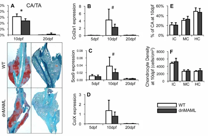
4.3.2 dnMAML Decreases Cartilage Formation During Fracture Healing ...53
4.3.3 dnMAML Inhibits Expression of Vascular Endothelial Cell Markers During Fracture Healing...54
4.3.4 dnMAML Alters Bone Remodeling During Fracture Healing ...55
4.3.5 dnMAML Prolongs Inflammation During Fracture Healing ...59
4.3.6 dnMAML Does Not Alter Cell Proliferation or Apoptosis During Fracture Healing ....61
4.4 Discussion ...62
4.5 References ...66
CHAPTER 5 ...71
The Role of the Notch Ligand Jagged1 During Bone Development and Aging...71
5.1 Introduction...71
5.2 Methods...74
5.2.1 Generation of Mice ...74
5.2.2 Experimental Design...75
5.2.3 Micro-computed Tomography (µCT) ...75
5.2.4 Quantitative Real-Time Polymerase Chain Reaction (QPCR)...76
5.2.5 Statistical Analysis ...76
5.3.1 Jag1 Deletion During Early and Late Differentiation Inhibits Trabecular Bone
Formation...77
5.3.2 Jag1 Deletion During Early and Late Differentiation Promotes Periosteal Expansion and Endosteal Resorption of Cortical Bone...79
5.3.3 Jag1 regulation of Notch Pathway, Osteoblast, Osteoclast, and Proliferation Gene Expression ...81
5.4 Discussion ...86
5.5 References ...89
CHAPTER 6 ...93
Activation of Notch Signaling by Jagged1 Immobilization to a Poly(
β
-amino
ester) Polymer Induces Osteoblastogenesis ...93
6.1 Introduction...93
6.2 Methods...95
6.2.1 Macromer Synthesis and Photopolymerization ...95
6.2.2 Jagged1 Immobilization Strategies...96
6.2.3 In vitro Experimental Design...96
6.2.4 Quantitative Real-Time Polymerase Chain Reaction (QPCR)...97
6.2.5 Alamar Blue Assay ...97
6.2.6 Alkaline Phosphatase (AP) Histochemical Staining...98
6.2.7 Alizarin Red S Staining ...98
6.2.8 ELISA...98
6.2.9 In vivo Analysis ...99
6.2.10 Statistical Analysis ...99
6.3 Results ...101
6.3.1 Direct Jagged1/A6 Is Most Effective at Activating Canonical Notch Signaling ...101
6.3.2 More Jagged1 is Successfully Immobilized to A6 via the Direct Method...101
6.3.4 Direct Jagged1/A6 is More Effective at Promoting an Osteogenic Phenotype...104
6.3.5 Direct Jagged1/A6 Induces Osteoblast Differentiation and Calcified Mineral Deposition...108
6.3.6 In vivo Evaluation of Jagged1/A6 ...111
6.4 Discussion ...113
6.5 References ...117
CHAPTER 7 ...121
Summary, Limitations and Future Directions, Conclusions...121
7.1 Summary ...121
7.1.1 Specific Aim I (Chapter 3)...122
7.1.2 Specific Aim II (Chapter 4)...123
7.1.3 Specific Aim III (Chapter 5)...124
7.1.4 Specific Aim IV (Chapter 6) ...126
7.2 Limitations and Future Directions...127
7.2.1 Specific Aim I (Chapter 3)...127
7.2.2 Specific Aim II (Chapter 4)...128
7.2.3 Specific Aim III (Chapter 5)...131
7.2.4 Specific Aim IV (Chapter 6) ...132
7.3 Conclusion...134
LIST OF TABLES
Table 4.1.µCT morphometric analysis of WT and dnMAML fractures at 10 and 20dpf. ...57
Table 4.2. Osteoblast and osteoclast density in immature bone at 10 and 20dpf and mature bone
Figure 1.1. Schematic of the temporal progression of endochondral fracture repair...3
Figure 1.2. Schematic of the Notch signaling pathway...4
Figure 1.3. General poly(β-amino ester) polymerization schematic and chemical structures. ...8
Figure 3.1. Radiographs of closed, transverse, mid-diaphyseal bilateral fractures with
intramedullary pin stabilization taken at the time of injury (A), and 1.5 mm diameter bilateral
calvarial defects taken at the time of harvest (B)...22
Figure 3.2. Col2 and Ocn gene expression during TF and CD...25
Figure 3.3. Gene expression of Notch ligands (left), receptors (middle) and target genes (right)
during TF (white bars) and CD (grey bars)...27
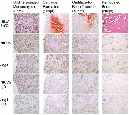
Figure 3.4. Jag1 and NICD2 are expressed in identical cell populations that participate in
endochondral bone repair during TF. ...31
Figure 3.5. Jag1 and NICD2 are expressed in vascular endothelial cells invading the cartilage
matrix, as well as terminal hypertrophic chondrocytes adjacent to the invading vasculature...31
Figure 3.6. Another example of Jag1 and NICD2 immunolocalization during TF with IgG control
sections (related to Figure 3.4)...32
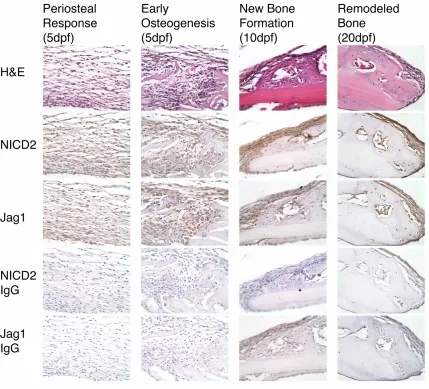
Figure 3.7. Jag1 and NICD2 expression in uninjured tibial growth plate and calvarium...33
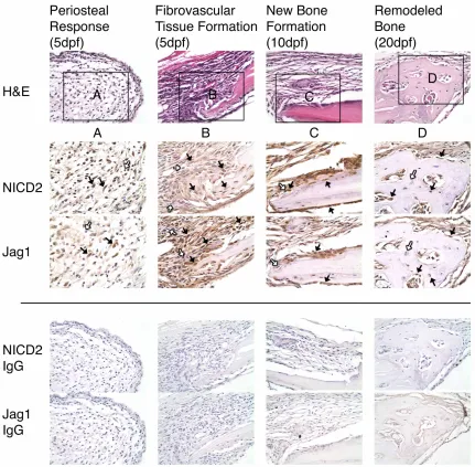
Figure 3.8. Jag1 and NICD2 are expressed in identical cell populations that participate in
intramembranous bone repair during CD. ...34
Figure 3.9. Another example of Jag1 and NICD2 immunolocalization during CD with IgG control
sections (related to Figure 3.8)...35
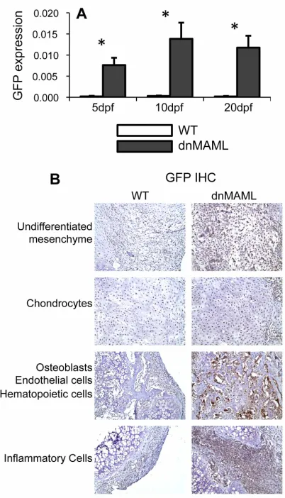
Figure 4.1. GFP-tagged dnMAML is expressed in dnMAML mice during fracture healing...52
Figure 4.2. dnMAML decreases cartilage formation during fracture...54
Figure 4.3. dnMAML inhibits expression of vascular endothelial cell markers during fracture
healing. ...55
Figure 4.4. dnMAML alters bone remodeling during fracture healing...56
Figure 4.5. dnMAML decreases callus size at 20dpf but not bone mass during fracture healing.57
Figure 4.6. dnMAML decreases bone mass during calvarial defect healing. ...59
Figure 4.8. dnMAML does not alter cell proliferation or apoptosis during fracture healing...61
Figure 5.1. Schematic depicting Prx1 and Col2.3 expression during osteochondral lineage
differentiation. ...74
Figure 5.2.µCT analysis of trabecular bone in Prx1 mice at 8 weeks (females and males) and 9
months of age (males)...77
Figure 5.3. µCT analysis of trabecular bone in Col2.3 mice at 8 weeks (females and males) of age...78
Figure 5.4. µCT analysis of cortical bone in Prx1 mice at 8 weeks (females and males) and 9 months of age (males)...79
Figure 5.5.µCT analysis of cortical bone in Col2.3 mice at 8 weeks (females and males) of age
...80
Figure 5.6. Gene expression of Notch pathway components Jag1, Hes1 and Hey1. ...82
Figure 5.7. Osteogenic gene expression of osteocalcin (Ocn), osterix (Osx) and collagen type I
(Col1a1)...82
Figure 5.8. Linear correlation of Notch components (Jag1, Hes1, Hey1) with osteogenic markers
(Col1a1, Ocn) for whole bone (WB only) or whole bone and cortical bone combined (WB and
CB). ...83
Figure 5.9. Gene expression of osteoblast mediators of osteoclast activity – RankL,
osteoprotogerin (OPG) and the ratio between the two (OPG:RankL) – and osteoclast marker
TRAP. ...84
Figure 5.10. Gene expression of proliferation markers Cyclin D1 and PCNA. ...85
Figure 6.1. Notch target Hey1 gene expression of hMSCs cultured on direct and indirect
Jagged1/A6 in SGM ...102
Figure 6.2. Relative surface density of successfully immobilized Jagged1 to A6 via Direct and
Indirect strategies, and the release kinetics profile...103
Figure 6.3. Cell number of hMSCs cultured on direct and indirect Jagged1/A6 in SGM ...104
Figure 6.4. Osteogenic gene expression of hMSCs cultured on direct and indirect Jagged1/A6 in
Figure 6.5. There is a significant positive linear correlation between Hey1 and bone sialoprotein
(BSP) gene expression...107
Figure 6.6. Alkaline Phosphatase (AP) enzymatic activity of hMSCs cultured on direct and
indirect Jagged1/A6 in SGM at day 7 ...107
Figure 6.7. Cell number of hMSCs cultured on direct Jagged1/A6 in OGM ...109
Figure 6.8. Alkaline Phosphatase (AP) enzymatic activity of hMSCs cultured on direct
Jagged1/A6 in OGM ...109
Figure 6.9. Calcified mineral deposition of hMSCs cultured on direct Jagged1/A6 in OGM with
[0/TCPS]. ...110
Figure 6.10. Evaluation of Jagged1/A6 porous scaffolds in murine calvarial defects...112
CHAPTER 1
Introduction to Bone Fracture Healing and the Notch Signaling Pathway
1.1 Clinical Significance of Bone Fractures
It is estimated that approximately 7.9 million fractures occur in the United States each
year, and although the majority of fractures heal with standard care, approximately 10-13% have
reported to exhibit delayed healing or develop into non-unions [1, 2]. Direct treatment costs are
approximately $3,400-$5,300 per fracture. However, the total financial burden to society is
approximately $12,500-17,300 per fracture when including associated costs such as lost
productivity [3]. Direct and societal costs are of course much higher for fractures that experience
delayed healing due to increased medical visits and continued loss of productivity. Furthermore,
fractures in an elderly population have increased costs upwards of $81,300 per injury, of which
nursing facility expenses account for nearly half, as well as result in an increased mortality rate
[4].
To treat severe injuries, therapeutic approaches have focused on delivery of
osteoinductive (biological cues to stimulate osteoblast activity) and osteoconductive (scaffold or
other cue to support bone formation) signals. Autologous bone grafts often harvested from the
patient’s iliac crest are considered the gold standard of care, but can result in significant donor
site morbidity and post-surgical pain, and yield only a limited amount of graft material [5].
Demineralized bone matrix, a common allograft therapeutic, is more readily available but has
limited osteoinductive potential and can induce immunogenic reactions [5]. More recently, growth
factor-based therapies have been developed to promote bone formation. Use of bone
morphogenetic proteins (BMPs) has become one of the more common treatments [6, 7].
However, recent reports suggest that BMPs lack the clinical efficiency and safety that has been
widely demonstrated in pre-clinical animal models [8, 9]. Furthermore, gene-based therapeutics
[10] or induced significant immunogenic responses [11]. Therefore, a clinical need persists for the
development of new methods to enhance bone fracture healing.
1.2 Bone Fracture Healing
Bone fracture healing occurs through a series of carefully regulated spatiotemporal
events that recapitulate many aspects of embryological bone development (Figure 1.1) [12-14].
Endochondral bones such as the tibia and femur heal primarily through endochondral ossification.
Following injury, inflammation and hematoma formation mediate an influx of undifferentiated
mesenchymal cells to the site of injury that rapidly proliferate to produce the initial fibrovascular
callus. These cells then condense and undergo chondrogenic differentiation to produce an
avascular cartilaginous callus. Terminal chondrocyte hypertrophy and cartilage matrix
mineralization are then followed by apoptosis and resorption, which allows for vascular invasion
of the callus. During this cartilage-to-bone transition, the vascular network mediates an influx of
osteoprogenitor cells that undergo differentiation and produce immature bone on top of the
resorbing cartilage matrix. Concomitantly, periosteal-derived osteoblasts form a mineralized bony
shell surrounding the callus. Over time, the callus matures and is remodeled through
osteoblast-mediated bone formation and osteoclast-osteoblast-mediated bone resorption, ultimately restoring the
structure and function of the original bone. Alternatively, intramembranous bones such as the
calvarium as well as other bones that are rigidly fixed during repair heal through
intramembranous ossification, which involves direct bone formation without a cartilage precursor
[15, 16].
Although physiological mechanisms of fracture healing are well-characterized, molecular
signals that control the complex temporal progression of events required for healing are poorly
understood, with most investigations limited to understanding the role(s) of the BMP [17] and Wnt
[18, 19] signaling pathways. Elucidating the significance of novel signaling pathways that regulate
Figure 1.1. Schematic of the temporal progression of endochondral fracture repair. Figure adapted from Gerstenfeld et al. [20]
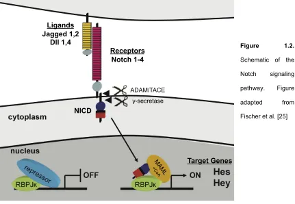
1.3 Notch Signaling Pathway
The Notch signaling pathway is a developmentally conserved cell-to-cell signaling
pathway that regulates cell proliferation, differentiation, fate determination and apoptosis [21].
Activation of the pathway occurs when a Notch ligand (Jagged 1,2 and Delta-like 1,4) expressed
on the surface of a signaling cell interacts with a Notch receptor (Notch 1-4) expressed on the
surface of a receiving cell. The Notch intracellular domain (NICD) is released via a two-stage
proteolytic event mediated first by the ADAM family metalloproteinase tumor necrosis factor α
conversion enzyme (ADAM/TACE), and then by the γ-secretase complex comprised of
Presenilins 1 and 2. Once released, NICD translocates to the nucleus where it binds to
Recombination Signal Binding Protein For Immunoglobulin Kappa J Region (RBPjκ), converting it
from a transcriptional repressor into an activator. Mastermind-like protein (MAML) then binds to
create the NICD-RBPjκ-MAML complex and serves as a scaffold to recruit other co-activators
necessary to initiate transcription of canonical Notch target gene families Hes and Hey (Figure
1.2) [22-24].
Initial Injury Cartilage Formation Cartilage-to-Bone Transition Secondary Bone Formation
Inflammation Callus Vascularization Bone remodeling
STAGES OF FRACTURE REPAIR
Figure 1.2.
Schematic of the
Notch signaling
pathway. Figure
adapted from
Fischer et al. [25]
1.4 Notch Signaling and Bone Formation
The Notch signaling pathway regulates mesenchymal cell lineage behavior and
embryological bone formation [26-33]. Deletion of Notch components in undifferentiated
mesenchymal progenitor cells stimulates osteoblast differentiation and early bone formation,
which is ultimately lost during aging due to depletion of the progenitor pool [26]. Constitutive
activation of Notch in committed but not completely mature osteoblasts promotes proliferation
while inhibiting differentiation, resulting in osteosclerotic immature bone formation that does not
properly mature [27]. These results demonstrate that activation of Notch signaling maintains
osteoprogenitor cells in an undifferentiated state. Deletion of Notch components in the same
committed osteoblast population or in mature osteoblasts does not alter early bone formation, but
instead results in osteopenia during aging due to increased osteoclast activity [26-28], Ligands
Jagged 1,2 Dll 1,4
Receptors Notch 1-4
!-secretase ADAM/TACE
NICD
demonstrating that activation of Notch signaling in mature osteoblasts promotes net bone gain by
secondarily inhibiting osteoclast activity.
Transient activation of Notch signaling in progenitor cells is required to initiate
chondrogenesis [34]. However, constitutive expression of Notch components prevents
differentiation from occurring [29, 30, 34], whereas sustained inhibition in undifferentiated
mesenchymal progenitor cells or committed chondrogenic cells results in the pathological
overproduction of chondrocytes [29, 30]. Reactivation of Notch is then required for proper
terminal hypertrophic chondrocyte maturation [31]. These results demonstrate that while transient
activation is required to initiate chondrogenesis, constitutive activation of Notch signaling inhibits
differentiation, but must be reactivated to complete terminal differentiation.
Since many aspects of bone development are recapitulated during repair, these results
collectively suggest that Notch signaling also regulates bone fracture healing.
1.5 Notch and Vasculogenesis
Bone formation during development and fracture repair is dependent upon proper
vascularization, which mediates an influx of osteogenic cells to sites of new bone formation.
Various gain-of-function and loss-of-function models have demonstrated that Notch signaling is a
critical regulator of vascular development. With regards to ligand activity, homozygous Jagged1
deletion [35] as well as Dll4 haploinsufficiency [36, 37] results in embryonic lethality due to
vascular defects. With regards to receptor activity, conditional Notch4 gain-of-function in
VEGFR-expressing cells results in embryonic lethality due to a restricted and disorganized vascular
network [38]. Interestingly, homozygous Notch4 deleted mice develop normally, but homozygous
Notch1 deleted mice as well as double homozygous Notch1 and Notch4 deleted mice show
vascular remodeling defects that result in embryonic lethality [39]. Furthermore, Notch1 deletion
in Tie2-expressing endothelium-specific cells also produce vascular abnormalities that result in
embryonic lethality [40]. The fact that gain-of-function and loss-of-function of Notch components
both result in embryonic lethality suggests that the proper spatiotemporal expression of Notch
Notch signaling has also been shown to regulate postnatal angiogenesis and
vasculogenesis. Use of various tissue-specific and inducible Jagged1 gain- and loss-of-function
mouse models have demonstrated that Jagged1 promotes angiogenesis and vessel sprouting by
antagonizing Dll4-Notch interactions, which are inhibitory [41]. Jagded1 expression also promotes
endothelial cell proliferation, differentiation and migration, whereas Dll1 has no effect [42].
1.6 The Notch Ligand Jagged1 (Jag1)
Clinically, loss-of-function mutations to Jagged1 are primarily responsible for Alagille
Syndrome (ALGS) [43, 44]. ALGS incorporates a wide range of developmental defects, including
chronic liver cholestasis, bile duct paucity, cardiovascular disease, kidney and pancreatic
disease, craniofacial development alterations and other musculoskeletal defects [45]. ALGS
patients present with decreased bone mass [46] and increased risk of fracture [47], which is often
assumed to be secondary to chronic liver cholestasis, where the resulting malabsorption of fat
soluble vitamins and minerals is believed to be primarily responsible for impaired skeletal
development. However, liver transplantations, which are common treatments for ALGS patients,
have not been able to recover normal bone growth [46, 48]. A recent study demonstrated a direct
role for Jagged1 in craniofacial development [49] and a SNP at the Jagged1 locus is associated
with bone mass. Furthermore, Jagged1 is the mostly highly expressed Notch ligand in
mesenchymal cells during skeletal development [30], (as stated above) enhances vasculogenesis
by promoting endothelial cell proliferation, differentiation and migration [41, 42], and its
expression in mesenchymal lineage cells promotes hematopoietic stem cell expansion [50, 51]
and inhibits osteoclast differentiation [52]. These results suggest that Jagged1 activity in
mesenchymal lineage cells directly regulates bone formation.
1.7 Notch and Regeneration
Notch signaling is upregulated following injury to many tissues including skin [53], retina
[54], brain [55, 56], heart [57], intestine [58], kidney [59, 60] and pancreas [61]. Activation of
Manipulations to Notch signaling can also enhance tissue regeneration. Specifically, transient
upregulation of Notch signaling via adenoviral transfection of NICD significantly improved
myocardial function in infarcted hearts [57]. These results identify Notch as a potential therapeutic
target for other injuries as well where the pathway is endogenously active.
Interestingly, in vivo delivery of soluble Notch ligands Jagged1 or Dll4 through an osmotic
pump did not improve healing following ischemic-induced brain injuries [55, 63]. However,
previous studies have demonstrated a requirement for Notch ligands to be immobilized to a
substrate in order to active NICD cleavage and downstream Notch signaling, such that
free-floating soluble ligands are also able to bind to receptors but instead effectively inhibit the
pathway [64-69]. It has been hypothesized that the naturally-occurring immobilized state of a
membrane-bound Notch ligand is required to apply a pulling force on the extracellular domain of
the Notch receptor, which precedes cleavage of the intracellular domain (NICD) [70]. These
results demonstrate the requirement for ligand immobilization to activate the Notch signaling
pathway for therapeutic applications.
1.8 Use of Poly(β-amino ester)s for Therapeutic Applications
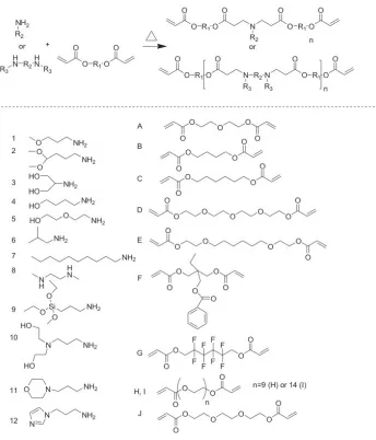
A combinatorial library of degradable, photocrosslinkable, acrylate-terminated poly(β
-amino ester)s (PBAE) comprised of amines and diacrylates was developed for the rapid
screening and design of biomaterials for a variety of therapeutic applications (Figure 1.3) [71].
Polymerization occurs through step-growth with resulting linear macromers containing ester and
tertiary amines in their backbones. Following addition of a photoinitiator and exposure to UV light,
crosslinking occurs between the functionalized acrylate groups. After photocrosslinking, PBAE
networks degrade via hydrolysis to their backbone esters into small molecule bis(β-amino acid)s,
diol products, and poly(acrylic acid) kinetic chains. PBAEs are clinically advantageous polymers
to use as therapeutics because they are simple to synthesize with no byproducts formed, thus
eliminating the need for multiple purification steps, and are inexpensive and commercially
available. Mechanical properties and degradation rates of PBAEs can be controlled by altering
PBAEs have successfully been used for a variety of therapeutic applications. PBAEs
have been used as gene-delivery vehicles for cardiovascular therapeutics [73] and as nonviral
DNA vectors for cancer therapeutics [74]. PBAE nanoparticles have also shown to be effective
drug delivery vehicles for targeting cancerous cells [75]. Importantly, one PBAE in particular,
diethylene glycol diacrylate combined with isobutylamine, has demonstrated osteoconductive
properties when used as a scaffold carrier for BMP delivery [76].
Figure 1.3. General poly(β-amino ester) polymerization schematic and chemical structures. Synthesis of
amines and diacrylates (top). Monomers depicting the 12 amines and 10 diacrylates (bottom). Figure from
1.9 Conclusions
This chapter provides an overview of the Notch signaling pathway and bone fracture
healing. The Notch signaling pathway has been shown to regulate embryological bone
development. Since many aspects of development are recapitulated during repair, Notch
signaling may also regulate bone fracture healing. Furthermore, activation of the pathway has
been shown to promote regeneration of other tissues, identifying it as a potential therapeutic to
also enhance regeneration of bone. The work described in this thesis will report on the
comprehensive assessment of Notch signaling during fracture. The significance of Notch
signaling will be determined by blocking canonical Notch signaling during fracture healing. The
role of Jagged1 specifically during bone development and remodeling will also be assessed by
deleting the gene in the osteoblast lineage, and finally, we will develop a biomaterial by delivering
1.10 References
1. Praemer, A., et al., Musculoskeletal Conditions in the United States1999: American
Academy of Orthopaedic Surgeons.
2. Audige, L., et al., Path analysis of factors for delayed healing and nonunion in 416
operatively treated tibial shaft fractures. Clin Orthop Relat Res, 2005. 438: p. 221-32.
3. Busse, J.W., et al., An economic analysis of management strategies for closed and open
grade I tibial shaft fractures. Acta Orthop, 2005. 76(5): p. 705-12.
4. Braithwaite, R.S., N.F. Col, and J.B. Wong, Estimating hip fracture morbidity, mortality
and costs. J Am Geriatr Soc, 2003. 51(3): p. 364-70.
5. Dimitriou, R., et al., Bone regeneration: current concepts and future directions. BMC Med,
2011. 9: p. 66.
6. Friedlaender, G.E., et al., Osteogenic protein-1 (bone morphogenetic protein-7) in the
treatment of tibial nonunions. J Bone Joint Surg Am, 2001. 83-A Suppl 1(Pt 2): p.
S151-8.
7. Govender, S., et al., Recombinant human bone morphogenetic protein-2 for treatment of
open tibial fractures: a prospective, controlled, randomized study of four hundred and fifty
patients. J Bone Joint Surg Am, 2002. 84-A(12): p. 2123-34.
8. Einhorn, T.A., Clinical applications of recombinant human BMPs: early experience and
future development. J Bone Joint Surg Am, 2003. 85-A Suppl 3: p. 82-8.
9. Carragee, E.J., et al., Future directions for The spine journal: managing and reporting
conflict of interest issues. Spine J, 2011. 11(8): p. 695-7.
10. Partridge, K.A. and R.O. Oreffo, Gene delivery in bone tissue engineering: progress and
prospects using viral and nonviral strategies. Tissue Eng, 2004. 10(1-2): p. 295-307.
11. Hannallah, D., et al., Retroviral delivery of Noggin inhibits the formation of heterotopic
ossification induced by BMP-4, demineralized bone matrix, and trauma in an animal
12. Vortkamp, A., et al., Recapitulation of signals regulating embryonic bone formation during
postnatal growth and in fracture repair. Mech Dev, 1998. 71(1-2): p. 65-76.
13. Ferguson, C., et al., Does adult fracture repair recapitulate embryonic skeletal formation?
Mech Dev, 1999. 87(1-2): p. 57-66.
14. Gerstenfeld, L.C., et al., Fracture healing as a post-natal developmental process:
molecular, spatial, and temporal aspects of its regulation. J Cell Biochem, 2003. 88(5): p.
873-84.
15. Einhorn, T.A., The cell and molecular biology of fracture healing. Clin Orthop Relat Res,
1998(355 Suppl): p. S7-21.
16. McKibbin, B., The biology of fracture healing in long bones. J Bone Joint Surg Br, 1978.
60-B(2): p. 150-62.
17. Tsuji, K., et al., BMP2 activity, although dispensable for bone formation, is required for
the initiation of fracture healing. Nat Genet, 2006. 38(12): p. 1424-9.
18. Minear, S., et al., Wnt proteins promote bone regeneration. Sci Transl Med, 2010. 2(29):
p. 29ra30.
19. Komatsu, D.E., et al., Modulation of Wnt signaling influences fracture repair. J Orthop
Res, 2010. 28(7): p. 928-36.
20. Gerstenfeld, L.C. and T.A. Einhorn, Developmental aspects of fracture healing and the
use of pharmacological agents to alter healing. J Musculoskelet Neuronal Interact, 2003.
3(4): p. 297-303; discussion 320-1.
21. Artavanis-Tsakonas, S., M.D. Rand, and R.J. Lake, Notch signaling: cell fate control and
signal integration in development. Science, 1999. 284(5415): p. 770-6.
22. Iso, T., L. Kedes, and Y. Hamamori, HES and HERP families: multiple effectors of the
Notch signaling pathway. J Cell Physiol, 2003. 194(3): p. 237-55.
23. Zanotti, S. and E. Canalis, Notch and the skeleton. Mol Cell Biol, 2010. 30(4): p. 886-96.
24. Engin, F. and B. Lee, NOTCHing the bone: insights into multi-functionality. Bone, 2010.
25. Fischer, A. and M. Gessler, Delta-Notch--and then? Protein interactions and proposed
modes of repression by Hes and Hey bHLH factors. Nucleic Acids Res, 2007. 35(14): p.
4583-96.
26. Hilton, M.J., et al., Notch signaling maintains bone marrow mesenchymal progenitors by
suppressing osteoblast differentiation. Nat Med, 2008. 14(3): p. 306-14.
27. Engin, F., et al., Dimorphic effects of Notch signaling in bone homeostasis. Nat Med,
2008. 14(3): p. 299-305.
28. Zanotti, S., et al., Notch inhibits osteoblast differentiation and causes osteopenia.
Endocrinology, 2008. 149(8): p. 3890-9.
29. Mead, T.J. and K.E. Yutzey, Notch pathway regulation of chondrocyte differentiation and
proliferation during appendicular and axial skeleton development. Proc Natl Acad Sci U S
A, 2009. 106(34): p. 14420-5.
30. Dong, Y., et al., RBPjkappa-dependent Notch signaling regulates mesenchymal
progenitor cell proliferation and differentiation during skeletal development. Development,
2010. 137(9): p. 1461-71.
31. Kohn, A., et al., Cartilage-specific RBPjkappa-dependent and -independent Notch signals
regulate cartilage and bone development. Development, 2012. 139(6): p. 1198-212.
32. Tao, J., et al., Osteosclerosis owing to Notch gain of function is solely Rbpj-dependent. J
Bone Miner Res, 2010. 25(10): p. 2175-83.
33. Salie, R., et al., Ubiquitous overexpression of Hey1 transcription factor leads to
osteopenia and chondrocyte hypertrophy in bone. Bone, 2010. 46(3): p. 680-94.
34. Oldershaw, R.A., et al., Notch signaling through Jagged-1 is necessary to initiate
chondrogenesis in human bone marrow stromal cells but must be switched off to
complete chondrogenesis. Stem Cells, 2008. 26(3): p. 666-74.
35. Xue, Y., et al., Embryonic lethality and vascular defects in mice lacking the Notch ligand
Jagged1. Hum Mol Genet, 1999. 8(5): p. 723-30.
36. Krebs, L.T., et al., Haploinsufficient lethality and formation of arteriovenous malformations
37. Gale, N.W., et al., Haploinsufficiency of delta-like 4 ligand results in embryonic lethality
due to major defects in arterial and vascular development. Proc Natl Acad Sci U S A,
2004. 101(45): p. 15949-54.
38. Uyttendaele, H., et al., Vascular patterning defects associated with expression of
activated Notch4 in embryonic endothelium. Proc Natl Acad Sci U S A, 2001. 98(10): p.
5643-8.
39. Krebs, L.T., et al., Notch signaling is essential for vascular morphogenesis in mice.
Genes Dev, 2000. 14(11): p. 1343-52.
40. Limbourg, F.P., et al., Essential role of endothelial Notch1 in angiogenesis. Circulation,
2005. 111(14): p. 1826-32.
41. Benedito, R., et al., The notch ligands Dll4 and Jagged1 have opposing effects on
angiogenesis. Cell, 2009. 137(6): p. 1124-35.
42. Kwon, S.M., et al., Specific Jagged-1 signal from bone marrow microenvironment is
required for endothelial progenitor cell development for neovascularization. Circulation,
2008. 118(2): p. 157-65.
43. Li, L., et al., Alagille syndrome is caused by mutations in human Jagged1, which encodes
a ligand for Notch1. Nat Genet, 1997. 16(3): p. 243-51.
44. Oda, T., et al., Mutations in the human Jagged1 gene are responsible for Alagille
syndrome. Nat Genet, 1997. 16(3): p. 235-42.
45. Krantz, I.D., D.A. Piccoli, and N.B. Spinner, Clinical and molecular genetics of Alagille
syndrome. Curr Opin Pediatr, 1999. 11(6): p. 558-64.
46. Olsen, I.E., et al., Deficits in size-adjusted bone mass in children with Alagille syndrome.
J Pediatr Gastroenterol Nutr, 2005. 40(1): p. 76-82.
47. Bales, C.B., et al., Pathologic lower extremity fractures in children with Alagille syndrome.
J Pediatr Gastroenterol Nutr, 2010. 51(1): p. 66-70.
48. Quiros-Tejeira, R.E., et al., Does liver transplantation affect growth pattern in Alagille
49. Humphreys, R., et al., Cranial neural crest ablation of Jagged1 recapitulates the
craniofacial phenotype of Alagille syndrome patients. Hum Mol Genet, 2012. 21(6): p.
1374-83.
50. Calvi, L.M., et al., Osteoblastic cells regulate the haematopoietic stem cell niche. Nature,
2003. 425(6960): p. 841-6.
51. Weber, J.M., et al., Parathyroid hormone stimulates expression of the Notch ligand
Jagged1 in osteoblastic cells. Bone, 2006. 39(3): p. 485-93.
52. Bai, S., et al., NOTCH1 regulates osteoclastogenesis directly in osteoclast precursors
and indirectly via osteoblast lineage cells. J Biol Chem, 2008. 283(10): p. 6509-18.
53. Chigurupati, S., et al., Involvement of notch signaling in wound healing. PLoS One, 2007.
2(11): p. e1167.
54. Hayes, S., et al., Notch signaling regulates regeneration in the avian retina. Dev Biol,
2007. 312(1): p. 300-11.
55. Oya, S., et al., Attenuation of Notch signaling promotes the differentiation of neural
progenitors into neurons in the hippocampal CA1 region after ischemic injury.
Neuroscience, 2009. 158(2): p. 683-92.
56. Tatsumi, K., et al., Transient activation of Notch signaling in the injured adult brain. J
Chem Neuroanat, 2010. 39(1): p. 15-9.
57. Gude, N.A., et al., Activation of Notch-mediated protective signaling in the myocardium.
Circ Res, 2008. 102(9): p. 1025-35.
58. Okamoto, R., et al., Requirement of Notch activation during regeneration of the intestinal
epithelia. Am J Physiol Gastrointest Liver Physiol, 2009. 296(1): p. G23-35.
59. Kobayashi, T., et al., Expression and function of the Delta-1/Notch-2/Hes-1 pathway
during experimental acute kidney injury. Kidney Int, 2008. 73(11): p. 1240-50.
60. Gupta, S., et al., Effect of Notch activation on the regenerative response to acute renal
failure. Am J Physiol Renal Physiol, 2010. 298(1): p. F209-15.
61. Su, Y., et al., Pancreatic regeneration in chronic pancreatitis requires activation of the
62. Conboy, I.M., et al., Notch-mediated restoration of regenerative potential to aged muscle.
Science, 2003. 302(5650): p. 1575-7.
63. Androutsellis-Theotokis, A., et al., Notch signalling regulates stem cell numbers in vitro
and in vivo. Nature, 2006. 442(7104): p. 823-6.
64. Varnum-Finney, B., et al., Immobilization of Notch ligand, Delta-1, is required for
induction of notch signaling. J Cell Sci, 2000. 113 Pt 23: p. 4313-8.
65. Vas, V., et al., Soluble Jagged-1 is able to inhibit the function of its multivalent form to
induce hematopoietic stem cell self-renewal in a surrogate in vitro assay. J Leukoc Biol,
2004. 75(4): p. 714-20.
66. Beckstead, B.L., D.M. Santosa, and C.M. Giachelli, Mimicking cell-cell interactions at the
biomaterial-cell interface for control of stem cell differentiation. J Biomed Mater Res A,
2006. 79(1): p. 94-103.
67. Beckstead, B.L., et al., Methods to promote Notch signaling at the biomaterial interface
and evaluation in a rafted organ culture model. J Biomed Mater Res A, 2009. 91(2): p.
436-46.
68. Goncalves, R.M., et al., Induction of notch signaling by immobilization of jagged-1 on
self-assembled monolayers. Biomaterials, 2009. 30(36): p. 6879-87.
69. Nobta, M., et al., Critical regulation of bone morphogenetic protein-induced osteoblastic
differentiation by Delta1/Jagged1-activated Notch1 signaling. J Biol Chem, 2005.
280(16): p. 15842-8.
70. Kramer, H., RIPping notch apart: a new role for endocytosis in signal transduction? Sci
STKE, 2000. 2000(29): p. pe1.
71. Anderson, T., Hossain, Navarro, Brey, Van Vliet, Langer, Burdick, A Combinatorial
Library of Photocrosslinkable and Degradable Materials. Advanced Materials, 2006.
72. Brey, D.M., I. Erickson, and J.A. Burdick, Influence of macromer molecular weight and
chemistry on poly(beta-amino ester) network properties and initial cell interactions. J
73. Brito, L.A., et al., In vitro and in vivo studies of local arterial gene delivery and transfection
using lipopolyplexes-embedded stents. J Biomed Mater Res A, 2010. 93(1): p. 325-36.
74. Anderson, D.G., et al., A polymer library approach to suicide gene therapy for cancer.
Proc Natl Acad Sci U S A, 2004. 101(45): p. 16028-33.
75. Shen, Y., et al., Degradable poly(beta-amino ester) nanoparticles for cancer cytoplasmic
drug delivery. Nanomedicine, 2009. 5(2): p. 192-201.
76. Brey, D.M., et al., Identification of osteoconductive and biodegradable polymers from a
CHAPTER 2
Specific Aims and Hypotheses
Bone fractures can exhibit delayed healing or develop into non-unions. Autologous bone
grafts and growth factor therapies such as bone morphogenetic proteins are common therapeutic
strategies to treat such severe injuries. However, they have well-documented limitations and
safety concerns. Furthermore, although the physiological mechanisms of fracture healing are well
characterized, the molecular mechanisms that regulate the complex spatiotemporal progression
of events required for healing are poorly understood. Therefore, a need persists for the
identification of novel signaling pathways that regulate fracture healing, and the development of
new therapies targeting these pathways to enhance bone regeneration.
Notch signaling regulates mesenchymal cell behavior and embryological bone formation,
and many aspects of bone formation are recapitulated during bone fracture healing. Furthermore,
Notch signaling has been shown to be required for successful wound healing, and targeting the
pathway can promote tissue regeneration. However, the role of Notch signaling during bone
fracture healing and the ability of the pathway to enhance regeneration has not been investigated.
Therefore, the overall objective of this thesis is to determine the role of Notch signaling
during bone fracture healing, and to create a clinically translatable therapy targeting the pathway
to enhance healing.
2.1 Specific Aim I (Chapter 3)
Characterize and compare activation of the Notch signaling pathway during endochondral and
intramembranous fracture healing using murine tibial fracture healing as a model of endochondral
bone repair and murine calvarial defect healing as a model of intramembranous bone repair.
2.1.1 Hypothesis I
Gene and protein expression of Notch signaling components will be quantified and
localized to specific cell populations.
2.2 Specific Aim II (Chapter 4)
Determine the importance of Notch signaling in regulating bone fracture healing by using a
temporally controlled inducible transgenic mouse model to impair canonical Notch signaling in all
cells during murine tibial fracture and calvarial defect healing
2.2.1 Hypothesis II
Inhibition of Notch signaling will alter murine tibial fracture and calvarial defect healing.
A floxed GFP-tagged dnMAML transgene will be activated in all cell types just prior to
injury using the inducible Mx1-Cre model. dnMAML expression inhibits the Notch signaling
pathway just prior to transcription of target genes. Multiple stages of healing will be evaluated,
including cartilage formation, callus vascularization, bone formation and remodeling, and
inflammation, as well as other cell behaviors such as proliferation and apoptosis.
2.3 Specific Aim III (Chapter 5)
Determine the direct role of Jagged1 during bone formation.
2.3.1 Hypothesis III
Jagged1 expression in the mesenchymal lineage regulates bone formation through paracrine
cell-to-cell signaling.
Jagged1 will be conditionally deleted in two skeletal-specific mouse models; first in a
mesenchymal progenitor cell population by using the Prx1-Cre model and then in a committed
osteoblast population by using the Col2.3-Cre model. Trabecular and cortical bone formation will
be analyzed as well as gene expression of Notch components and markers of osteoblast and
osteoclast differentiation and proliferation.
Develop a clinically translatable biomaterial construct comprised of Jagged1 and an
osteoconductive scaffold, and evaluate its ability to induce bone tissue formation.
2.4.1 Hypothesis IV
Delivery of Jagged1 immobilized to a poly(B-amino ester) polymer will activate the Notch
signaling pathway and promote osteoblast differentiation.
The ability of direct and indirect Jagged1 immobilization strategies to activate the Notch
signaling pathway and promote an osteogenic phenotype will be evaluated in standard growth
media. Then, the ability of the ideal immobilization strategy to induce osteoblast differentiation
and calcified mineral deposition will be evaluated in osteogenic media. Finally, translatable
biomaterial constructs will be evaluated in murine calvarial defects and tibial fractures.
This thesis aims to uncover the role of the Notch signaling pathway during bone fracture
healing, and to develop a clinically translatable therapy targeting the pathway to improve bone
repair. In all, this thesis serves as the foundation for Notch signaling-based translational research
in regenerative orthopaedic medicine, and represents a model approach to uncover additional
novel signaling pathways that regulate – and therefore could potentially enhance – bone fracture
CHAPTER 3
Notch Signaling Components Are Upregulated During Endochondral and
Intramembranous Bone Regeneration
3.1 Introduction
Bone regeneration occurs through a series of spatiotemporal events that recapitulate
many aspects of embryological development [1, 2]. Long bones such as the tibia develop and
heal primarily through endochondral ossification (indirect bone formation on a cartilage
intermediate), whereas bones such as the calvarium develop and heal through intramembranous
ossification (direct bone formation) [3]. A number of growth factor pathways, including bone
morphogenetic protein (BMP) and Wnt signaling, have been widely demonstrated to be required
for fracture healing and have also been shown to promote regeneration [4-9]. However, despite
the importance of these pathways, the significance of other growth factor pathways that regulate
bone healing is not as well described.
Notch signaling is a developmentally conserved pathway that mediates the development
of stem and progenitor cell populations in many tissues. Activation of the canonical Notch
signaling pathway occurs through direct cell-to-cell contact. When one of four Notch ligands,
Jagged (Jag) 1,2 and Delta-like (Dll) 1,4, interacts with one of four Notch receptors, Notch1-4, a
two-stage proteolytic event liberates the Notch intracellular domain (NICD) which then
translocates to the nucleus and binds with co-activators to initiate transcription of Notch target
gene families Hes and Hey.
Notch gain of function mutations in the murine mesenchymal lineage result in enhanced
cell proliferation while inhibiting differentiation, which prevents mature endochondral and
intramembranous bone development [10, 11]. Alternatively, loss of Notch signaling in the
mesenchymal lineage results in enhanced osteoprogenitor differentiation and early endochondral
Notch signaling in osteoblasts has also been shown to negatively regulate osteoclast behavior
[10, 13-15]. Collectively, these studies demonstrate that the Notch signaling pathway regulates
endochondral and intramembranous bone formation.
Although Notch signaling has been shown to regulate tissue repair in a variety of tissues
[16-21], an extensive characterization of Notch signaling during bone fracture healing has not
been reported. Therefore, the objective of this study was to rigorously characterize and compare
activation of the Notch signaling pathway during endochondral and intramembranous bone
regeneration, using tibial fracture healing (TF) as a model of endochondral bone repair and
calvarial defect healing (CD) as a model of intramembranous bone repair. We hypothesize that
Notch signaling components are active during murine tibial fracture and calvarial defect healing.
3.2 Methods
3.2.1 Experimental Design
All in vivo protocols were approved by the IACUC. Bilateral tibial fractures or bilateral
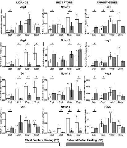
calvarial defects were created in 8-11 week old male C57Bl/6 mice to evaluate Notch signaling
during endochondral and intramembranous bone healing, respectively. Specimens were
harvested at 0, 5, 10 and 20 days post-fracture (dpf). Quantitative real-time polymerase chain
reaction (QPCR) was used to quantify gene expression of Notch pathway components including
ligands (Jag1,2, Dll1,4), receptors (Notch1-4), and target genes (Hes1, Hey1,2,L) (n=4-5).
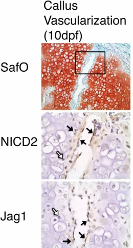
Immunohistochemistry (IHC) was used to identify cell types that express the Jag1 ligand and the
activated form of the Notch2 receptor, called the Notch2 intracellular domain (NICD2).
3.2.2 Tibial Fracture (TF) Procedure
Closed, transverse, mid-diaphyseal bilateral tibial fractures were created similar to
previously published methods [22]. Briefly, under isoflurane anesthesia, a small incision was
made medial to the tuberosity. A canal was punctured through the cortex using a 26-gauge
needle, and a 0.009-inch diameter rod was inserted through the length of the intramedullary
three-point bending apparatus. Radiographs were generated to verify correct pin placement and
fracture location (Faxitron X-Ray) (Figure 3.1A). 0.05 mg/kg of buprenorphine was administered
subcutaneously once after surgery. Mice recovered on heating pads and were fed ad libitum.
3.2.3 Calvarial Defect (CD) Procedure
Bilateral 1.5 mm diameter calvarial defects were created similar to previously published
methods [23]. Under isoflurane anesthesia, the mouse was placed into stereotaxic equipment
(Stoelting) and a sterile tegaderm drape (3M Health Care) was applied to the cranium after hair
removal (Nair, Church & Dwight). A midline incision exposed the parietal bones, and a 1.5 mm
diameter biopsy punch (Premier) was used to create a defect in the central portion of each
parietal bone, leaving the surrounding periosteum intact (Figure 3.1B). PBS was used to hydrate
the tissue. The incision was closed with 5-0 prolene non-absorbable sutures (Ethicon). 0.05
mg/kg of buprenorphine was administered subcutaneously once after surgery. Mice recovered on
heating pads and were fed ad libitum.
Figure 3.1. Radiographs of closed, transverse, mid-diaphyseal bilateral fractures with intramedullary pin stabilization taken at the time of injury (A), and 1.5 mm diameter bilateral calvarial defects taken at the time
of harvest (B). Radiographs were acquired at 15 sec with 25 kV.
3.2.4 Quantitative Real-Time Polymerase Chain Reaction (QPCR)
Fractured tibial calluses were dissected from the surrounding soft tissue at 5, 10 and 20
dpf. Uninjured diaphyseal bone, flushed of marrow, served as 0 dpf controls. Calvarial defects
surrounding bone tissue. Uninjured calvarial bone was similarly dissected for 0 dpf controls.
Tissue was placed in Qiazol lysis reagent (Qiagen) and homogenized using the Tissue Tearor
(BioSpec Products). mRNA was extracted using the Qiagen miRNeasy Mini Kit with DNase
digestion to remove DNA contamination. RNA yield was determined spectrophotometrically. 1 µg
of mRNA was reverse transcribed into cDNA using the Applied Biosystems High Capacity
RNA-to-cDNA Kit. Gene expression was quantified from 0.5 µl of cDNA in 10 µl of Power SYBR Green
PCR Master Mix (Applied Biosystems) using a 7500 Fast Real-Time PCR system (Applied
Biosystems). For each gene of interest, samples were run in duplicate with several controls per
primer set to verify that the measured signal was not due to DNA contamination or primer dimer
binding. Proper amplicon formulation was confirmed by melt curve analysis.
Fracture healing involves a temporally changing profile of cells derived from different
lineages. Although there is no ideal housekeeping gene for normalization across different cell
types, a series of genes were identified that show minimal variation in expression [24]. We
included three of those genes, run in duplicate and averaged together, as our housekeeping
control: β-actin, which regulates cell motility; ornithine decarboxylase antizyme (OAZ1), which
regulates polyamine synthesis; and 40S ribosomal protein 29 (RPS29), a component of the 40S
ribosomal subunit that regulates protein synthesis. QPCR data is presented as relative gene
expression to housekeeping control, calculated using the formula 2-ΔC(t), where ΔC(t) is the
difference in C(t) values between the gene of interest and the average of all three housekeeping
genes.
3.2.5 Histology and Immunohistochemistry (IHC)
Tissue was fixed in 4% paraformaldehyde at 4°C for 2-3 days, decalcified in a 4%
hydrochloric acid 4% formic acid solution, paraffin embedded, and sectioned into 5 µm
longitudinal slices. For Jag1 and NICD2 IHC, sections were deparaffinized and gradually
hydrated. Sections were treated with blocking serum (5% donkey, 4% BSA, 0.1% Triton-X 100,
sc-6011, 1:100) and rabbit cleaved NICD2 (Millipore 07-1234, 1:100) were incubated in a dilution
buffer (2% BSA, 0.25% Triton-X 100) overnight at 4°C in a humidified chamber. Control sections
were treated with goat IgG (Santa Cruz sc-2028, 1:200) or rabbit IgG (Santa Cruz sc-2027,
1:200) to match the concentration of the appropriate antibody. Sections were then treated with
3% H2O2 for 30 minutes at room temperature, followed by biotinylated secondary antibody
donkey anti-goat (Santa Cruz sc-2043, 1:200) or donkey anti-rabbit (Santa Cruz sc-2089, 1:200)
for 30 minutes at room temperature, and finally streptavidin-HRP (Abcam ab7403, 1:500) for 30
minutes at room temperature. Sections were developed with DAB (Vector SK-4100) and
counterstained with Hematoxylin. Additional sections were stained with Hematoxylin and Eosin
(H&E) for 15 and 2.5 minutes, respectively, or 0.1% Safranin O and 0.03% Fast Green (SafO) for
5 minutes each to visualize tissue structure and cell morphology. Slides were imaged in
brightfield with an Olympus BX51. Color images were acquired with a Spot RT3 2 megapixel
camera.
3.2.6 Statistical Analysis
Significance was assessed by one-way ANOVAs comparing the effect of time on gene
expression during TF and CD separately, followed by Tukey’s post-hoc test. Pairwise t-tests were