Original Research Article.
Study of Nutrient Foramina in the Long Bones of Human Upper Limb in
Ajmer, Rajasthan
Samata Goyal
1*, Mahima Shrivastava
21*PG Resident (IIIrd Year), 2Senior Professor,
Department of Anatomy, J. L. N. Medical College Ajmer, Rajasthan, India.
ABSTRACT
Introduction: The nutrient foramina are cavities that conduct the nutrient arteries and the peripheral nerves. The major blood supply for long bones originate from the nutrient arteries, mainly during the growing period and during the early phases of ossification.
Materials and Methods: The material of the present study consisted of 105 adult human cleaned and dried bones of the upper limb. They were divided into three groups, 35 bones of each, for determination of the number, position, size, direction and obliquity of the nutrient foramina in the human upper limb long bones. They were obtained from the osteology collection held in the Department of Anatomy, Jawahar Lal Nehru Medical College, Ajmer, Rajasthan. Only well-defined foramina on the diaphysis were accepted. Foramina at the ends of the bone were ignored.
Results: The majority of nutrient foramina in all bones studied were single in number and were secondary in size. Most of the nutrient foramina were concentrated in the middle third and were mostly located on the anterior surface of the shaft of bones. Most of the long bones follow the dictum “Towards the elbow I go, away from the knee I flee” and the direction of
nutrient foramen is opposite to growing end i.e. away from the elbow.
Conclusion: The study confirmed previous reports regarding the number and position of the nutrient foramina in the long bones of the limbs. Exact position and distribution of the nutrient foramina in bone diaphysis is important to avoid damage to the nutrient vessels during surgical procedures.
Key Words: Nutrient Foramina, Nutrient Arteries, Diaphysis.
*Correspondence to:
Dr. Samata Goyal,
PG Resident (IIIrd Year), Department of Anatomy,
J. L. N. Medical College Ajmer, Rajasthan, India.
Article History:
Received: 21-01-2018, Revised: 15-02-2018, Accepted: 19-03-2018
Access this article online
Website:
www.ijmrp.com
Quick Response code
DOI:
10.21276/ijmrp.2018.4.2.046
INTRODUCTION
The nutrient artery is the principal source of blood supply to a long bone and is particularly important during its active growth period in the embryo and fetus, as well as during the early phase of ossification.1 During childhood, the nutrient arteries provide
70-80% of the interosseous blood supply to long bones: when this supply is compromised, medullary bone ischemia occurs with less vascularization of the metaphysis and growth plate.2
The diaphyseal nutrient arteries obliquely penetrate in the diaphysis of the long bones, their entrance point and angulations being relatively constant, dividing in ascending and descending branches, once they reach the medullary cavity.3
It has been suggested that the direction of the nutrient foramina is determined by the growing end of the bone. The growing end is supposed to grow at least twice as fast as the other end. As a characteristic, the diaphyseal nutrient vessels move away from the growth extremity dominant in the bone.4
A considerable interest in studying nutrient foramina resulted not only from morphological, but also from clinical aspects. Nutrient foramina reflect to a certain degree the bone vascularization.
Some pathological bone conditions such as developmental abnormalities, fracture healing or acute hematogenic osteomyelities are closely related to the vascular system of the bone.5
MATERIALS AND METHODS
The material of the present study consisted of 105 adult human cleaned and dried bones of the upper limb. They were divided into three groups, 35 bones of each, for determination of the number, position, size, direction and obliquity of the nutrient foramina in the human upper limb long bones. They were obtained from the osteology collection held in the Department of Anatomy, Jawahar Lal Nehru Medical College, Ajmer, Rajasthan.
Inclusion Criteria
105 dry adult human upper limb bones irrespective of sex and race.
Exclusion Criteria
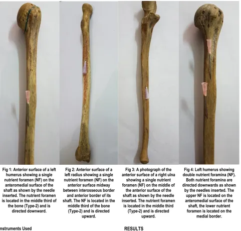
Fig 1: Anterior surface of a left humerus showing a single nutrient foramen (NF) on the
anteromedial surface of the shaft as shown by the needle inserted. The nutrient foramen is located in the middle third of
the bone (Type-2) and is directed downward.
Fig 2: Anterior surface of a left radius showing a single nutrient foramen (NF) on the anterior surface midway between interosseous border
and anterior border of its shaft. The NF is located in the
middle third of the bone (Type-2) and is directed
upward.
Fig 3: A photograph of the anterior surface of a right ulna
showing a single nutrient foramen (NF) on the middle of
the anterior surface of the shaft as shown by the needle inserted. The nutrient foramen
is located in the middle third (Type-2) and is directed
upward.
Fig 4: Left humerus showing double nutrient foramina (NF).
Both nutrient foramina are directed downwards as shown
by the needles inserted. The upper NF is located on the anteromedial surface of the shaft, the lower nutrient foramen is located on the
medial border.
Instruments Used
1. Hand lens: used to locate nutrient foramina.
2. Osteometric board: used to measure the length of long bones. 3. Vernier caliper: used to measure diameter of the long bones. Bones were examined for the number, position, size, direction and obliquity of nutrient foramina. The position of all nutrient foramina was determined by calculating a foraminal index (FI) using the formula:
FI = (DNF/TL) x 100 (Hughes6; Shulman7).
DNF = The distance from the proximal end of the bone to the nutrient foramen.
TL = Total bone length.
All measurements were taken to the nearest 0.1 mm using an INOX sliding caliper.8
Nutrient foramina smaller than the size of 24 hypodermic needle (0.56 mm in diameter) were considered as being secondary nutrient foramina (S.F) while those equal or larger were accepted as being dominant nutrient foramina (D.F).8
A fine stiff wire was used to confirm the direction and obliquity of the foramen.
The results were analyzed and tabulated using the Statistical Package of Social Sciences (SPSS) 8.0 windows. The range, mean and standard deviation of FI were determined.
RESULTS
In the whole series of 35 humeri examined, 18(51.4%) had a single foramen, 15 (42.9%) had double foramina and 2 (5.7%) had three foramina. The nutrient foramina were located along the whole middle third of the humerus with the foramen index ranging between 30.2% and 69% of the bone length. Of the total 54 foramina, 3 (5.6%) were in the proximal third (Type-1), 49 (90.7%) in the middle third (Type-2), and 2 (3.7%) were in the distal third (Type-3). Of the 54 foramina, 23 (42.6%) were dominant and 31 (57.4%) were secondary foramina. The nutrient foramina in all humeri examined were directed distally.
foramina. The nutrient foramina of all ulnae examined, were directed proximally. The direction of nutrient foramina in human long bones is directed away from the growing end. This is due to
one end of long bone is growing faster than the other end.4 There
was no change in the obliquity of the nutrient foramina, whether they were in the centre of the bone or nearer to the ends.
Table 1: Number of nutrient foramina observed in the long bones of the upper limb.
Bone Number of bone Number of foramina Percentage
Humerus (n=35) 18
15 2
1 2 3
51.4% 42.9% 5.7%
Radius (n=35) 35 1 100%
Ulna (n=35) 31
4
1 2
88.6% 11.4%
Table 2: Position and number of dominant (DF) and secondary (SF) nutrient foramina observed in the humerus.
Position Total No. Of
Foramina % Single Number Of Foramina Two Three
DF SF DF SF DF SF
Anteromedial surface 30 55.5 8 6 4 10 - 2
Posterior surface (in the middle of surface) 4 7.4 1 - 1 2 - -
Posterior surface (close to medial border) 2 3.7 - - - 1 - 1
Posterior surface (close to lateral border) 5 9.3 - - 1 2 - 2
Medial border 13 24.1 3 - 5 4 - 1
Table 3: Position and number of dominant (DF) and secondary (SF) nutrient foramina observed in the radius.
Position Total No. Of
Foramina % Single Number Of Foramina Two Three
DF SF DF SF DF SF
Anterior surface (midway between
interosseous and anterior borders) 7 20 5 9 2 - - -
Anterior surface (close to interosseous
border) 11 31.4 2 5 9 - - -
Anterior surface (close to anterior border) 13 37.1 5 7 8 - - -
Posterior surface (close to interosseous
border) 4 11.4 1 3 3 - - -
Table 4: Position and number of dominant (DF) and secondary (SF) nutrient foramina observed in the ulna.
Position Total No. Of
Foramina % Single Number Of Foramina Two Three
DF SF DF SF DF SF
Anterior surface ( in the middle of surface) 6 15.4 3 1 1 1 - -
Anterior surface (close to interosseous
border) 10 25.6 1 8 - 1 - -
Anterior surface (close to anterior border) 23 59 9 9 2 3 - -
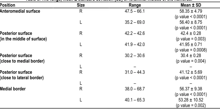
Table 5: The range, mean ± standard deviation (SD) of foraminal indices of the humerus.
Position Size Range Mean ± SD
Anteromedial surface R
L
47.5 – 66.1
35.2 – 69.0
58.35 ± 4.79 (p value < 0.0001)
56.40 ± 8.75 (p value < 0.0001)
Posterior surface
(in the middle of surface) R
L
42.2 – 42.6
41.9 – 42.0
42.4 ± 0.28 (p value = 0.003)
41.95 ± 0.71 (p value = 0.0008)
Posterior surface
(close to medial border) R
L
30.2 – 30.6
–
30.4 ± 0.28 (p value = 0.004)
–
Posterior surface
(close to lateral border) R
L
31.0 – 44.3
–
41.12 ± 5.69 (p value < 0.0001)
–
Medial border R
L
38.0 – 68.7
40.1 – 65.3
56.37 ± 9.38 (p value < 0.0001)
Table 6: The range, mean ± standard deviation (SD) of foraminal indices of the radius.
Position Side Range Mean ± SD
Anterior surface
(in the middle of surface) R L 29.1 – 41.3 – 33.87 ± 4.06 –
(p value < 0.0001)
Anterior surface
(close to interosseous border)
R
L
30.8 – 40.2
34.4 – 45.0
34.91 ± 3.10 (p value < 0.0001)
38.55 ± 4.71 (p value = 0.0005)
Anterior surface
(close to anterior border)
R
L
28.9 – 39.4
28.9 – 32.1
34.82 ± 3.55 (p value < 0.0001)
30.38 ± 1.18 (p value < 0.0001)
Posterior surface
(close to interosseous border) R
L
34.8 – 40.0
36.8 – 48.4
37.40 ± 3.68 (p value = 0.0442)
42.60 ± 8.20 (p value = 0.0861)
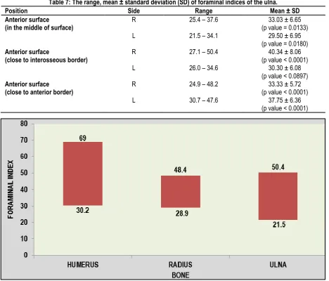
Table 7: The range, mean ± standard deviation (SD) of foraminal indices of the ulna.
Position Side Range Mean ± SD
Anterior surface
(in the middle of surface) R
L
25.4 – 37.6
21.5 – 34.1
33.03 ± 6.65 (p value = 0.0133)
29.50 ± 6.95 (p value = 0.0180)
Anterior surface
(close to interosseous border) R
L
27.1 – 50.4
26.0 – 34.6
40.34 ± 8.06 (p value < 0.0001)
30.30 ± 6.08 (p value < 0.0897)
Anterior surface
(close to anterior border) R
L
24.9 – 48.2
30.7 – 47.6
33.33 ± 5.72 (p value < 0.0001)
37.75 ± 6.36 (p value < 0.0001)
Graph 1: Localization of the nutrient foramina (NF), independent of the surface in each bone, based on the range of the Foraminal Index (FI) of the humerus, radius and ulna.
DISCUSSION
In the present study, a single nutrient foramen has a higher percentage (51.4%) in the humeral bones, compared to that of double (42.9%) and triple foramina (5.7%) respectively. Many studies reported a percentage approximately similar to that of the present result (Forriol Campos et al.2; Mysorekar4;
Lutken9; Carroll10).
In this study, 90.7% of the nutrient foramina were located along the whole middle third of the humerus, with the foramen index ranging between 30.2% and 69% of the bone length. In accordance with the present results, previous studies reported the position of the nutrient foramina within the middle third of the bone (Forriol Campos et al.2; Mysorekar4; Kizilkanat et al.8; Carroll10;
In the present study, all the radii examined had a single nutrient foramen. The same finding was reported by Forriol Campos et al.2
and Nagel12. In other studies, the majority of radii (more than 90%)
were found to possess a single nutrient foramen (Mysorekar4;
Shulman7; Kizilkanat et al.8; Longia et al.11).
In the present study, 65.7% of the total nutrient foramina were distributed most often in the middle third of the radius and 34.3% were in the proximal third, with the foramen index ranging between 28.9% and 48.4% of the bone length. The ratios of the present study were close to those reported by Mysorekar4 who
found 62% of foramina located in the middle third of the bone and 36% in the proximal end. In the present study, 88.6% of ulnae examined had a single nutrient foramen. Double nutrient foramina were observed in the rest of the ulnae examined. With the exception of Nagel12 who recorded a single nutrient foramen in all
specimens examined, other authors reported a single nutrient foramen in more than 91% of ulnae (Forriol Campos et al.2;
Mysorekar4; Shulman7; Kizilkanat et al.8; Longia et al.11).
Regarding the ulna, the majority of nutrient foramina (59%) were in the middle third while 41% were in the proximal third of the bone, with the foramen index ranging between 21.5% and 50.4% of the bone length. No nutrient foramina were detected in the distal third of the ulnae. Reviewing the literatures, some authors reported that the majority of nutrient foramina were located in the middle third4 while others stated that most of foramina were in the
proximal third7,11. However, all authors agreed that there were no
nutrient foramina in the distal third of the ulna.
The majority of nutrient foramina in all bones studied were single in number and were secondary in size. These results were in agreement with those of Carroll10 and Longia et al.11 who reported
that about two third of the nutrient foramina were secondary. The present results contradicted with those of Kizilkanat et al.8 who
stated that most foramina were of the dominant type.
Direction and obliquity of nutrient canal shows the general pattern i.e away from the elbow. There was no change in the obliquity of the canal when the foramina were situated in the centre of the bone compared to when they were nearer the ends.
CONCLUSION
The material of the present study consisted of 105 adult human cleaned and dried bones of the upper limb. They were obtained from the osteology collection held in the Department of Anatomy, Jawahar Lal Nehru Medical College, Ajmer, Rajasthan. For each bone, the number, position, size, direction and obliquity of their nutrient foramina were studied. This anatomical study of nutrient foramina in shaft of long bones is of paramount importance in medico-legal aspect and also important in surgical procedures like bone grafting and microsurgical bone transplantation. Accordingly, a well understanding of the characteristic morphological features of the nutrient foramina by orthopaedic surgeons is recommended. Exact position and distribution of the nutrient foramina in bone diaphysis is important to avoid damage to the nutrient vessels during surgical procedures.
Investigations on the vascular anatomy of long bones are important to human because it is relevant to fracture treatment (Bridgeman and Brookes13; Al-Motabagani14).
Position of the fracture relative to the nutrient foramen of the long bone and the patterns of edema are the secondary signs in the key of the diagnosis of this type of fracture (Craig et al.15).
REFERENCES
1. Lewis OJ. The blood supply of developing long bones with special reference to the metaphyses. Bone & Joint Journal. 1956 Nov 1;38(4):928-33.
2. Campos FF, Pellico LG, Alias MG, Fernandez-Valencia R. A study of the nutrient foramina in human long bones. Surgical and Radiologic Anatomy. 1987 Nov 1;9(3):251-5.
3. Collipal E, Vargas R, Parra X, Silva H, del Sol M. Diaphyseal nutrient foramina in the femur, tibia and fibula bones/Foramenes nutricios diafisarios de los huesos femur, tibia y fibula. International Journal of Morphology. 2007 Jun 1;25(2):305-9. 4. Mysorekar VR. Diaphysial nutrient foramina in human long bones. Journal of anatomy. 1967 Sep;101(Pt 4):813.
5. Skawina A, Wyczółkowski M. Nutrient foramina of humerus, radius and ulna in human fetuses. Folia morphologica. 1986 Dec;46(1-2):17-24.
6. Hughes H. The factors determining the direction of the canal for the nutrient artery in the long bones of mammals and birds. Cells Tissues Organs. 1952;15(3):261-80.
7. Shulman SS. Observations on the nutrient foramina of the human radius and ulna. The Anatomical Record. 1959 Aug 1;134(4):685-97.
8. Kizilkanat E, Boyan N, Ozsahin ET, Soames R, Oguz O. Location, number and clinical significance of nutrient foramina in human long bones. Annals of Anatomy-Anatomischer Anzeiger. 2007 Feb 1;189(1):87-95.
9. Lütken P. Investigation into the position of the nutrient foramina and the direction of the vessel canals in the shafts of the humerus and femur in man. Cells Tissues Organs. 1950;9(1-2):57-68. 10. Carroll SE. A study of the nutrient foramina of the humeral diaphysis. Bone & Joint Journal. 1963 Feb 1;45(1):176-81. 11. Longia GS, Ajmani ML, Saxena SK, Thomas RJ. Study of diaphyseal nutrient foramina in human long bones. Cells Tissues Organs. 1980;107(4):399-406.
12. Nagel A. The clinical significance of the nutrient artery. Orthopaedic review. 1993 May;22(5):557-61.
13. Bridgeman GE, Brookes MU. Blood supply to the human femoral diaphysis in youth and senescence. Journal of anatomy. 1996 Jun;188(Pt 3):611.
14. Al-Motabagani MA. The arterial architecture of the human femoral diaphysis. J Anat Soc India. 2002;51:27-31.
15. Craig JG, Widman D, van Holsbeeck M. Longitudinal stress fracture: patterns of edema and the importance of the nutrient foramen. Skeletal radiology. 2003 Jan 1;32(1):22-7.
Source of Support: Nil. Conflict of Interest: None Declared.
Copyright: © the author(s) and publisher. IJMRP is an official publication of Ibn Sina Academy of Medieval Medicine & Sciences, registered in 2001 under Indian Trusts Act, 1882. This is an open access article distributed under the terms of the Creative Commons Attribution Non-commercial License, which permits unrestricted non-commercial use, distribution, and reproduction in any medium, provided the original work is properly cited.
Cite this article as: Samata Goyal, Mahima Shrivastava. Study of Nutrient Foramina in the Long Bones of Human Upper Limb in Ajmer, Rajasthan. Int J Med Res Prof. 2018 Mar; 4(2):207-11.