Acute Pancreatitis: CT Imaging Features
Santosh N. PawarA, Bharati P. ChavanB
A-Assistant Professor, Dept. of Radio diagnosis, S.R.T.R.Govt., Medical College, Ambajogai, Beed, Maharashtra, India
B-Resident, Dept. of Pediatric, S.R.T.R.Govt.Medical College, Ambajogai, Beed, Maharashtra, India
Abstract:
Background: The evolving role of CECT in the noninvasive study of pancreas is undebatable . Its ability to define the presence of an abnormality surpasses the other imaging modalities in being able to demonstrate the extent of the disease and its spread to contiguous areas by virtue of its being non-organ specific investigation. The ability of CT to image the pancreas adequately regardless of the bowel gas and fat gives it an advantage over ultrasound.
Aim: To assess acute pancreatitis by computed tomography (CT) and classify and grade pancreatitis with the help of CT imaging features.
Methodology: This was prospective study. This study comprises 30 of different age groups in whom there was clinical suspicion of pancreatitis, of which 16 patients found to be of acute pancreatitis by CT features are taken in our study. Each patient had been studied by using plain and contrast computed tomography.
Results: Maximum no. of patients’ age was from middle age group. Acute pancreatitis were more commonly found in males than in females. It is also significantly seen association with alcohol abuse. Acute Pancreatitis was graded according to Balthazar CT severity grading system.
Conclusion: In present study an attempt has been made to study CT imaging features of acute pancreatitis. CT signs of acute pancreatitis are very useful for grading acute pancreatitis in respect to CT severity index for clinical management of patents. Therefore, CT is an excellent noninvasive imaging modality in diagnosing and further management of acute pancreatitis when used judiciously in good clinical settings.
Key words: acute pancreatitis, computed tomography, non invasive imaging
Radio-diagnosis
Corresponding Address
Dr. Santosh N. Pawar Assistant Professor, Dept. of Radio diagnosis, S.R.T.R. Govt. Medical College, Ambajogai, Beed, Maharashtra Email: santoshnpawar@gmail. com
Introduction:
Computed Tomography (CT) is a highly accurate, non-invasive imaging modality of choice in evaluating the pancreas.1 CT enables the imaging of the entire pancreas easily from the surrounding fat and bowel air together with simultaneous imaging of other abdominal organs.2 It also enables detection of unsuspected additional or ancillary abnormalities which may be responsible for clinical manifestations.
pancreas and various CT appearances in acute pancreatitis with regards to sizes, shape, position, margins (contour) volume, density characteristics, enhancement patterns, vascular landmarks, pancreatic and common bile ducts and the surrounding organs.
Materials & Methods:
Study design: This was the prospective analytical study.
Place of research: The work was carried out in the Department of Radio diagnosis, Dr. V.M.Govt. Medical College and Shri Chatrapati Shivaji Maharaj General Hospital, Solapur. Ethics committee clearance was obtained for the present study. Informed consent of patients also taken from each patient.
This study comprises 30 unselected patients of different age groups in whom there was clinical suspicion of pancreatitis, of which 16 patients found to be of acute pancreatitis by CT features are taken in our study. Clinical symptom of acute pain in epigastric region radiating to back is considered .The each patient was initially gone through the routine clinical examination, lab diagnosis (BSL, Serum Amylase, and Serum Bilirubin). Ultrasonographic findings suggestive of acute pancreatitis were also included in this study.
Patients showing normal ultrasonography report and laboratory investigations were excluded from this study.
Methodology:
Each patient had been studied by using plain and contrast computed tomography on: Third Generation spiral CT- Philips Company (CT Model – CT vision, CT-secura). The following CT findings seen : Contour (Regular / irregular, Nodular), Size (Focal enlargement, Diffuse enlargement, Diffuse atrophy, Focal atrophy), Attenuations (Plain, Arterial, Venous, Pancreas, Aorta, IVC), Density (Homogenous/ heterogenous, Focal Hypodense Areas, Focal Isodense areas, Focal hyperdense areas, Necrosis), Calcification (Parenchymal, Ductal, Both), Pancreatic duct (Size, Calculi), Commone Bile duct (Size, Calculi), Pancreatic abscess, Pancreatic gas, Peripancreatic fat stranding, Phlegmonous changes (Mesentery, Transverse mesocolon, Anterior pararenal fascia, Lesser sac, Pelvis), Acute fluid Accumulations (Intrapancreatic/
Extrapancreatic), Psuedocyst, Ascites, Pleural effusion, Vascular structures, Varices, Fat plane around the vessels, Liver (Normal / Obliterated, IHBR: Normal / Dilated).
Results:
Table 1:
Age and sex distribution of acute pancreatitis (n=16)
Age
(Years) Male % Female % Total %
21-30 1 6.25 1 6.25 2 12.50
31-40 5 31.25 2 12.5 7 43.75
41-50 3 18.75 -- -- 3 18.75
51-60 1 06.25 -- -- 1 6.25
61-70 2 12.50 -- --- 2 12.50
>70 -- -- 1 6.25 1 6.25
Total 12 75% 4 16 100%
Comments: Acute pancreatitis was more common in males than in females in this study. The commonest age group affected was between 30-50 yrs.
Table 2:
Table showing the CT signs of acute pancreatitis
Signs No. %
Gland
Normal 0 0
Diffuse
Enlargement 11 68.75
Focal Enlargement 5 31.25
Contour Irregular 10 62.5
Regular 6 37.5
Density HomogenousHeterogeneous 115 31.2568.75
Necrosis <30%30-50% 31 18.756.25
>50% 2 12.50
Phlegmonous
changes 7 43.75
Fluid accumulation
Intrapancreatic 3 18.75 Extrapancreatic 4 25.00
Both 2 12.50
Presence of gas/Abscess 0 0
Pseudocyst 3 18.75
Ascites 3 18.75
Pleural effusion 8 50.00
Table 3:
Distribution of patients of acute pancreatitis according to grade of pancreatits (n=16)
Grade No. of patients %
A 0
--B 4 25.00
C 3 18.75
D 7 43.75
E 2 12.50
Grade A: Normal pancreas, Grade B: Focal or diffuse enlargement of the gland, including contour irregularity, non homogenous attenuation of gland, dilatation of the pancreatic duct, foci of small fluid collections within the gland. Grade C: Intrinsic pancreatic abnormality associated with haziness and streaky densities representing inflammatory changes in the peripancreatic fat. Grade D: single ill defined fluid collection. Grade E: Two or multiple poorly defined fluid collections as presence of gas in or adjacent to pancreas.
Table 4:
Distribution of Necrosis in various grades of Pancre-atitis (n=6)
Grade No. of patients %
A --
--B --
--C 2 33.33
D 3 50.00
E 1 16.66
Total 6 100%
Comments: Necrosis is the non enhancing areas of pancreas on dynamic contrast CT. Necrosis is identified in 6 patients in this study.
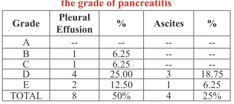
Table 5:
Distribution of pleural fluid and Ascites according to
the grade of pancreatitis
Grade EffusionPleural % Ascites %
A -- -- --
--B 1 6.25 --
--C 1 6.25 --
--D 4 25.00 3 18.75
E 2 12.50 1 6.25
TOTAL 8 50% 4 25%
Comments: Ascites and pleural effusions were noted in patients with more severe grade of pancreatitis.
Table 6:
Distribution of phlegmonous changes and pseudocysts in various anatomical compartments
Anatomical
comparatment Phlegmon (7) % Psendocyst (3) %
Intrapancreatic
spaces -- -- 2 66%
Lesser sac 3 42% 1 33%
Perinephsic
spaces 4 57% --
--Mesenteric root 5 71% --
--Paraconal
spaces 2 28% --
--Pelvis 1 14% --
--Comments: Total number of patients does not correlate with the number of anatomical sites, as more than one anatomical site was involved in a patient.
Discussion:
In our two and half years experience with 30 patients referred for CT scanning of abdomen for suspected pancreatitis, of which 16 patients found to be of acute pancreatitis by CT features are taken in our study. We had a highly selected group of patients for CT study, because of the availability of US in the hospital and strongly clinically suspected patients were taken for CT examination.
CT examination9,10
In our study 16 patients were diagnosed as having acute pancreatitis. (32%). 12 patients (75%) were of the male sex and this was correlated with the high incidence of alcohol abuse in these patients as being the commonest cause of acute pancreatitis. Brooke Jeffery et al (1982 )11 found the cause of acute pancreatitis in 24 of 36 patients to be due to alcohol abuse, as was also noted by Gaston Mendez et al (1980).12
Peak age of incidence was noted in the 30-50 years age range. In B Jeffery study (1982)11 the mean age was 41 years. In our study, 11 of 16 patients (68.75%) had diffuse enlargement of the pancreas, with focal enlargement of the pancreas seen in the 5 patients (31.25%). This correlated with Brooke Jeffery et al (1982)11 study in which 31 of 36 patients showed diffuse enlargement and 2 patients showed focal enlargement. This also compared with Mendez et al (1980)12 in which 32 patients showed gland enlargement.
In this study, peripancreatic phlegmonous changes were noted in 7 patients (43.75%) with involvement of mesenteric root in 5(71%), perinephric spaces in 4 (57%) lesser sac in 3(42%), paraconal spaces in 2(28%) and pelvis in 1 (14%) patient. Out of 7 patients 85.7% (6 patients) were of necrotizing pacreatitis and 14.28% (1 patient) of acute edematous pancreatitis. This correlated with Hill et al (1982)5 in which phlegmonous changes were reported in 11% of acute edematous pancreatitis and 89% of necrotizing pancreatitis.
In our study, 9 patients (56.25%) had acute fluid accumulations, of which 3 patients (18.7%) had intrapancreatic, 4 patients (25%) had extrapancreatic fluid accumulations, and 2 patients (12.50%) had both extra and intrapancreatic fluid collections. Seigleman Stanley et al (1980)13 also reported pancreatic and extrapancreatic fluid accumulations in 54% cases with 16% having intrapancreatic and 42% having extrapancreatic collections. Balthazar E J et al (1994)11 also reported acute fluid collections in 40% of patients early in the course of acute pancreatitis of which 50% resolved spontaneously. In our study, the natural history of acute fluid collections could not be followed up, as our patients could not afford rescans.
In our study we had 3 cases of pseudocyst, 2 in intrapancreatic locations and one in lesser sac. The commonest site of pseudocyst; a late sequlae of the
disease, in our study was intrapancreatic location (66%) in acute pancreatitis. CT is a better investigation than US for detection of remote pseudocysts.10 Kresses says that CT has 100% sensitivity while US has 50% in detection of extrapancreatic predocysts.
In our study, no patient had Grade A, 25% had Grade B, 18.75% Grade C, 43.75% Grade D and 12.50% Grade E pancreatitis. The patients who developed two or multiple poorly defined fluid collections were of Grade E pancreatitis. Further pleural effusion in 50% cases and ascites in 25% were found in more sever grades, Grade D and E pancreatitis. The patients of Grade A, B, C had no or less number of complications like pleural effusion and ascites. Balthazar E. J (1985)14 reported the following: Grade A 14.5%, Grade B 22.9%, Grade C 25%, Grade D 14.5%, Grade E 27.7%. Our study correlated with the study of Balthazar E J (1985)14 for the presence of pleural effusion and ascites like complications occurs more in Grade D and E pancreatitis. Pancreatic necrosis described as focal nonenhancing low attenuation areas was noted in 6 patients (37.5%) in our study. Necrosis was not found in Grade A and B pancreatitis, but was found 33.33% in Grade C, 50% in Grade D and 16.66% in Grade E pancreatitis. These findings correlated with Balthazar E J et al (1990)15 noted total necrosis being 20.4%. Necrosis was not found in Grade A and B pancreatitis, but was found 25% in Grade C, 50% in Grade D and 25% in grade E. Most patients with Grade D and E pancreatitis exhibited higher incidence of pancreatic necrosis detected in our study could be attributed to spiral acquisition of data during peak pancreatic parenchymal enhancement, thus allowing good discrimination between necrosed and viable portions of the gland.
Conclusion:
References:
1. Brooke Jeffery R, Federle Michael P, Jeffery Brooke R, Cello John P: Early computed Tomographic scanning in Acute Severe Pancreatitis; Surgery, Gynecology & Obstetrics; February 1982, Volume 154.
2. Mendez Gaston Jr., Isikoff Michael B, Hill Michael C: CT of Acute Pancreatitis: Interim Assessment; American Journal of Roentgenology 135 : 463-469, September 1980.
3. Balthazar Emil J, Ranson J H C, Naidich David P, Megibow Alec J et al : Acute pancreatitis : Prognostic value of CT; Radiology 1985 ; 156-767-772.
4. Alexander Elizabeth S, Clark Robert A, Federle Michael P : Pancreatic Gas : Indication of pancreatic Fistula; American Journal of Roentgenology 139 : 1089-1093, December 1982.
5. Hill Michael C, Barkin Jamie, Isikoff Michael B, Silverstein William, Kaster Martin: Acute pancreatitis: Clinical vs. CT findings; American Journal of Roentgenology 139: 263-269, August 1982.
6. Alpern M.B., MA Sandler , G.M. Kellman, B.L. Madrazo: Chronic pancreatitis: U.S. features, Radiology ;155:215, 1985.
7. Levitt R., Geisse, S. Sagel, R. Stanley: Complementary use of US and CT in studies of the pancreas and kidney. Radiology 126 : 149-152, 1978.
8. Silverstein, Isikoff, Hill , Barkin: Diagnostic Imaging of Acute Pancreatitis – Prospective study using CT & US, American Journal of Roentgenology – 137: 497, 1981.
9. Kolmannskog F, A. Kolbenstvedt, T. Aakhas: CT in inflammatory mass lesions following acute pancreatitis. Radiology 141: 872, 1981
10. Margulis A., Kressel, Gooding, Filly, Moss, Kerobkin: CT scanning and US in the evaluation of pancreatic preudocyst – A preliminary comparison, Radiology, 126:53-157, 19778.
11. Brooke Jeffery R, Federle Michael P, Jeffery Brooke R, Cello John P: Early computed Tomographic scanning in Acute Severe Pancreatitis; Surgery, Gynecology & Obstetrics; February 1982, Volume 154.
12. Mendez Gaston Jr., Isikoff Michael B, Hill Michael C: CT of Acute Pancreatitis: Interim Assessment; American Journal of Roentgenology 135 : 463-469, September 1980.
13. Seigelman Stanley S; Copeland Bruce E, Saba George P, et al: CT of fluid collections Associated with Pancreatitis; American Journal of Roentgenology 134 : 1121-1132, June 1980.
14. Balthazar Emil J, Ranson JHC, Naidich David P, Megibow Alec J et al: Acute pancreatitis : Prognostic value of CT; Radiology 1985; 156-767-772.