DEVELOPMENT OF BRAIN SULCI IN FETUSES: A MORPHOLOGICAL STUDY
Jyotsna Singh, Kanchan Kapoor*, Joseph Abraham, Amrutha K.
Department of Anatomy, Government Medical College and Hospital, Chandigarh. 160047, India A R T I C L E I N F O
INTRODUCTION
The development of cerebral sulci & gyri is a result of an extensive infolding of the cerebral cortex due to increase of its outer dimensions. The detailed knowledge of their appearance, form & extent acts as a guide for microneuro
sulcal approaches (Ribass 2010).
Although the arrangement of sulci & gyri is not uniform in two individuals, yet a regular pattern could be classified. Ono et al. (1990) have classified the sulci in four main types: large primary sulci; short primary sulci; short sulci and short, supplementary sulci. Usually the side branches of sulci connect either neighbouring sulci or same sulcus from end to end.
Embryologically the formation of sulci starts with the development of fissures, followed by primary, secondary and tertiary sulci (Broca1877; Chi et al. 1977). The longitudinal interhemispheric fissure is estimated to appear by 10
gestation. However the main primary sulci make their appearance during 4th – 5th weeks of IU life; while some of them develop after birth. The appearance of each sulcus follows a specific pattern in terms of gestational age (Nishikuni and Ribass 2013).
International Journal of Current Advanced Research
ISSN: O: 2319-6475, ISSN: P: 2319-6505,
Available Online at www.journalijcar.org
Volume 8; Issue 09 (D); September 2019
DOI: http://dx.doi.org/10.24327/ijcar.2019
Copyright©2019 Jyotsna Singh et al. This is an open access article distributed under the Creative Commons Attribution License, which permits unrestricted use, distribution, and reproduction in any medium, provided the original work is properly cited.
Article History:
Received 06thJune, 2019
Received in revised form 14th July, 2019 Accepted 23rd August, 2019
Published online 28th September, 2019
Key words:
Fetal study, cerebral cortex, sulci, functional areas.
*Corresponding author: Kanchan Kapoor
Department of Anatomy, Government Medical College and Hospital, Chandigarh. 160047, India
DEVELOPMENT OF BRAIN SULCI IN FETUSES: A MORPHOLOGICAL STUDY
Jyotsna Singh, Kanchan Kapoor*, Joseph Abraham, Amrutha K. Valapil
Department of Anatomy, Government Medical College and Hospital, Chandigarh. 160047, India A B S T R A C T
Introduction: The development of sulci and gyri is the result of folding of cerebral cortex to accommodate in a limited space. Although the morphology is variable, yet a regular pattern could be classified. Any gross variation from this pattern can be indicative of certain neuropsychiatric disorders. Further, the shape, form and time of appearance can aid in microneurosurgery in trans-sulcal approach. Therefore it was considered worthwhile to observe the pattern of development of sulci and gyri in different gestationa
Materialand methods:The study was conducted in 50 fetuses, sent by the department of
obstetrics and gynaecology for routine autopsy. Brains were removed from skull and the time of appearance of various fissures and sulci were observed. T
into four gestational age groups.
Observations: The longitudinal fissure was the first to appear in 10 Other sulci appear in response to the development of specific function
sulcus on superolateral surface, callosal sulcus on medial surface were among the earlier developing sulci. The sulci on the inferior surface developed much later. The present observations are discussed in light of the available literature.
Conclusion: These findings will be helpful in providing baseline data to assess the normal development as well as any variation in foetuses.
cerebral sulci & gyri is a result of an extensive infolding of the cerebral cortex due to increase of its outer dimensions. The detailed knowledge of their appearance, form & extent acts as a guide for microneuro-surgery in
trans-Although the arrangement of sulci & gyri is not uniform in two individuals, yet a regular pattern could be classified. Ono et al. (1990) have classified the sulci in four main types: large primary sulci; short primary sulci; short sulci and short, supplementary sulci. Usually the side branches of sulci connect either neighbouring sulci or same sulcus from end to
Embryologically the formation of sulci starts with the development of fissures, followed by primary, secondary and oca1877; Chi et al. 1977). The longitudinal interhemispheric fissure is estimated to appear by 10th week of gestation. However the main primary sulci make their weeks of IU life; while some of rance of each sulcus follows a specific pattern in terms of gestational age
Knowledge of normal pattern of sulcul development and ability to discriminate the appearance of different sulci at their respective gestational ages allows early suspicion of lissencephaly and other neuropsychiatric disorders (Toi et al. 2004). Most of the available lite
from ultrasonography or MRI scans (Monteagudo and Timor 1997; Garel et al. 2001). Owing to the two dimensional aspect of USG and the extent of resolution, these studies lag behind by 2-8 weeks from their actual appearance. The
established, can be used as a reliable guide to estimate the gestational age during fetal autopsy of unknown gestational age. Also this knowledge can be applied to compare the normal versus retarded intrauterine growth. The present study is an attempt to define the morphological development of sulci and gyri with relation to gestation age.
MATERIAL AND METHODS
The present study was conducted on 100 cerebral hemispheres in 50 fetuses of varying gestational age. The fetuses were obtained from department of obstetrics and gynaecology of the same institute for routine autopsy. The consent was taken from the parents as well as from the ethics committee of the institute to carry on further research. The exclusion criteria included congenitally malformed fetuses. The specimens were kept in 10% formalin for one month; direct formalin was injected through fontenelle to preserve the brain.
International Journal of Current Advanced Research
6505, Impact Factor: 6.614
www.journalijcar.org
2019; Page No.19946-19952
//dx.doi.org/10.24327/ijcar.2019.3880.19952
This is an open access article distributed under the Creative Commons Attribution License, which permits unrestricted use, distribution, and reproduction in any medium, provided the original work is properly cited.
Department of Anatomy, Government Medical College and Hospital,
DEVELOPMENT OF BRAIN SULCI IN FETUSES: A MORPHOLOGICAL STUDY
Valapil and Jessy JP
Department of Anatomy, Government Medical College and Hospital, Chandigarh. 160047, India
The development of sulci and gyri is the result of folding of cerebral cortex to accommodate in a limited space. Although the morphology is variable, yet a regular pattern could be classified. Any gross variation from this pattern can be indicative of , form and time of appearance can aid sulcal approach. Therefore it was considered worthwhile to observe the pattern of development of sulci and gyri in different gestational age groups.
The study was conducted in 50 fetuses, sent by the department of obstetrics and gynaecology for routine autopsy. Brains were removed from skull and the time of appearance of various fissures and sulci were observed. The fetuses were divided into four gestational age groups.
The longitudinal fissure was the first to appear in 10-11 weeks gestation. Other sulci appear in response to the development of specific functional areas. The central sulcus on superolateral surface, callosal sulcus on medial surface were among the earlier developing sulci. The sulci on the inferior surface developed much later. The present
ature.
These findings will be helpful in providing baseline data to assess the normal
rmal pattern of sulcul development and ability to discriminate the appearance of different sulci at their respective gestational ages allows early suspicion of lissencephaly and other neuropsychiatric disorders (Toi et al. 2004). Most of the available literature includes observations from ultrasonography or MRI scans (Monteagudo and Timor 1997; Garel et al. 2001). Owing to the two dimensional aspect of USG and the extent of resolution, these studies lag behind 8 weeks from their actual appearance. The fact, if established, can be used as a reliable guide to estimate the gestational age during fetal autopsy of unknown gestational age. Also this knowledge can be applied to compare the normal versus retarded intrauterine growth. The present study empt to define the morphological development of sulci and gyri with relation to gestation age.
MATERIAL AND METHODS
The present study was conducted on 100 cerebral hemispheres in 50 fetuses of varying gestational age. The fetuses were department of obstetrics and gynaecology of the same institute for routine autopsy. The consent was taken from the parents as well as from the ethics committee of the institute to carry on further research. The exclusion criteria included ormed fetuses. The specimens were kept in 10% formalin for one month; direct formalin was injected through fontenelle to preserve the brain.
Research Article
Brains were removed from skull as per standard protocol mentioned in (Romanes 2018). It was very difficult to dissect out the full brain from calvaria as fetal brain is very soft and fragile. A few specimens were lost during dissections. Each specimen was weighed and photographed. A careful examination was done using a hand lens if necessary and a Performa was filled regarding the appearance, development and completion of various sulci on all the three surfaces of cerebral hemispheres.
The fetuses were divided into four groups according to different gestational age. The appearance of various sulci was tabulated and an effort was made to pinpoint the exact time (week wise) for the appearance of a particular sulcus. A correlation was also calculated between fetal body weight and brain weight.
RESULTS
The findings were grouped according to gestational age. (Table 1)
Table 1 Distribution of fetuses in different gestational age groups and mean body and brain weight
Gestational age Number of
cases
No of males
No of females
Body weight (gm)
Brain weight(gm)
11-15 weeks 3 2 1 60-74 8-10
15+ -20 weeks 27 14 13 230-329 23-51 20+-25 weeks 16 10 6 359-593 49-93
25+weeks
onwards 4 1 3 771-1607 92-183
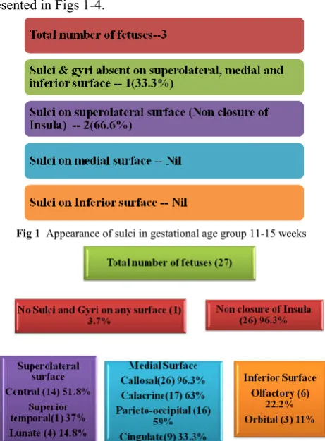
It was observed that longitudinal cerebral fissure was present in 49 (98%) cases. A longitudinal cerebral sulcus was visible in one case of 11 weeks of gestation (Fig 7 ).The appearance of sulci was observed in superolateral, medial and inferior surfaces of each cerebral hemisphere in all gestational age groups. The percentages were calculated and the results are presented in Figs 1-4.
Fig 1 Appearance of sulci in gestational age group 11-15 weeks
Fig 2 Appearance of sulci in gestational age group of 15+- 20 weeks
Fig 3 Appearance of sulci in gestational age group of 20+-25 weeks
A first glance reveals that the appearance of various sulci are more visible on the medial surface of the cerebrum, followed by the superolateral while the inferior surface is the last one.
Insula: the involution of insular cortex starts very early
(before 11 weeks of gestation) in fetal life, probably along with the appearance of longitudinal fissure. However the three opercula develop gradually and till the age of 20 weeks, the insula is defined as open with nonclosure of opercula and thereby incomplete formation of lateral sulcus. Initially the angle is obtuse and later acute angle became evident at 23 weeks.
Superlateral surface
Longitudinal fissure appeared at 11 week of gestation. Lateral sulcus and insular depression were present by the 14th week in 100 % of cases. Operculisation of insula was observed upto 22 weeks and closure of lateral sulcus was complete by 28weeks. Central sulcus appeared in 40.7% at 18 weeks. Central sulcus appeared parasaggitally over the convexity of superolateral surface in 50.8% of fetuses in 15-20week gestation and was completely present in 100% fetal brains by 25weeks. The central sulcus finally abuts the superomedial border at 30 weeks to appear on medial surface. Lunate sulcus made its appearance at 18+2 weeks and was observed in 15% cases in 15-20 weeks gestation group, 12.5% in 21-25 weeks gestation group and in 50% in 26-31 week gestation group. Precentral sulcus was observed in 43.7% fetal brains in 21-25 weeks and in all fetal brains above 25 weeks. Superior temporal sulcus was observed on the right side at the middle of the cerebral hemisphere however it was completely present on the corresponding left hemisphere at 20 weeks in a single case. It was observed in 18% of fetal brains at 20-25weeks gestation and 50% in 25-31 weeks. Inferior temporal sulcus made its first appearance at 25+4 weeks gestation and was complete by 31 weeks. Intraparietal sulcus and superior frontal sulci were observed in 50% of fetal brains at 25+5 weeks . Post central sulcus appeared at a later gestation with first appearance at 26+
5
Medial surface: The medial surface did not show any sulcus in the age group of 11-15 weeks. From the beginning of 15-20 weeks, the primary sulci including callosal, calcarine, parieto-occipital and cingulate made their appearance. Callosal sulcus was the first to appear on the medial side of cerebral hemisphere visible in 96.3% of fetal brains in 15-20week age group. The cingulate sulcus was the last one which is visible in only 33% brains in the same week age group. In the age group of 20-25 weeks, the callosal and calcarine sulci were present in all (100%), while cingulate and parieto-occipital were present in 68.75%. Smaller secondary sulci such as paracentral lobule started appearing in 25-31 weeks, these were present in 75% specimens. Therefore all the sulci and gyri had appeared and were complete by the age of 31st week of gestation. The cingulate sulcus and calcarine sulcus are tortuous due to development of secondary sulci. Cerebral asymmetery was observed in appearence of sulci on medial surface. There was rapid development on left side in 21-25 weeks gestation. Thereafter the sulci developed on both cerebral hemispheres equally in 25-31 weeks of gestation.
Fig 4 Appearance of sulci observed in gestational age group of 25-31 weeks
Inferior surface: Olfactory sulcus has been observed to appear
at 17 weeks and was detectable thereafter in all fetal brains. Orbital sulcus made its appearance at 20 weeks in 11% brains was present in 31% in 21-25 weeks gestation and 100% in 25-31 week gestation. Orbital sulcus initially was observed developing on left side at 20 weeks and later was bilaterally present. Collateral and rhinal sulci appeared at 21+2 week gestation in 18.7% and were fully developed by 31 weeks. Occipitotemporal sulcus first appeared at 25 weeks fetal brain and was present thereafter in subsequent gestational age groups.
Fig 5 Correlation of brain weight with gestational age
As is evident, there is a gradual increase in brain weight with advancing gestational age. However rapid increase in brain weight was observed after 25 weeks of gestation.
Fig 6 Correlation between brain weight and body weight of fetus
Gradual increase of brain weight in correlation with increasing body weight (graph does not depict any specific peak period for brain growth).
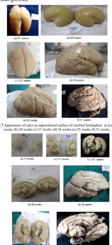
Fig 7 Appearance of sulci on superolateral surface of cerebral hemisphere at (a) 18+2
weeks (b) 20 weeks (c) 21 weeks (d) 24 weeks (e) 25 weeks (f) 31 weeks.
Fig 8 Appearance of sulci on Medial surface of cerebral hemisphere at (a) 11weeks (b) 15+4 weeks (c) 18+4 weeks (d) 20 weeks (e) 24 weeks (f) 26+5 weeks (g) 31 weeks
0 2000 4000 6000 8000 10000 12000 14000 16000 18000 20000
11
wks
16
wks
17
wks 18+2 18+6 19+3 20 20+3 20+6 21+2 22 25+4
B
rain weight (m
g)
Correlation of brain weight with gestational age
Brain weight in milligrams
0 0.02 0.04 0.06 0.08 0.1 0.12
0 500 1000
Brain Weight (gm)
Brain Weight (gm)
Fig 9 Appearance of sulci on Inferior surface of cerebrum of cerebral hemisphere at (a) 11weeks (b) 20 weeks (c) 24 weeks (d) 26+5 weeks
DISCUSSION
The cerebral cortex is divided into gyri and sulci the morphology is variable among different individuals (Ono et al.1990). The anatomical organisation is related to its cortical functional organisation and may reflect pathology as specific abnormalities in sulcul pattern are observed in neuropsychiatric disorders such as autisim, William syndrome and schizophrenia (Dubios J 2008). These observations are seen in adults, but structural abnormalities might be present long before the appearance of functional symptoms since cortical folding occurs during early intrauterine life (Chi et al, 1977; Garel et al, 2001).
For accommodation in a limited space within the rigid cranial cavity, the cerebral cortex is folded. The sulci evolve as the brain undergoes a process of circular curvature, wrapping the thalamus in its morphological centre. The development of sulcul pattern on superolateral and inferior surface is influenced by the lateral ventricle especially evident in coronal slices of MR imaging studies. This disposition of sulci is absent on medial surface of the hemisphere. The medial surface sulci development is influenced by corpus callosum and the sulci tend to be arranged in parallel with the commisure. Congenital absence of corpus callosum is linked with radial pattern of medial surface sulci and absence of an arched cingulate gyrus (Nishikuni and Ribas 2013).
Based on MR images of 20 healthy patients, Regis et al. (2005) proposed the arrangement of cortical sulci and gyri is dependent on the variable development of connecting gyri buried within the depth of sulci as a result from trade off between various folding pressures occurring during brain growth. Chaotic behaviour in the burying process as seen in the occasional interruption of central sulcus leads to a prominent middle frontoparietal connection of Broca.
The formation of cortical sulci is considered a good marker of fetal maturation and is currently of significant interest to calculate gestational ages using USG studies (Ribaas 2010).
In the last two decades, the advancement in transabdominal and transvaginal sonography have enabled the clinicians to visualize the developing human fetus with increasing clarity. However discrepancies exist concerning the time of appearance of the cerebral sulci between various authors. This has increased the demand for a normative anatomical data to assess the specific time period for the appearance of cortical sulci. Lack of standardisation inspired us to observe the morphological cerebral sulci development in different gestational age groups.
The development of main sulci according to their appearance is being discussed below. (Table2)
Table 2 Gestation wise time of appearance of sulci on different surfaces of cerebral hemisphere
Gestationa
l age Superolateral surface Medial surface Inferior surface
26 onwards
Post central sulcus Inferior temporal sulcus Closure of Lateral sulcus
Transverse Occipital
Marginal sulcus Paracentral sulcus
Paraolfactory
Occipitotemporal sulcus
21-25
Precentral sulcus Superior Frontal sulcus
Superior Temporal Intraparietal sulcus
Orbital sulcus Rhinal sulcus Collateral sulcus
15-20 Central sulcus Lunate sulcus
Callosal Calcarine sulcus Cingulate sulcus Parieto occipital
Hippocampal sulcus Olfactory sulcus
10-14
Longitudanal cerebral fissure Lateral sulcus
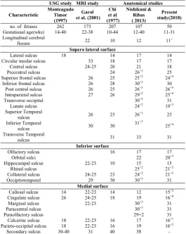
Table 3 Comparison of Prenatal cerebral sulci development between ultrasonographic, magnetic resonance imaging and
anatomical studies
USG study MRI study Anatomical studies
Characteristic
Monteagudo Timor (1997)
Garol et al. (2001)
Chi et al (1977)
Nishikuni & Ribas ( 2013)
Present study(2018)
no. of fetuses 262 173 207 107 50
Gestational age(wks) 14-40 22-38 10-44 12-40 11-31
Longitudinal cerebral
fissure 22 10 12 11
+
Supero lateral surface
Lateral sulcus 18 14 17 14
Circular insular sulcus 33 18 17 17
Central sulcus 24-25 20 21 18
Precentral sulcus 24 26+3 25
Superior frontal sulcus 26 25 25+2 24+4
Inferior frontal sulcus 26 28 30+3 30
Post central sulcus 26 25 26+3 26+5
Intraparietal sulcus 27 26 29+2 25+4
Transverse occipital 30+3 31
Lunate sulcus 24+2 19+3
Superior Temporal
sulcus 26 23 26
+3 23
Inferior Temporal
sulcus 30 30
31+3
25+4
Transverse Temporal
sulcus 31 33 31
Inferior surface
Olfactory sulcus 16 17 17
Orbital sulci 22 20+3
Hippocampal sulcus 22-23 10 15 15
Rhinal sulcus 25+2 21+2
Collateral sulcus 24-25 23 24+2 21+2
Occipitotemporal 29 30 30+3 31
Medial surface
Callosal sulcus 14 22-23 14 12 15+2
Cingulate sulcus 26 24-25 18 19 16+4
Marginal sulcus 22-23 30+3 31
Paracentral sulcus 30+3 31
Paraolfactory sulcus 29+2 31
Calcarine sulcus 18 22-23 16 17 16+1
Parieto-occipital sulcus 18 22-23 16 19 16+2
Longitudinal cerebral fissure: The time of appearance of fissure is estimated between 10-12 weeks of IU life as observed by (Nishikuni and Ribas 2006) and Lan et al.( 2000) . In the present study, the fissure was observed as early as 11 weeks fetus and was present in all the ages subsequently.
Sulci of the superolateral cerebral surface
Insular gyral and sulcul development
Appearance of first sulcus at 14weeks with development of periinsular sulci upto 19 weeks and operculisation of insula in 19-22 weeks was observed. From 24-26 weeks the covering of posterior insula was observed and closure of lateral sulcus was complete by 28 weeks from posterior to anterior direction.
Phylogenetically the lateral sulcus develops as a result of invagination process accompanied by progressive enlargement of frontal and temporal lobes (Nishikuni and Ribass 2013). USG studies document the appearance of lateral sulcus by 18 weeks (Montaegudo and Timor 1997). The presence of sylvian fissure at 23weeks gestation in all fetuses has been seen on MRI by Garol (2001). By 33 weeks of gestation insular sulci are recognisable and surface of sylvian fissure is indented on magnetic resonance imaging techniques.
Central sulcus: The central sulcus developed as a groove
parasagitally separating the motor from sensory cortex superiorly towards the longitudinal cerebral fissure and inferiorly towards sylvian fissure. Its presence was evident in 40% fetuses in 15-20 weeks gestation and 50% of fetuses of 20 -25weeks gestation and 100% fetuses at 25 weeks in the present study. MRI appearance was detected at 24-25 weeks by Garol et al. (2001) and 2weeks later by (Leviene and Barness 1999).
Precentral sulcus: The precentral sulcus developed anterior to
the central sulcus parallel to it and its appearance was detected in 11% of fetal brains of 20 weeks gestation and 45% between 21-25 week and 100% at 25weeks in accordance with Chi et al (1997). The present study differs from (Nishikuni and Ribas 2006, Dorovini Zis and Dolman;1977) and Larroche (1981) who detected the sulcus at 26 weeks and 28 weeks respectively. The MRI studies observed its appearance at 27-29 weeks Lan et al. (2000).
Superior temporalsulcus: The superior temporal sulcus runs
parallel to the lateral sulcus. According to Larroche (1981), the superior temporal sulcus is reliable morphological criterion of gestational age at 28 weeks gestation, when it was visible. In the present study it was observed at 23+4 weeks similar to Chi et al(1997). MRI studies have observed it posteriorly at 26 weeks gestation and its extension anteriorly at 30 weeks.
Superior Frontal sulcus: At 24 +4 weeks the superior frontal
sulci were visible at the frontal pole and initially appeared posteriorly. A complete sulcus was present in more than 50% of fetal brains at 25+4 weeks in the present study earlier to other anatomical studies and MRI studies by 1 week.
Intra parietal sulcus: This sulcus was distinguished at 25+4
weeks similar to Chi et al (1977), but (Nishikuni and Ribas 2013) observed it at 29+2 weeks.
Inferior temporal sulcus: The sulcus appeared at the temporal
pole at 25+4 week gestation in the present study and complete by 31 weeks similar to other neuroanatomic studies
Transverse occipital sulcus: This sulcus was observed at 31
week fetal brain similar to (Nishikuni and Ribas 2013) who observed it in 30+3 week fetal brain on autopsy.
Inferior frontalsulcus: Its appearance was detected at 30week
gestation similar to (Nishikuni and Ribas 2013) and Levine & Barnes (1999) on MRI imaging. Our studies had few cases between 27 weeks and 30 weeks gestation, so might be the reason of dissimilarity with Chi et al.(1977) who observed it at 28 weeks.
The outer convex surface of cerebral hemisphere is difficult to explore by USG hence except superior temporal sulcus no data concerning the sulci on superolateral surface is available (Monteagudo and Timor 1997, Bernard et.al 1988, Toi et al. 2004).
Differential growth of sulci in right and left sides of hemispheres was observed especially on superolateral surface. On right side, rapid growth was evident in almost 10% of cases till 20 weeks. In 21 – 25weeks gestation, left side of cerebral hemisphere (in 12.5%) showed early appearance of sulci. However after 25 weeks, both the hemispheres had well developed sulci and gyri. The present study is in accordance with Gilmore et al. (2007) and Kivilevitch et al.(2010) who observed cerebral asymmetry in fetal brains in second half of gestation.
Sulci on the medial surface
Callosal sulcus: the callosal sulcus separates the corpus
callosum from the cingulate gyrus; it was the first sulcus to appear on the medial surface observed in a 15+2week fetus in accordance with Chi et al (1997)but in difference with (Nishikuni and Ribas 2006)who observed its presence earlier by 2 weeks. Transvaginal sonographic study by (Monteagudo & Timor 1997) also observed its appearance at 14 weeks, however Toi et al.(2004) observed it earliest at 18.5 weeks. MRI studies by Garel et al. (2001) have detected it at 22-23 weeks.
Cingulate sulcus: Sonographically cingulate sulcus is
identifiable at 26 weeks but anatomical specimens have appreciated it at 18-19 weeks (Nishikuni and Ribas 2013). The present study observed its appearance in a 16+4 week fetus. Above 17+4 weeks of gestation it was observed in 100% of cases.USG studies and MRI lagged behind in identifying this sulcus. Toi et al( 2004)observed it to be always present after 24.3weeks, (Monteagudo and Timor 1997) at 26 weeks, while Garel et al. (2001) documented its presence at 25 weeks respectively. Pathological delay in the development of cingulate sulcus has been described by (Pape and Wigglesworth 1979) due to its proximity to an area affected by hemmorrhage and infarction.
Parieto occipital sulcus: This sulcus separates the precuneus
Calcarine sulcus: In accordance with Chi et al. (1997) and (Nishikuni and Ribas 2013) appearance of the sulcus was observed at 16+1weeks gestation concomitantly with parieto occipital sulcus in the present study. Complete sulcus in 100 % of fetal brains was detected at 18weeks similar to the observations of (Monteagudo and Timor 1997). MRI studies could only detect these sulci at 22-23 week of gestation.
The development of corpus callosum influences sulcul pattern on medial surface as its absence culminates in absence of arched cingulated gyrus and radical pattern of sulci on medial surface of cerebrum (Ono et al. 1990)
Sulci on inferior surface
Hippocampal sulcus separates archicortex from paleocortex. Rhinal sulcus separates paleocortex from neocortex
Olfactory sulcus: Olfactory sulcus developed on the medial
aspect of cerebral orbital surface in the 17 weeks of gestation in accordance to (Nishikuni and Ribas 2013) but in comparison to Chi et al. (1977) who reported it 16 weeks respectively. The developmental relationship between the sulcus and the olfactory tract and bulb is well documented by Dooling (1983).
Rhinal sulcus: The rhinal sulcus develops due to the ventral
displacement of the lateral olfactory area and separates the paleocortex from neocortex. In our study the rhinal sulcus separating the uncus appeared at 21+2 weeks gestation in 11% of the cases and at 25 weeks it was present in all specimens (100%). Rhinal sulcus appearance was reported by at 25+2 weeks by (Nishikuni and Ribas 2013). The collateral sulcus had a similar presentation in the present study.
Occipitotemporal sulcus: In the present study first appearance
starting at temporal pole was evident at 24+4 weeks gestation on the left side in one case. The complete development of the sulcus was seen at 31 week similar to Chi et al. (1977). However (Nishikuni and Ribas 2013) reported its appearance at 33 weeks.
The developmental maturation of fetal brain follows a predictable time table but in some high risk situations there may be an accelerated or predictable developmental delay. According to Slagle et al.(1989) in presence of brain insults like intraventricular haemorrhage there is significant delay in continuity and cobblestone pattern of cingulate sulcus.
On careful examination of a particular sulcus, it is evident that some sulci appear earlier and are completed earlier while a few others have not made their appearance. For example central sulcus on superolateral surface, callosal sulcus on medial surface are fully developed within 15-25 weeks of gestation. Whereas the olfactory and orbital sulci on inferior surface develop between 25-31 weeks, much later in this region. The present observations are in full agreement with Ribass (2010) who attributed the differential time plan to be directly correlated with the functional importance of the areas.
Another point of discussion is the disparity in the time plan between right and left side of cerebrum. Chi et al. (1977) stated that right hemisphere develops earlier and this asymmetry can influence the learning capabilities regarding speech and language. The asymmetrical development in the present study was observed first on right side between 15-20 weeks whereas from 21-25 weeks left side showed more growth. However by the end of 26-33 weeks both the
hemispheres were equally developed apparently with limited scope to influence any functional disability.
CONCLUSION
The present observations of the development of specific cerebral sulcus at a particular gestational age provides a baseline data to assess the normal development and towards better understanding of cortical maturation in fetuses. The folding of cortical surface starts as early as 11week of gestation and is mostly completed by 34th week. The development of the cerebral sulci and gyri is gradual and directly proportional to the increasing brain weight, body weight and gestational age. The appearance and completion of sulci is according to a hierarchal pattern where the development of functionally important areas is earlier. Also, the asymmetry in the development of right and left cerebral cortex is established.
It provides standards of reference that can be used to asses normalcy of development of fetal sulci to pathological delay. USG imaging cannot comment on superolateral as well as inferior surface of cerebral hemisphere while MRI studies lag behind the neuroanatomic time table. The key points are useful during intra operative sulcul identification and trans-sulcul approaches, in neonatal surgeries. The deviation from normal time of development, if diagnosed can be interpreted as pathology and allows genetic counselling and option to exercise obstetric management.
Conflict of interest: The authors declare that they have no
conflict of interest.
Ethical approval The method were carried out in accordance
with the 1964 Declaration of Helsinki and the routine fetal autopsy being carried out in department of anatomy is approved by the Government Medical College and Hospital, Chandigarh.
References
1. Broca P: Sur la cinconvolution limbique et al scissure limbique Bull Soc d’ Anth .12:646-657,1877,cited in Finger S(1994).Origins of Neuroscience. New York: Oxford university press,
2. Chi JG, Dooling EC,Gilles FH(1977) Gyral Development of the human brain. Ann Neurol ;1: 86-92,
3. Dooling E. (1983) Telencephalic development, changing gyral patterns, In Gilles (ed). The developing human brain. Boston : Wright-PSG. pp94
4. Dorovini Zis K and Dolman CL (1977) Gestational development of brain. Arch Pathol Lab Med 101:192-95 5. Dubios J, Benders M, Borradori TC, Cachia A, Lazeyras F, Havinh LR et al . (2008) Primary cortical folding in human newborn : an early marker of later functional development. Brain 131 :2028-41. Epub 2008
6. G.J. Romanes (2018). Cunningham Manual of Practical Anatomy, Head and neck and Brain. In The cranial cavity ( 16th eds) Oxford University press pp81, 346 7. Garel C, Emmanuel C, Herve B,Monique E. Dominique
8. GilmoreJh, Lin W, Prastawa MW, et al.( 2007) Regional gray matter growth, sexual dysmorphism and cerebral asymmetery in the neonatal brain. J Neurosci 27:1255-60
9. Kivilevitch Z, Achiron R, Zalel Y (2010) Fetal brain asymmetry : in utero sonographic study of normal foetuses. Am Obstet Gynecol 202:359. el-8
10. Lan LM,Yamashita Y, Tang Y, Sugahara T, Takahashi M, Ohba T et al .(2000) Normal fetal brain development :MR Imaging with a half –Fourier rapid acquisition with relaxation enhancement sequence. Radiology 215 :205-210
11. Larroche JC (1966) Morphological criteria of Central nervous system development during intrauterine life, in Falkner F(ed): Human Development .Philadelphia: WB Saunders ,pp257-276
12. Levine D, Barnes PD (1999) Cortical Maturation in normal and abnormal foetuses as assessed with prenatal MR imaging. Radiology 210:751-758
13. Monteagudo A and Timor Tritsch(1997) Development of fetal gyri, sulci and fissures: a transvaginal sonographic study. Ultrasound Obstet. Gynecol. 9:222-228
14. Nishikuni K and Ribas G(2013). Study of fetal and postnatal morphological development of brain sulci. J Neurosurg Pediatrics. 11:1-11
15. Ono M,Kubik S, Abernathy CD.(1990) Atlas of Cerebral Sulci. Stuttgart . Thieme,
16. Pape,K,and Wigglesworth J.S.(1979) Blood supply to developing brain. In Haemmorrhage, Ischemia and Perinatal brain, Lavenham :International Medical Publication ,pp11-38.
17. Regis J,Mangin JF, Ochiai T et al.(2005) Sulcul root generic model: a hypothesis to overcome the variability of the human cortex folding patterns. Neurol. Med. Chir. 45(1):1-17
18. Ribas GC (2010) The cerebral sulci and gyri. Neurosurg Focus 28(2):1-24
19. Slagle T.A., Oliphant M, and Gross S(1989).Cingulate sulcus development in preterm infants. Pediatr.Res 1989;26:598-602
20. Toi A,Lister WS, Fong KW( 2004) How early are fetal cerebral sulci visible at prenatal ultrasound and what is the the normal pattern of early fetal sulcul development. Ultrasound Obstst Gynaecol 24(7):706-15
How to cite this article:
Jyotsna Singh et al (2019) 'Development of Brain Sulci in Fetuses: A Morphological Study', International Journal of Current
Advanced Research, 08(09), pp.19946-19952. DOI: http://dx.doi.org/10.24327/ijcar.2019.3880.19952