International Journal of Current
Medical and Pharmaceutical
Research
Available Online at http://www.journalcmpr.com
DOI: http://dx.doi.org/10.24327/23956429.ijcmpr20170147
RESEARCH ARTICLE
COMPARATIVE EVALUATION OF THE DIAGNOSTIC EFFICACY OF CONVENTIONAL FILM
RADIOGRAPHY AND DIRECT DIGITAL RADIOGRAPHY FOR
DETECTION OF PERIAPICAL LESIONS
Anuj Bhattacharya., Jyothi S Kumar., NeelkantPatil.,
Ankita Bohra and Santanu Podder
Department of Oral Medicine and Radiology, Vyas Dental College & Hospital, Jodhpur, Rajasthan
ARTICLE INFO ABSTRACT
Purpose: The study evaluated the accuracy of diagnosing periapical lesions through conventional
radiography (CR) and digital radiography (DR) technique.
Material and methods: 250 patients (125 with tenderness and 125 without tenderness) in the age
range of 20-39 yrs with clinically suspected periapical pathosis and 50 normal subjects as control were included in the study. Both the conventional and digital images were taken with same exposure parameters keeping the film without lead foil and sensor simultaneously, to standardize the images. One endodontist and two oral radiologists evaluated all conventional and digital images and gave their final diagnosis for each technique separately. The diagnostic accuracy of each observer and image mode was calculated as the areas under receiver operating characteristic (ROC) curves. The mean values were statistically compared with the Wilcoxon’s signed rank test.
Results: The intraobserver variation and interobserver variation were high with conventional
radiographic technique in diagnosing initial periapical lesions. The difference between the conventional radiography and digital radiography in accurately diagnosing periapical lesion was non-significant.
Conclusion: Conventional radiography has more diagnostic value as compared to digital radiography
in diagnosing periapical lesions and hence, can be employed in routine practice owing to its advantages over conventional one.
Copyright © 2017 Anuj Bhattacharya et al. This is an open access article distributed under the Creative Commons Attribution License, which permits unrestricted use, distribution, and reproduction in any medium, provided the original work is properly cited.
INTRODUCTION
Conventional radiographs (CR) are routinely employed in diagnosis, treatment procedures and follow-up in case of periapical lesions. However, lesions in cancellous bone cannot be detected radiographically because of the bone density and randomness of cancellous bone structure.1-3 Thus research has led to development of alternative imaging techniques that provide dental professionals with more accurate information, while at the same time reducing radiation exposure.
One such imaging technique, Direct Digital Radiography (DDR), is the direct replacement of an X-ray film with an electronic image receptor or sensor and an image displayed on a computer.4 The most significant advantage to the Direct Digital Radiography style devices is the near instantaneous (a few seconds) availability of the images after exposure without removing the sensor from the mouth. This allows multiple angles to be taken to help in location of canals, identification of root curvatures and verification of periapical region. It has given the dentist the ability to perform radiographic examination with up to 80% reduction of radiation dose when
compared with conventional plain film radiography.5 In addition, digital radiography offers software controlled image enhancement.
Accuracy of imaging techniques in diagnosing periapical lesions is a topic of debate. Literature search reveals many published studies which have been carried out to compare the accuracy of diagnosing periapical lesions through conventional and direct digital radiographic technique. However, most of the studies are in vitro, only few in vivostudies have compared periapical lesions through CR and DDR.6-10 Hence considering these facts, an in-vivo study was conducted to compare the accuracy for diagnosing periapical lesions by conventional and direct digital radiography in Jodhpur city.
MATERIAL AND METHODS
A total number of 300 patients were selected from the OPD of Department of Oral Medicine and Radiology. Informed consent was obtained from them after explaining the aims and methodology of study. The approval from the ethical committee of the institution was obtained for the study. 300
Article History:
Received 18th April, 2017 Received in revised form 8th May, 2017
Accepted 6th June, 2017 Published online 28th July, 2017
Key words:
Diagnostic Efficacy, Radiography, Periapical Lesions
experimentally healthy subjects in the age group of 20-39 years of both the sexes were included. Out of 300 subjects, 250 individuals with clinically suspected periapical lesion comprised of study group and 50 individuals without any clinically suspected periapical lesion were included under the control group.
Selection criteria: Only mandibular first molars with caries were selected for the study. Two groups were divided as: a.Group 1 - Subjects with carious tooth without tenderness (Clinically suspected periapical lesions) b.Group 2 -Subjects with carious tooth with tenderness (Clinically suspected periapical lesions) c.25 controls in each of the above group (no clinically suspected periapical lesion)
Subjects with previous history of dental pain, grossly carious teeth, orofacial pain other than odontogenic pain, large periapical radiolucent lesions, and root canal treated tooth were excluded from the study.
Armamentarium for radiographic analysis was X-ray machine (Gnatus, Brazil)with the setting of 70 kVp, 7 mA, CEI-BOLOGNA, Italy x-ray tube, focus of 0.8mm x 0.8mm, provided with inherent filtration 2.81 mm aluminium (AL) equivalent, total filtration 3.81 mm AL equivalent and 0.8 second of exposure time with Kodak Ekta speed intraoral radiographic film. Planmeca digital charged couple device (CCD) intra oral sensor (size#1, 23.1 mm x 40.8 mm, Pixel Size 38 µm normal resolution and 19 µm enhanced resolution; Planmeca, Finland) was used for the study, with plastic covering sheet, transparent film packets, lighted view box was there.
METHOD
In the present study we employed bisecting angle technique for acquiring Conventional Radiography and Digital Radiography image (Exposure time of 0.8 sec) because of its preference over paralleling angle technique to detect periapical lesions (Forsberg and Tyndall).11 The lead foil of the film was taken out in the dark room and the film and digital sensor were kept together in elastic covering.12
All radiographs were taken by the examinee with bisecting angle technique. The conventional films were processed manually with Kodak X-ray developing and fixing chemicals (Kodak India Ltd.). The volume of the developing tank was 13.5 litres and it was placed in a thermostatically controlled bath containing circulating water. The films of each manufacturer were from the same batch. The processed radiographs were mounted in transparent frames. They were examined using a viewing box with constant light intensity. The digital radiographs were stored in a computer without any manipulation and enhancement of the image. Fixed settings were used for Digital radiography, because improperly used ‘‘enhancements’’ would decrease diagnostic performance.13As per Kullendorff et al. image, processing did not improve the observer’s performance in diagnosing periapical lesions.14 Fixed settings gave an equal chance of comparable performance to those observers who are less experienced with digital format images.
The stored images of Conventional Radiography and Digital Radiography were allocated a number and shown to three different observers (two Oral and Maxillofacial Radiologists and one Endodontist). The observers had no information regarding the clinical signs and symptoms and were advised to
complete the reviews. The criteria of diagnosing the absence or presence of lesion were same for each observer as described by IB Bender and Seltzer.15 The default digital image was presented to the observers. The readings were made with standardized viewing conditions of subdued light. The observer to screen distance was about 60 cm.
Scoring Criteria: The images were presented in random order irrespective of presence or absence of lesions. Observers were asked to indicate on a 3 point rating scale.
1. Lesion absent
2. Not sure or cannot interpret the radiograph. 3. Lesion present.
The standard diagnosis was made by the examinees based on clinical findings, conventional radiograph and digital radiograph. This standard diagnosis served as gold standard for the study as described by MB Saunders et al.16 A consensus decision was reached by negotiation of disparities. A set of 8 digital images were presented on laptop monitor, and a set of 8 conventional radiographs were kept in a transparent sheet to minimize bias that an observer might have towards one type of imaging technique in preference to another. All the observers reexamined the conventional and digital images after a period of at least 1 week to test for intraobserver and interobserver agreement.
Statistical analysis
The diagnostic efficacy of both the conventional radiography and digital imaging (RVG) were assessed through ROC curve. Karl Pearson correlation coefficient test was performed for each observer for each time with respect to standard values. Diagnostic validity tests listed below were performed to determine the accuracy of both the imaging modalities. A p value of ≤ 0.05 was considered as significant. Sensitivity, specificity, positive predictive value (PPV), negative predictive value (NPV), diagnostic accuracy (DA) of both the modalities in both the groups was calculated.
RESULTS
Out of 300 subjects in the present study, 179 (59.7%) were males and 121 (40.3%) were females. The majority of the subjects comprised of from the age group of 20-30 (70.6%).
The deviation between the first and second observations was minimum with observer 3 followed by observer 1, and deviation was found more in observer 2 in group 1; whereasit was minimum with observer 1 followed by observer 3, and deviation was found more in observer 2 in group 1.(Table 1)
The intraobserver reliability was more for digital (RVG) radiographic technique (mean 0.516 ± 0.15) while it was comparatively less through conventional technique (mean 0.412 ± 0.13). The kappa value was high with digital imaging (0.418), suggesting that the observers were more consistent
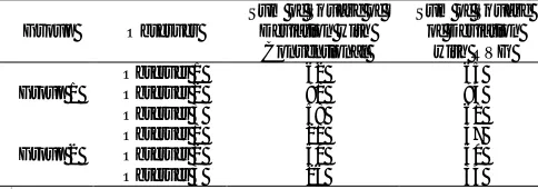
Table 1 Sum of Squares of Difference between first and
second observation by each observer for each technique
Group Observer
Sum of Square of Deviation with
Conventional
Sum of Square of Deviation
with RVG
Group 1
Observer 1 62 65
Observer 2 91 94
Observer 3 58 61
Group 2
Observer 1 21 37
Observer 2 50 50
with digital imaging when observing same digital image at two different times. Karl Pierson’s Correlation coefficient test was performed for each observer for each time in each group with respect to the standard accurate values. Higher the value of correlation indicated more closeness of the observations to accuracy. A significant increase in the accuracy among observers was observed with both techniques in second observation.(Table 2) Observations in group 1 were more or less same where a slight difference of observations was included in the group 2. Hence, the accuracy of diagnosing the presence or absence of lesion was significantly increased when each observer evaluated the same image second time.
The study determined the percentage of correct diagnosis, sensitivity, specificity and reliability of both the techniques in either of the groups. Sensitivity was high (67% and 70% in group1, 61% and 64% in group 2); however there is still a slight difference between both the techniques regarding the sensitivity with digital imaging being slightly more sensitive.(Table 3) Both the techniques showed that they have
higher specificity than sensitivity. The specificity in group 2 (95% and 98%) was to some extent more when compared to group1 (90% and 92%). The digital imaging had a better specificity as compared to conventional imaging even though it was statistically insignificant. (Table 3) The PPV (97.8% and 98%.8 in group 1, 97% and 98% in group 2), and the NPV (36.9% and 39% in group 1 and 29% and 34% in group 2)
values were correlating to their sensitivity and
specificity.(Table 3)
The value of the probability of Diagnostic accuracy was measured by Receiver Operating Characteristic (ROC) Curve and these were obtained using SPSS software (Statistical Package for the Social Sciences, IBM, USA). The values of DA are given in Table 4 and the curves plotted in graph 1 and graph 2. The DA value is the area under a ROC curve where points representing the true-positive fraction (sensitivity) and false-positive fraction (specificity) are plotted on linear probability scales. The AZ value represents the area under a straight ROC graph plotted on a binormal scale. The DA values were calculated for each observer and each imaging technique (Graph 1 and Graph 2).
The area under the curve shows slightly more area with the digital technique for both the groups (0.832 for RVG & 0.816 for CR in group 1 and 0.808 for RVG & 0.760 for Cr in group 2) (Table 5), but almost both the lines are overlapping each other in the end, which signifies that there is not much difference in diagnosing periapical lesions through digital and conventional radiographic techniques.
The mean of six values for conventional radiographic technique and direct digital radiographic technique was
Table 2 Karl Pearson Correlation Coefficients with final
diagnosis of each observer for each technique
Group Observer Conventional RVG
1stobsvn. 2ndobsvn. 1stobsvn. 2ndobsvn.
Group 1
Observer 1 0.451 0.642 0.468 0.666
Observer 2 0.424 0.629 0.436 0.654
Observer 3 0.483 0.642 0.502 0.674
Combined 0.530 0.556
Group 2
Observer 1 0.397 0.442 0.437 0.511
Observer 2 0.339 0.415 0.396 0.477
Observer 3 0.377 0.442 0.429 0.511
Combined 0.456 0.525
RVG: radiovisiography; Obsvn.: observation
‘
Graph 1 ROC Curve for both T1 (Conventional) and T2 (RVG)
Group 2 ROC Curve for both T1 (Conventional) and T2 (RVG)
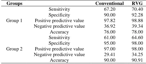
Table 3 Sensitivity, specificity and PPV, NPV and
Diagnostic accuracy of conventional and digital imaging
Groups Conventional RVG
Group 1
Sensitivity 67.20 70.40
Specificity 90.00 92.28
Positive predictive value 97.82 98.88
Negative predictive value 36.92 39.34
Accuracy 76.00 78.00
Group 2
Sensitivity 61.00 64.60
Specificity 95.00 98.00
Positive predictive value 97.00 98.00
Negative predictive value 29.41 34.25
Accuracy 90.00 90.91
Table 4 Value of the probability of accuracy was
measured by Receiver Operating Characteristic Curve
Group Observer Conventional RVG
1stobsvn. 2ndobsvn. 1stobsvn. 2ndobsvn.
Group 1
Observer 1 0.821 0.920 0.831 0.928
Observer 2 0.800 0.914 0.808 0.923
Observer 3 0.839 0.920 0.852 0.932
Combined 0.869 ± 0.055 0.879 ± 0.056
Group 2
Observer 1 0.779 0.810 0.806 0.852
Observer 2 0.735 0.792 0.778 0.832
Observer 3 0.764 0.810 0.801 0.852
Combined 0.782 ± 0.029 0.820 ± 0.030
Table 5 Mean value of CR and RVG for detecting
periapical lesions null hypothesis: true area = 0.5
Groups Test Variables Area Std. Error
Asymptomatic 95% Confidence Interval
Lower Bound
Upper Bound
Gr 1 T1 (Conventional) 0.816 0.038 0.741 0.891
T2 (RVG) 0.832 0.037 0.760 0.904
Gr 2 T1 (Conventional) 0.760 0.040 0.681 0.839
calculated, in which there is a slight increase in the mean value for DDR in the accuracy of diagnosing the periapical lesions as seen in Graph 1 and graph 2 (mean of 0.879 ± 0.056 as compared to 0.869 ± 0.055 in group 1 and 0.820 ± 0.03 to 0.782 ± 0.029 in group 2), but using Wilcoxon’s signed rank test (Table 6) the Z-value was 1.156 and 3.464 in group 1 and 2 respectively and P-value 0.248 and 0.001(significant) in group 1 and 2. These values suggest that statically there is slight significant difference found between the accuracy for diagnosing periapical lesions through digital imaging as compared to Conventional imaging. RVG was more accurate in assessing the presence of lesions as compared to absence of the lesion.
DISCUSSION
Radiographical visualization with conventional radiography has been the main stay for diagnosis the periapical lesions, since they have been introduced into the medical field in the early 20th century. In the last two decades, digital radiography has gained popularity as alternative to conventional radiography, as it has given the dentist the ability to perform radiographic examination with a significant reduction in radiation exposure up to 50% - 80%. However, the literature is not conclusive on whether the digital radiographic method is more efficient than the conventional radiographic method.17-19 The results of the present study for highintraobserver reliability are in accordance with the in-vivo studies conducted by Bohay RN (2000),20 Parihar A et al. (2010)12 and the in-vitrostudies conducted by Wallace JA et al. (2001),21 Goga and Chandler et al.(2004)22 which might be attributed to vast experience of the observers in diagnosing periapical lesions through both conventional and digital technique. Digital radiography showed higher interobserver aggrement as compared to conventional radiography. It was better with observer 1 and observer 3 as compared to observer 2. For digital radiography fair to good reliability was observed in all the three observers. Interobserver agreement varied from 0.4 to 0.6 (Group 1) and 0.3 to 0.4 (Group 2) for CR while in digital it varied from 0.4 to 0.7 (Group 1) and 0.4 to 0.5 (Group 2). Our results are in contrast to the study conducted by Tirrell et al (1996)9 where authors found a very high interobserver agreement of 85.6% because of inclusion of small as well as large periapical lesions of different sizes, which were very easy to detect by observers with any technique.
Sensitivity and specificity are routinely used and easily understood measures of diagnostic performance and they can be assessed separately unlike percent accuracy. Sensitivity is the ability of a test to correctly identify those with the disease. Specificity is the ability of a test to correctly identify those without the disease. The results of the present study were in contrast with an in-vitro studies carried out by Tirrell et al
(1996)9 and Yokota et al. (1994)7 where CR was more accurate in identifying specimens without a lesion as compared to digital radiography, thus making digital radiography more specific. The results also showed that digital imaging tends to be more sensitive and specific than conventional imaging for the diagnosis of initial periapical lesions.
For ROC curve analysis technique, our results were in accordance with an in vivostudy by Parihar et al.12 where ROC curve was used to check the diagnostic accuracy of the different techniques. The overall DA value was also found very close to our values. However, difference in the Wilcoxon’s signed rank test values in both the studies might be because of the difference in the sample sizes. In the present study of the DA values generated by ROC curve were compared in the pair wise testing. In the pair wise testing, Digital imaging was slightly better than Conventional imaging in group 2 patients as compared to group 1. Similar results were seen in the study conducted by Parihar et al.12
Most of the studies reported in literature are in-vitro studies as compared to in-vivo studies. The results of in-vitro studies can’t be compared with that of in-vivo studies as the lesions in in-vitro studies are created by a round bur or by placing acid, which do not accurately recreate the anatomical shapes of inflammatory periapical lesions. The burs often can create an image of a lesion with a well-defined round outline, possibly making detection of lesion an easier task relative to the same task in a clinical situation, like bone loss which would be more diffuse and difficult to detect. The results of the present study support those prior studies of Mistak et al.23 and Holtzmann et al.24 and wherein no differences in diagnostic accuracy between conventional and digital techniques were found in diagnosing periapical lesions.
The inconsistency in assessing and interpreting radiographic may contribute to the variation in diagnostic accuracy of identifying bony lesions. Although the intraobserver agreement was fair in our study, there was an improvement in the 2nd observation for each observer. This is because of learning tendency that occurs during each observation which results in retaining the image in the observer’s memory and comparing it with images that are subsequently viewed. Hence, in the present study, the authors meticulously followed the methodology given by Parihar et al.12 to obtain unbiased predictions. Although the design of this study cannot entirely eliminate the risk of this bias, the method of sampling and the use of the entire clinical and radiographic record in the determination of the “true” periapical status should minimize it. The present study found that CCD based images were clinically equal to Ektaspeed Plus film radiographs in the diagnosis of periapical lesions. As advances and innovations occur with digital imaging, more in vivo studies will be needed to make accurate comparisons of new imaging technologies.
CONCLUSION
Digital images had a slight advantage (negligible) in diagnostic value according to the sensitivity and specificity when compared to conventional. However, digital imaging is not superior to film based radiographs in the initial depiction of periapical bone lesions. More clinical studies are needed to determine the relative diagnostic efficacy of these imaging modalities, with respect to detection of periapical lesions.
References
1. Torabinejad M, Eby WC, Naidorf IJ. Inflammatory and
immunological aspects of the pathogenesis of human periapical lesions, J. Endod 1985; 11:479.
2. Wood N, Goaz P. Differential diagnosis of oral and maxillofacial lesions, 5th ed, USA, Mosby, 1997. p. 252-58.
Table 6 Comparing final Conventional Diagnosis with
RVG diagnosis using Wilcoxon signed Rank Test
Group Z value P value Significance
Group 1 1.155 0.248 Not significant
3. Bender IB, factors influencing the radiographic appearance of bony lesions, J Endod, 1982; 8:161. 4. Van Der Stelt PF. Filmless imaging: the uses of digital
radiography in dental practice. J Am Dent Assoc. 2005 Oct; 136(10):1379-87.
5. Gundappa M, Ng SY, Whaites EJ. Comparison of
ultrasound, digital and conventional radiography in differentiating periapical lesions. Dentomaxillofacial Radiology 2006 35:5, 326-333.
6. NicopoulouKarayianni K, Bragger U, Burgin W,
Nielsen PM, Lang NP. Diagnosis of alveolar bone
changes with digital subtraction images and
conventional radiographs- an in vitro study. Oral Surg
Oral Med Oral Pathol. Aug. 1991; 72(2):251-56.
7. Yokota ET, Miles DA, Newton CW, Brown CE (Jr).
Interpretation of periapical lesions using
RadioVisioGraphy. J Endod. Oct. 1994; 20(10):490-94.
8. Farman AG, Scarfe WC, Schick DB, Rumack PM.
Computed dental radiography: Evaluation of a new charge coupled device based intraoral radiographic system. Quintessence International 1995; 26:399-404.
9. Tirrell BC, Miles DA, Brown CE Jr, Legan JJ.
Interpretation of chemically created lesions using direct digital imaging. J Endod. 1996 Feb; 22(2):74-8.
10. Kullendorff B, Nilsson M, Rohlin M. Diagnostic
accuracy of direct digital dental radiography for the detection of periapical bone lesions: overall comparison between conventional and direct digital radiography.
Oral Surg Oral Med Oral Pathol Oral RadiolEndod. 1996 Sep; 82(3):344-50.
11. Tyndall DA, Ludlow JB, Platin E, Nair M. A
comparison of Kodak Ektaspeed Plus film and Siemens Sidexis digital imaging system for caries detection using receiver operative characteristic analysis. Oral Surg Oral Med Oral Pathol Oral RadiolEndod. 1998; 85:113-18.
12. Parihar A, Keluskar V, Bagewadi A, Shetti A.
Comparing the Accuracy in Diagnosing Periapical
Lesions by Conventional and Direct Digital
Radiography. J Indian Acad Oral Med Radiol. 2010; 22(4):185-189.
13. Forsberg J, Halse A. Radiographic simulation of a periapical lesion comparing the paralleling and the bisecting angle techniques. IntEndod J. 1994; 27:133-38.
14. Kullendorff B, Petersson K, Rohlin M. Direct digital radiography for the detection of periapical bone lesions: A clinical study. Endod Dent Traumatol Aug. 1997; 13(4):183-89.
15. Bender IB, Seltzer S. Roentgenographic and direct observation of experimental lesions in bone I. J Am Dent Assoc.1961a; 62, 152-60.
16. Saunders MB, Gulabivala K, Holt R, Kahan RS.
Reliability of radiographic observations recorded on a proforma measured using inter- and intra-observer variation: a preliminary study. IntEndod J. 2000 May; 33(3):272-8.
17. Krajczár K, Marada G, Gyulai G, Tóth V. Comparison
of radiographic and electronical working length determination on palatal and mesio-buccal root canals of extracted upper molars. Oral Surg Oral Med Oral
Pathol Oral RadiolEndod. 2008; 106:e90-3.
18. Pratten DH, McDonald NJ. Comparison of radiographic
and electronic working lengths. J Endod. 1996; 22:173-6.
19. Sanderink GCH, Huiskens R, van der Stelt PF,
WelanderUS, Stheeman E. Image quality of direct intraoral x-ray sensors in assessing root canal length. The Radio VisioGraphy, Visualix/VIXA, Sens-A-Ray, and Flash Dent systems compared with Ektaspeed films.
Oral Surg Oral Med Oral Pathol. 1994; 78:125-32.
20. Bohay RN. The Sensitivity, Specificity, and Reliability of Radiographic Periapical Diagnosis of Posterior Teeth. Oral Surg Oral Med Oral Pathol Oral Radiol
Endod. 2000 May; 89(5):639-42.
21. Wallace JA, Nair MK, Abomr D, Colaco MF, Kapa SF.
A comparative evaluation of the diagnostic efficacy of film and digital sensors for detection of simulated periapical lesions. Oral Surg Oral Med Oral Pathol
Oral RadiolEndod 2001; 92(1):93-97.
22. Goga R, Chandler NP, Love RM. Clarity and diagnostic
quality of digitized conventional intraoral radiographs.
Dentomaxillofacial Radiology 2004; 33:103-07.
23. MistakEJ, Loushine RJ, Primack PD, West LA, Runyan
DA. Interpretation of periapical lesions comparing
conventional, direct digital and telephonically
transmitted radiographic images. Journal of
Endodontics 1998 Apr; 24(4):262-66.
24. Holtzmann DJ, Johnson WT, Southard TE, Khademi
JA, Chang PJ, Rivera EM. Storage phosphor computed radiography versus film radiography in the detection of pathologic periradicular bone loss in cadavers. Oral Surg Oral Med Oral Pathol Oral RadiolEndod 1998; 86:90-97.
25. Kaffe, Gratt BM. Variations in the radiographic interpretation of the periapical dental region. I. Journal of Endodontics 1988; 14: 330-35.