Original Research Article.
Diagnostic Role of Immunohistochemistry in Mimics of
Prostatic Adenocarcinoma
Gayatri Behera
1, Lity Mohanty
2*, Sujata Pujari
3, Sambit Kumar Behera
4,
Jyoti Ranjan Behera
5, Shushruta Mohanty
11Post graduate, 2*Associate Professor, 3Assistant professor,
Department of Pathology, S.C.B. Medical College, Cuttack, Odisha, India.
4DM Resident, Department of Gastroenterology, S.C.B. Medical College, Cuttack, Odisha, India. 5Assistant Professor, Department of Paediatrics,
Kalinga Institute of Medical Sciences (KIMS), Bhubaneswar, Odisha, India.
ABSTRACT
Objectives: To study and recognize different morphologic patterns of benign mimics of prostatic adenocarcinoma and couple it with use of immunohistochemistry for correct diagnosis and avoid false positive carcinoma interpretation.
Materials and Methods: Prostatic biopsy specimens were evaluated by histopathology using haematoxylin and eosin (H/E) staining. In suspicious cases for malignant lesions, immunohistochemistry (IHC) analysis using basal cell markers
p63 and HMWCK 34βE12 were performed to confirm the final diagnosis.
Results: One hundred and ninety three prostatic biopsy specimens were received in department of pathology, 92(47.66%) were benign hyperplasia while carcinoma constituted 39(20.2%) cases. Among the remaining 62 specimens, 30 showed features of mimics while 32 specimens were suspicious of carcinoma. Of these 62 specimens, IHC analysis using basal cell markers like p63 and 34bE12 were performed. All the mimics were found negative of malignancy. But among 32 suspicious specimen of carcinoma, 8 cases were confirmed as prostate carcinoma while 24 cases were diagnosed as mimics of prostatic carcinoma.
Conclusion: In morphologically equivocal glandular
architectural patterns and cytological features,
immunohistochemistry highlighting the basal cell marker can help the pathologist to arrive to a correct diagnosis.
Key Words: Mimics, Prostate Adenocarcinoma,
Immunohistochemistry, p63, HMWCK.
*Correspondence to:
Dr. Lity Mohanty,
Associate Professor, Department of Pathology,
S.C.B. Medical College, Cuttack, Odisha, India.
Article History:
Received: 28-09-2017, Revised: 18-10-2017, Accepted: 01-11-2017
Access this article online
Website:
www.ijmrp.com
Quick Response code
DOI:
10.21276/ijmrp.2018.4.1.014
INTRODUCTION
Prostate cancer is the second most frequently diagnosed cancer in men worldwide.1 It is the most frequently diagnosed cancer among men in more developed countries, where about two-thirds
of all prostate cancer cases occur among just 17% of the world’s
male population.1 With the shift towards a greater proportion of men diagnosed with localized disease, the proportion living more than 5 years after diagnosis has increased.2 So correct diagnosis
has a definite role to play. In case of smaller biopsies, diagnosis can be difficult due to the presence of either a small focus of cancer or due to the many benign mimickers of malignancy.3
Chronic prostatitis, atrophy, partial atrophy, basal cell hyperplasia, clear cell hyperplasia, post atrophic hyperplasia, atypical adenomatous hyperplasia (adenosis), seminal vesicle, colonic mucosa, squamous metaplasia, prostatic intraepithelial lesions all can mimic the malignancy.3 False-positive cancer diagnosis,
although uncommon, may be rendered in some cases and may
lead to serious clinical, psychological and medicolegal consequences.
Immunohistochemical (IHC) markers are often used as an aid in case of suspicious lesions to reach at a diagnosis.4 The diagnosis
is aided by IHC staining for basal cell layer markers, such as p63 (nuclear marker), cytokeratin 5/6 (CK 5/6), and high molecular weight cytokeratin (CK HMW) (cytoplasmic marker) as well as prostate-‘specific’ markers.5 The loss of basal cells in prostate
carcinomas is the most important diagnostic hallmark of malignancy, and basal cell markers has been the immunohistochemical cornerstone of prostate diagnostics for more than 25 years.6
MATERIALS AND METHODS
This prospective study was conducted in the Department of Pathology and Department of Urology, Shriram Chandra Bhanja Medical College and Hospital, Cuttack, Odisha from September 2013 to October 2015.
One hundred and ninety three patients with enlarged prostate diagnosed either by Digital Rectal Examination (DRE) or Ultrasonography (USG) were included in the study. Previously diagnosed cases of adenocarcinoma of prostate were excluded. Haematoxylin and Eosin staining was performed in all biopsy samples. Paraffin blocks in suspicious cases were chosen for performing IHC using p63 and 34βE12. PathNSitu ready to use
mouse monoclonal antibody (p63→4A4 clone, CKHMW-34βE12) were utilised for this purpose.
The process of IHC included a two-step process in which the tissue is treated with an unlabelled primary antibody, followed by application of a chromogen labelled secondary antibody (directed against the primary antibody). Its main advantage was an increased sensitivity.
Antibody concentration was optimized to obtain the strongest target staining with minimal background staining. The slides then were incubated sequentially with primary antibody, biotinylated secondary antibody, avidin-biotin complex. The primary antibodies were incubated for 45 minutes at room temperature and the
secondary biotin-labelled antibody for 30 minutes for 34βE12 and
45 minutes for p63. The streptavidin-labelled streptavidin-biotin amplification method (PathNsitu) was carried out for 30 minutes, followed by peroxidase substrate/ chromogen for 34βE12 and
p63. Slides were counterstained with hematoxylin.
The basic principle, as with any other special staining method was a sharp localization of target components in the cell & tissue, based on a satisfactory signal-to-noise ratio. Amplifying the signal, while reducing non-specific background staining (noise) was the major strategy to achieve a satisfactory & practically useful result. The Primary Ab (mouse monoclonal Ab) with specificity against the Ag in the test was added to the section & the excess antibody was washed off. The labelled secondary Ab which has specificity
against an antigenic determinant present on the primary mouse Ab was then added; it serves to label sites of tissue localization of Primary Ab. To overcome masking of the antigenicity of tissues by formalin fixation antigen retrieval was done before application of antibodies to expose the masked antigens in tissues. It was performed by steaming the slides for 15 minutes in a 10-mmol/L concentration of sodium citrate buffer, pH 6.0, in a microwave oven.
Non immunological binding of the applied antibody with the tissue under evaluation and presence of endogenous peroxidase in certain tissues which result in background staining that is minimized by pretreatment of tissues with hydrogen peroxide prior to application of primary antibody which eliminates endogenous peroxidise is called as blocking.
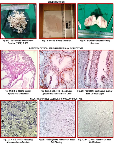
One positive control and negative control was set up with each batch of tissues undergoing IHC to validate the procedure and increase its specificity. In the present study, we used benign hyperplasia of prostate diagnosed in histopathology cases as positive control and for negative control, we used adenocarcinoma of prostate.
Differences in tissue processing & technical procedure may produce variable results. Hence controls used are fresh autopsy/surgical specimens processed in same manner as
patient’s sample. The controls slides were provided by PathnSitu
manufacturers. Presence nuclear staining is considered as positive while absence of staining is considered as negative for p63. For cytoplasmic marker 34βE12 presence of cytoplasmic staining is considered positive while negative staining indicated by absence of cytoplasmic staining.
IHC stain in all cases were evaluated independently by two expert histopathologists and in case of any discrepancy the slides were subjected to concurrent review under pentahead microscope by both of them. Positive staining in suspicious foci was considered evidence of benignity and negative staining of an entire suspicious focus was considered presumptive evidence of a malignant process as long as the morphologic features were in agreement. Statistical analysis was done in SPSS software.
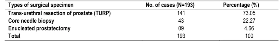
Table 1: Different Types of Surgical Specimens Received
Types of surgical specimen No. of cases (N=193) Percentage (%)
Trans-urethral resection of prostate (TURP) 141 73.05
Core needle biopsy 43 22.27
Enucleated prostatectomy 09 4.66
Total 193 100
Table 2: Distribution of Cases According To HP Diagnosis
HP DIAGNOSIS NO OF CASES (%)
Fibromusculoglandular hyperplasia 92(47.66%)
Mimics of prostatic adenocarcinoma 30(15.54%)
Carcinoma prostate 39(20.20%)
Suspicious of malignancy 32(16.58%)
Total 193(100%)
Table 3: Result of Suspicious of Malignancy Cases After IHC
FINAL DIAGNOSIS AFTER IHC ANALYSIS(n=32)
34βE12 (nuclear) P63(cytoplasmic)
Diagnosis before IHC analysis Carcinoma Mimics Carcinoma Mimics
Table 4: Comparison of the Association of IHC with Mimic and Suspicious Cases
HP DIAGNOSIS IHC RESULT TEST RESULTS
X2 =6.53 df=1 p value<0.01
POSITIVE NEGATIVE
MIMIC 30 00
Suspicious of Carcinoma 24 08
Table 5: Age Incidence in Different Mimics of Prostatic Adenocarcinoma
Age group Mimics of prostatic adenocarcinoma (%)
41-50 01 (1.85%)
51-60 12 (22.22%)
61-70 34 (62.96%)
71-80 06 (11.11%)
81-90 01 (1.85%)
Total 54 (100%)
Table 6: Presenting Features in Different Prostatic Mimics
CLINICAL PRESENTATION NO OF CASES (n=54) PERCENTAGE (%)
Retention of urine 44 81.48 %
Frequency 32 59.25 %
Urgency 14 25.92 %
Nocturia 09 16.66 %
Dysuria 01 2.63 %
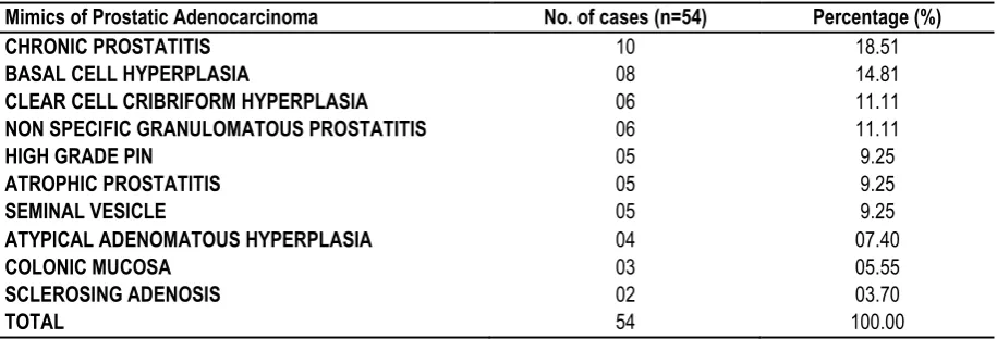
Table 7:Different Histological Mimics of Prostatic Adenocarcinoma
Mimics of Prostatic Adenocarcinoma No. of cases (n=54) Percentage (%)
CHRONIC PROSTATITIS 10 18.51
BASAL CELL HYPERPLASIA 08 14.81
CLEAR CELL CRIBRIFORM HYPERPLASIA 06 11.11
NON SPECIFIC GRANULOMATOUS PROSTATITIS 06 11.11
HIGH GRADE PIN 05 9.25
ATROPHIC PROSTATITIS 05 9.25
SEMINAL VESICLE 05 9.25
ATYPICAL ADENOMATOUS HYPERPLASIA 04 07.40
COLONIC MUCOSA 03 05.55
SCLEROSING ADENOSIS 02 03.70
TOTAL 54 100.00
Table 8: Correlation of Serum PSA with Different Mimics of Prostatic Adenocarcinoma
Range of serum PSA Mimics of Prostatic Adenocarcinoma (%)
<4 ng/ml 33(61.11%)
4-10 ng/ml 18(33.33%)
>10 ng/ml 3(5.55%)
Total 54(100%)
RESULTS
Transurethral resection of prostate (TURP) was the most common surgical procedure performed and hence TURP bits were the commonest type of prostatic specimen received which is about 73.05% followed by core needle biopsy which was 22.27% (Table 1). The core needle biopsy is slowly gaining popularity recently. Prostatectomy specimen comprising of only 4.66% of the total was the least common procedure resorted to.
Out of total 193 cases of clinically diagnosed BHP, after histopathological analysis using H/E stain, 92 (47.66%) cases are confirmed as fibromusculoglandular hyperplasia (Table 2), 30(15.54%) cases as mimics of prostatic adenocarcinoma, 39(20.20%) cases as carcinoma prostate. 32(16.58%) cases could not be resolved using H/E stain and were interpreted as
suspicious of malignancy and were subjected to immunohistochemical analysis. These 32 suspicious cases were subjected to immunohistochemical analysis using both p63 and HMWCK 34βE12. In 8 cases, both p63 and HMWCK 34βE12
were entirely negative in the atypical focus while positive in the adjacent surrounding benign glands, supporting the diagnosis of prostate carcinoma. Of the remaining 24 cases, both markers showed positive staining for basal cell layers confirming the diagnosis of the mimics of prostatic carcinoma. (Table 3)
Out of total 54 cases of prostatic mimics biopsies studied, 62.96% of cases were in the 7th decade & 22.22% of cases were in the 6th
decade followed by 11.11% in the 8th decade and only 1.85%
cases in the 5th and 9th decade (Table 5).
Overlapping of clinical features is observed in most of the cases studied. The usual presenting features were retention of urine, followed by frequency, nocturia, urgency, dysuria, etc. Retention of urine was the commonest i.e. 81.48 % of cases (Table 6).
Being the most common mimic, chronic prostatitis comprising of 10 (18.51 %) cases (Table 7). This is followed by basal cell
hyperplasia 8 (14.81%) cases, clear cell cribriform hyperplasia and nonspecific prostatitis each of 06 (11.11%) cases, high grade PIN, atrophic prostatitis and seminal vesicle each 5 (09.25%) cases, atypical adenomatous hyperplasia(adenosis) comprises 4 (7.40%) cases while colonic mucosa 3 (5.55%) and sclerosing adenosis being the least common one with 02 (03.70%) cases. From the 54 cases of mimics studied, 61.11% of cases had a serum PSA below <4 ng/ml & 33.33% of cases had a serum PSA in range of 4-10 ng/ml and only 5.55% had serum PSA >10ng/ml (Table 8).
GROSS PICTURES
Fig 1A: Transurethral Resection Of
Prostate (TURP) CHIPS Fig.1B: Needle Biopsy Specimen Fig 1C: Enucleated Prostatectomy Specimen
POSITIVE CONTROL- BENIGN HYPERPLASIA OF PROSTATE
Fig. 2A: H & E (100X): Benign
Hyperplasia Of Prostate Fig. 2B: 34bE12(400X) : Continuous Cytoplasmic Stain Of Basal Layer Fig. 2C: P63(400X): Continuous Nuclear Stain Of Basal Layer
NEGATIVE CONTROL- ADENOCARCINOMA OF PROSTATE
Fig. 3A: H & E (400X): Infiltrating
BASAL CELL HYPERPLASIA
Fig.4A- H & E (400X): Proliferation
Of The Basal Cell Layer Fig.4B- P63(100X): Positive Staining Of Basal Cell Nuclei Fig.4C- P63 (400X): Positive Staining Of Basal Cell Nuclei
Fig.4D: 34bE12(100X):Intense And Diffuse
Cytoplasmic Staining Of Basal Cell Layer Fig.4E: 34bE12 (400X): Intense And Diffuse Cytoplasmic Staining Of Basal Cell Layer ADENOSIS
Fig.5A- H & E(400X): Adenosis Showing
Proliferation Of Small Glands Fig.5B- P63 (100X): Positive Basal Cell Nuclear Staining Fig.5C- 34bE12 (100X): Positive Basal Cell Cytoplasmic Staining SQUAMOUS METAPLASIA
Fig 6A: H&E: Squamous Metaplasia Mimicking Squamous Carcinoma
Fig.6B- 34bE12 (400X): Positive Basal Cell Cytoplasmic Staining
SCLEROSING ADENOSIS
Fig.7A: H & E(100X): Proliferation Of Both Glands And Stroma Mimicking High Grade Invasive Adenocarcinoma
Fig.7B: 34bE12 (100X): Positive Basal Cell Cytoplasmic Staining
Fig.7C: P63 (100X): Positive Basal Cell Nuclear Staining
CLEAR CELL CRIBRIFORM HYPERPLASIA
Fig.8A- H & E (400X): Complex Papillary Cribriform Hyperplasia Of Clear Cells
Involving Acini
Fig.8B- 34bE12 (400X): Positive Basal
Cell Cytoplasmic Staining Fig.8C- P63 (400X): Positive Basal Cell Nuclear Staining
ATROPHIC PROSTATITIS
Fig.9A H & E: Atrophic Acini Lined By Small, Dark Nuclei, Scanty Cytoplasm.
Fig.9B- P63 (100X): Positive Basal Cell Nuclear Staining
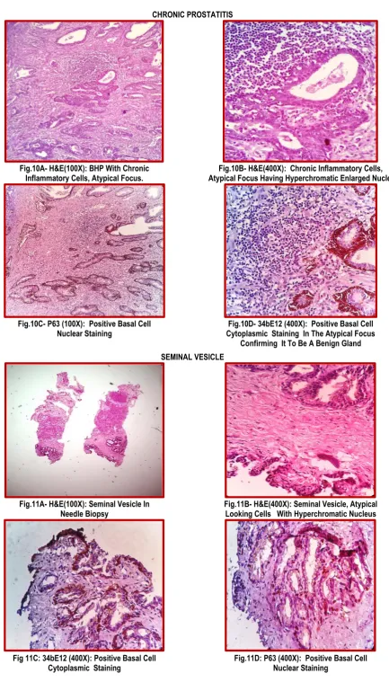
CHRONIC PROSTATITIS
Fig.10A- H&E(100X): BHP With Chronic
Inflammatory Cells, Atypical Focus. Atypical Focus Having Hyperchromatic Enlarged Nuclei Fig.10B- H&E(400X): Chronic Inflammatory Cells,
Fig.10C- P63 (100X): Positive Basal Cell Nuclear Staining
Fig.10D- 34bE12 (400X): Positive Basal Cell Cytoplasmic Staining In The Atypical Focus
Confirming It To Be A Benign Gland SEMINAL VESICLE
Fig.11A- H&E(100X): Seminal Vesicle In
Needle Biopsy Fig.11B- H&E(400X): Seminal Vesicle, Atypical Looking Cells With Hyperchromatic Nucleus
Fig 11C: 34bE12 (400X): Positive Basal Cell
COLONIC MUCOSA
Fig.12A- H&E (100X): Colonic Mucosa In Needle Biopsy Fig.12B- H&E(400X): Prostate Tissue And
Colonic Mucosa In Needle Biopsy
Fig.12C- 34bE12 (400X): Negative Basal Cell
Cytoplasmic Staining Fig.12D- P63 (100X): Negative Basal Cell Nuclear Staining
LOW GRADE PROSTATIC INTRAEPITHELIAL LESION
Fig.13A- H&E(400X): Low Grade Pin With Epithelial Cell Crowding &
Stratification
Fig.13B- 34bE12 (400X): Continuous Basal Cell Cytoplasmic Staining
Fig.13C- P63 (400X): Continuous Basal Cell Nuclear Staining
HIGH GRADE PROSTATIC INTRAEPITHELIAL LESION
Fig.14A- H&E(400X): High Grade Prostatic Intraepithelial Lesion, Cribriform Pattern Of Architecture, Enlarged Dark Nuclei, Prominent Nucleoli.
DISCUSSION
In our study, BHP constitutes almost half number of cases while that of mimics and carcinoma constitutes about one fourth each which is similar to the study by Angurana et al.7 In our hospital, transurethral resection of prostate (TURP) was the most common surgical procedure performed and hence TURP bits were the commonest type of prostatic specimen received which is about 73.05% followed by core needle biopsy which was 22.27% which is becoming more popular recently. Enucleated prostatectomy specimen comprising of only 4.66% of the total is performed mostly in very large glands and the cases are mostly coming from periphery and now-a-days it is being gradually replaced by the core needle biopsies.
Out of total 193 prostatic biopsy specimen, after histopathological analysis using H/E stain, 32(16.58%) cases could not be resolved using H/E stain and were diagnosed as suspicious of malignancy.
After IHC, both p63 and HMWCK 34βE12 were entirely negative in 8 out of 32 specimens with atypical focus supporting the diagnosis of prostate carcinoma.
Of the remaining 24 cases, both markers showed positive staining for basal cell layers confirming the diagnosis of the mimics of prostatic carcinoma. We observed no differences in staining pattern between the two nuclear and cytoplasmic markers. This study does not correlates with the study of Wu et al26 who observed that for diagnosing prostate carcinoma in the needle biopsies, p63 is as specific and sensitive as 34βE12 and therefore
can be used as a complementary basal cell-specific stain for
34βE12 in difficult cases. This may be due different manufactures
of the antibody and methods of antigen retrieval during immunohistochemical staining. We used the antibody from PathnSitu while Wu et al used that from DAKO.
Shah et al 27 used Laboratory vision anti-p63 antibody and a
microwave for antigen retrieval and observed that 34βE12 and
p63 are highly specific for basal cells and p63 is more sensitive
than 34βE12 in staining benign basal cells, particularly for TURP specimens, offering slight advantage over 34βE12 in
diagnostically challenging cases.
Mimics have a peak age incidence i.e. the 7th decade. The results
of our study tallies with Ro et al8 who stated that most of the mimics of cancer occurs in the same age as prostatic carcinoma. But the results of our study oppose with Harbitz et al 9 and Bostwick et al10. They noted a rise in incidence after the age of 40 years. This is due to lower life expectancy and negligent attitude towards very old people.
There was overlapping of clinical symptoms in most of the cases. They are retention of urine in 81.48% of cases, frequency of micturition in 59.25% of cases, urgency in 25.92%, nocturia in 16.66%, dysuria present in 2.63%. Similar findings were suggested by Steers et al11 andAnderson et al12. J.Y. Ro et al2 also noted that mimics do not have a specific clinical manifestation.
Being the most common mimic, chronic prostatitis comprising of 18.51% cases and is followed by basal cell hyperplasia in 14.81% cases. Our study is similar to Mittal et al15 who studied 185 consecutive prostate specimens and found chronic prostatitis was
frequently encountered (58%) cases associated with BHP. But
there were differences with Billis A et al16who concluded partial atrophy was the most common mimic studied. In our study, prostatic atrophy was found in only 9.25 % cases. As prostatic
atrophy is located in peripheral zone it is less common in TURP specimens which may be the reason for low percentage.
We got all cases of basal cell hyperplasia in TURP specimens only and none were found in needle biopsies which tallies with Hosler et al17 who recognized basal cell hyperplasia as a well-recognized entity in TURP specimens but an uncommon finding in prostatic needle biopsies. Nonspecific granulomatous prostatitis in our study was found in 6 (11.11%) cases. Only one of these cases
had serum PSA less than 4ng/ml while rest all had serum PSA ≥4
ngm/ml. This study is in correlation with the study of Pavlica P et al18 who studied 20 cases of nonspecific granulomatous prostatitis
and found that 19 cases had serum PSA level ≥4 ngm/ml & only
one case had less than 4(mean value was 13.3 ngm/ml). Another rare entity sclerosing adenosis which was seen in 2 (3.70%)
cases, compared with Sakamoto et al19who reviewed sections of
prostate from 263 patients and found only 5 cases of sclerosing adenosis with an incidence of 1.9%. This discrepancy is due to less number of cases in our study.
Clear cell cribriform hyperplasia had an incidence of 6(11.11%) cases in our study. All the 6 cases had positive basal cell layer staining with both the markers which correlates with the study of
Elizabeth et al20 who studied 15 cases of CCCH by
immunohistochemistry.
A basal cell layer was demonstrated in all 15 CCCHs by the use of the 34 beta E12 anti-high-molecular-weight keratin antibody that reacts with the basal cells but not with the acinar cells of the prostate. Atypical adenomatous hyperplasia which accounted for 7.40% cases in our study correlated with Helpap B et al21, who stated the incidence of AAH in TURP material between 4 to 15%.High grade PIN was found in 5(09.25%) cases in our study.
This figure tallies with Troncoso et al22 who noted 17.9% of PIN
without carcinoma.
But according to Goudin et al23, Pacelli A et al24 and Bostwick DG et al25the frequency of high grade PIN in needle biopsies range to 5% to 16% and in TURP specimens between 2.3-4.2%. The raised percentage in our study is due to inclusion of mostly mimics in our study and exclusion of other benign entities and frank carcinoma from our study.
In our study, 61.11% had a serum PSA below <4 ng/ml & 33.33% had a serum PSA in range of 4-10 ng/ml. Only 5.55% cases had serum PSA >10ng/ml. This result can be correlated with Robbins
et al13 who had stated serum PSA can be elevated in conditions
like chronic prostatitis, digital rectal examinations, transrectal ultrasonography, older age groups and in patients with enlarged hyperplastic prostate glands. Akhter et al14found higher levels of PSA (>20 ngm/ml) in 57.1% of patients of BHP with chronic prostatitis. Our study correlates with them, 2(50%) out of 10 cases of chronic prostatitis had serum PSA (>20ngm/ml).
CONCLUSION
REFERENCES
1. Torre LA, Bray F, Siegel RL, Ferlay J, Lortet‐Tieulent J, Jemal
A. Global cancer statistics, 2012. CA: a cancer journal for clinicians. 2015 Mar 1;65(2):87-108.
2. Altekruse SF, Kosary CL, Krapcho M, Neyman N, Aminou R, Waldron W, Ruhl J, Howlader N, Tatalovich Z, Cho H, Mariotto A.
SEER cancer statistics review, 1975–2007. Bethesda, MD:
National Cancer Institute. 2010 Aug 1;7.
3. Shen SS, Ro JY. Benign Mimickers of Prostate Cancer. Advances in Surgical Pathology: Prostate Cancer. 2012 Mar 14;1:59.
4. Farinola MA, Epstein JI. Utility of immunohistochemistry for α -methylacyl-CoA racemase in distinguishing atrophic prostate cancer from benign atrophy. Human pathology. 2004 Oct 31;35(10):1272-8.
5. Kusumi T, Koie T, Tanaka M, Matsumoto K, Sato F, Kusumi A, Ohyama C, Kijima H. Immunohistochemical detection of carcinoma in radical prostatectomy specimens following hormone therapy. Pathology international. 2008 Nov 1;58(11):687-94.5 6. Zink D, Fischer AH, Nickerson JA. Nuclear structure in cancer cells. Nature reviews cancer. 2004 Sep 1;4(9):6.
7. Neha Angurana in 2014, Ludhiana, India International Journal of Basic and Applied Medical Sciences ISSN: 2277-2103 An Open Access, International Journal http://www.cibtech.org/jms.htm 2014 Vol. 4 (3) September-December, pp. 163-167.
8. J.Y. Ro et al : Tumors an tumorous condition of the male genital tract. Diagnostic Histopathology of tumors, Edited by Christopher D.M. Fletcher, 2nd Edition, Vol. 1 2000.
9. Harbitz TB, Hugen OA: Histology of the prostate in elderly men. A study in an autopsy series. Acta. Patho. Microbial. Scand section A, 80:756-768,1972.
10. Bostwick DG, Cooner WH, Denis L et al : The association of benign prostate hyperplasia and cancer of the prostate, cancer 70, 291-1992.
11. Steers WB, Zorn B: Benign Prostate Hyperplasia Dis Mon. 41:437,1995.
12. Anderson’s pathology Edited by Ivan Damjonov and james Linder, 10th Edition, 1996, (C.V. Mosby Company).
13. Robbins and Cotrans Pathologic basis of disease edited by Kumar, Abbas and Frausto, 7th edition, 2004.
14. Rukhsana Akhter1 , Ruby Reshi1 , Zubair Ahmad Dar2 et al , Srinagar, India. academicjournals/IJMMS.Vol. 6(3), pp. 87-91, March 2014. 1014.
15. Mittal BV, Amin MB, Kinare SG. Spectrum of histological lesions in 185 consecutive prostatic specimens. Journal of postgraduate medicine. 1989 Jul 1;35(3):157.
16. Billis A. Prostatic atrophy: clinicopathological significance. International braz j urol. 2010 Aug;36(4):401-9.
17. Hosler GA, Epstein JI. Basal cell hyperplasia: an unusual diagnostic dilemma on prostate needle biopsies. Human pathology. 2005 May 31;36(5):480-5.
18. Pavlica P, Barozzi L et al of Italy 2005. Nonspecific granulomatous prostatitis. Ultraschall Med. 2005 Jun;26(3):203-8. 19. Sakamoto N, et al 1991 Sclerosing adenosis of the prostate. Histologic and immunohistochemical analysis, Am J Surg Pathol. 15:660-667.
20. Elizabeth E. Frauenhoffer, MD, et al. : Clear cell cribriform hyperplasia of the prostate. Am J Clin Pathol 1991 ; 95 : 446-453. 21. Helpap B: Observation on the number, size and localization of nucleoli in hyperplastic and neoplastic prostatic disease, Histopathology 13:203,1998.
22. Troncoso et al 1989 Prostate intraepithelial neoplasia and invasive prostatic adenocarcinoma in cystoprostatectomy specimens. Uro. 34:52-56.
23. Goudin et al 1997. Incidence and clinical significance of high grade prostatic intraepithelial neoplasia in TURP specimens Uro. 49:558-563.
24. Pacelli A et al 1997, clinical significance of high grade prostatic intraepithelial neoplasia in TURP specimens. Urology. 50:355-359.
25. Bostwick DG et al 1995, The incidence of high grade prostatic intraepithelial neoplasia in needle biopsies. J Urol. 154:1791-94. 26. Wu, Howard Her-Juing, Lapkus, Odeta, Corbin, Mykim HT ASCP. Comparison of 34[beta]E12 and P63 in 100 Consecutive Prostate Carcinoma Diagnosed by Needle Biopsies. Applied Immunohistochemistry & Molecular Morphology Issue: Volume 12(4), December 2004, pp 285-289
27. Shah, Rajal B, Zhou, Ming ; LeBlanc, Michele B.S.; Snyder, Matthew et al : Comparison of the Basal Cell-Specific Markers, 34[beta]E12 and p63, in the Diagnosis of Prostate Cancer. The American Journal of Surgical Pathology Issue: Volume 26(9), September 2002, pp 1161-1168
[
Source of Support: Nil.
Conflict of Interest: None Declared.
Copyright: © the author(s) and publisher. IJMRP is an official
publication of Ibn Sina Academy of Medieval Medicine &
Sciences, registered in 2001 under Indian Trusts Act, 1882. This is an open access article distributed under the terms of the Creative Commons Attribution Non-commercial License, which permits unrestricted non-commercial use, distribution, and reproduction in any medium, provided the original work is properly cited.
Cite this article as: Gayatri Behera, Lity Mohanty, Sujata Pujari, Sambit Kumar Behera, Jyoti Ranjan Behera, Shushruta Mohanty. Diagnostic Role of Immunohistochemistry in Mimics of Prostatic
Adenocarcinoma. Int J Med Res Prof. 2018 Jan; 4(1):61-70.