COMPARATIVE EVALUATION OF EFFICACY OF PHYSICS FORCEPS AND CONVENTIONAL
EXTRACTION FORCEPS IN EXTRACTION OF UPPER MOLARS-
A RANDOMISED CONTROLLED TRIAL
Ramakrishna Shenoi., Sapna K. Vadera and Nikhil Moriwal
Department of Oral and Maxillofacial Surgery, V.S.P.M’s Dental College and Research Centre, LataMangeshkar Hospital, Digdoh hills, Hingna road, Nagpur, Maharashtra
A R T I C L E I N F O A B S T R A C T
Introduction-Controlled force and finesse are required for simple tooth extraction. Various technological advances are available to improve outcome for patients with the aim of atraumatic extraction. Physics forceps is uniquely designed in such a way that the "bumper" on the buccal side will act as a fulcrum while flat palatal beak will be the effort arm. Thus it acts as a 1stclass lever providing a mechanical advantage which makes it very efficient. Unlike conventional forceps, physics forceps make one point contact with the tooth surface which reduces trauma at the surgical site and consequently the pain and inflammation which results in post-operative phase. Trial design-A randomized controlled, single-blind, prospective trial was carried out in the Dept. of OMFS and outcome variables (operating time, incidence of fractured alveolus and root fracture, inflammatory complications including post-operative pain) were compared. Method- Patients requiring maxillary molar extraction were divided into two groups, and extraction was carried out using Physics forceps and Conventional extraction forceps. Result-Statistically significant difference was noted in operative time and post-operative pain, both being less in physics forceps group.Conclusion- Physics forceps with a definite learning curve offers the clinician unique opportunity to undertake conventionally difficult extractions atraumatically so as to maintain the alveolar height thus facilitating immediate prosthetic rehabilitation.
INTRODUCTION
Background-Exodontia is the most common/routine surgical procedure to be performed in clinical dentistry, with a history which dates back to the days of Aristotle1 (384-322 BC). Simple tooth removal demands a controlled amount of force clubbed with finesse for delivering a tooth as atraumatically as possible.2 The conventional extraction forceps has undergone many design changes each one offering more advantage over the other. It basically has two long handles working as 1st class levers and united by a hinge acting as fulcrum to provide optimal mechanical advantage.
The classic design of extraction forceps sometimes may produce forces more than required and may result in excessive damage to the dentoalveolar housing. The stereotypical method of extraction may also lead to significant effects on ridge after healing.3,4 The increasing need of atraumatic tooth extraction for multitude of reasons such as lesser post-operative pain, inflammation, easier return to normalcy as well as orthodontic reasons and immediate implant placement
has led to the emergence of numerous designs and vis-a-vis - Physics forceps, Periotomes, Proximators, Powertomes, and Benex Extractors.5
Dr. Richard Golden (2004) has been credited for his simplistic yet revolutionary design of Physics forceps.6 The sweeping new concept of Physics forceps working on class 1 lever mechanism, provides a biomechanical advantage over conventional extraction forceps. The design of forceps has 2 handles; one connected to the bumper, placed at the level of mucogingival junction on the buccal / labial surface, acts as a fulcrum, which is the exclusive design providing advantage. The 2nd handle in the form of beak is positioned on the palatal/ lingual aspect in the sulcus at a lower level.7 Unlike the grasping movement of conventional extraction forceps, the handles are rotated as a single unit with a constant and fluid rotational force with wrist movement only. Once the tooth is luxated in the socket, it can be easily removed with extraction forceps.
This design is advantageous as chances of root fractures diminish while the buccal bone height is maintained. It is helpful in immediate implant placement or any other prosthetic rehabilitation.8,9
ISSN: O: 2319-6475, ISSN: P: 2319 – 6505, Impact Factor: SJIF: 5.995
Available Online at www.journalijcar.org
Volume 6; Issue 8; August 2017; Page No. 5467-5471
DOI: http://dx.doi.org/10.24327/ijcar.2017.5471.0729
Article History:
Received 16th May, 2017 Received in revised form 25th June, 2017 Accepted 6th July, 2017 Published online 28th August, 2017
Key words:
Physics Forceps; Extraction; Maxillary molars;
Research Article
Copyright©2017 Ramakrishna Shenoi et al. This is an open access article distributed under the Creative Commons Attribution License, which permits unrestricted use, distribution, and reproduction in any medium, provided the original work is properly cited.
*Corresponding author: Ramakrishna Shenoi
Randomised Controlled Trial
The purpose of the present study was to compare the efficacy of Physics forceps and Conventional extraction forceps in extraction of upper molars.
The objectives were to compare the Primary outcome including operative complications (incidence of incomplete removal of root or fractured alveolus) and duration of procedure,and Secondary outcome which includes inflammatory complications (dry socket, delayed healing,post-operative pain and infection) between Physics forceps and Conventional extraction forceps.
METHODS
Trial has been successfully submitted for review under the Clinical Trials Registry- India with trial acknowledgement number- REF/2017/01/013185.
Trial design and sample size-A prospective, parallel, single blinded randomized control trial was carried out in the department. 64 patients visiting the outpatient department of Oral and Maxillofacial surgery at a tertiary care setting were included in the study based on the inclusion and exclusion criteria. The allocation ratio was 1:1.
Material
Diagnostic instruments.
2 ml syringe with 25 G short needle.
2% lignocaine HCl with adrenaline 1:2,00,000.
Physics forceps for Maxillary molar
extraction.(figure.1 and 2)
Standard exodontia armamentarium for maxillary molar extractions.
Stop Watch.
Inclusion criteria
Patients of either sex, who can be assessed till 7th post-operative day.
Age group of 18-60 years.
Maxillary molar teeth indicated for extraction under
local anaesthesia with sound tooth structure of at least 3mm above gingival margin and 2 or more unflawed surface.
Patient in good health and not taking any
antimicrobials and medication that alters pain perception.
Exclusion criteria
Medically compromised patients.
Patient with known allergy to local anesthetic agent.
Mentally challenged patients.
Uncooperative & Unwilling patients.
Patient unable to give informed consent.
Procedure- The patients were categorized into two groups of 32 patients each. In group A patients, maxillary molars were extracted using conventional extraction forceps and in group B, physics forceps was used for extraction.
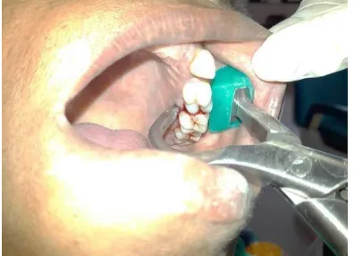
After explaining the procedure to the patient and taking consent, local anaesthesia with adrenaline 1:2,00,000 concentration was used to give PSA and GP nerve block. Buccal infiltration was used additionally whenever required. Extraction using conventional forceps was done in a traditional manner after proper reflection of the gingiva and use of elevator whenever required.(figure.3)
In Group B patients Physics forceps was used in the underneath manner.
1. The two beaks of the forceps were separated by opening the handles wide apart. The bumper which is guarded by the bumper guard (latex free surgical grade plastic) is positioned buccally at the level of mucogingival junction, right angled to the long axis of the tooth to be extracted.(figure 4)
2. The beak is placed on the palatal aspect as deep in the sulcus as possible to get a strong grip over the root surface which is very crucial for the removal of the entire tooth.
3. The beak is positioned to give a gentle and constant force directed towards the bumper. Care has to be taken to avoid compressing the handles, till the tooth starts becoming loose in the socket.
Fig 1 Armamentarium for extraction using Physics Forceps
Fig 2 Maxillary Molar Physics Forceps
A: Left Side; B: Right Side
4. Once the tooth gets luxated it is easily delivered using conventional extraction forceps.
In both the groups, after extraction of tooth socket was compressed using digital pressure and gauze was placed instructing the patient to bite on it for 45 minutes. Haemostasis was achieved and post-operative instructions and medications were given.
Primary Outcome
Duration of Extraction (Time required for extraction)
The time required for extraction was counted from the commencement of the extraction procedure (reflection of flap and/or use of elevator was included) till the time the tooth was delivered out of the socket (Measured in seconds).
Operative complications
Fracture of alveolar bone and incomplete removal of tooth root (present=1, absent=0).
It was assessed by an observer who was blinded regarding the type of forceps being used.
Secondary Outcome
Inflammatory complications
These include pain, delayed healing, dry socket and post-operative infection, which were assessed on 1st, 3rd and 7th post-operative day.
Pain- Pain was assessed by using Visual Analog Scale with ‘0’ indicating no pain and ‘10’ indicating maximum possible pain.
Extraction was also rated using Verbal response scale. The patients were queried regarding the procedure being acceptable or non-acceptable as well as if the pain was less than expected, as expected or more than expected.
Delayed Healing- Incomplete soft tissue coverage till 21 days post extraction was considered as delayed healing.
Erythema, Swelling, Pus discharge and Pain if present were considered to be signs of infection.
Sample Size- Based on the previous studies and using Open EPI system, a sample size of 64 was selected with the allocation ratio of 1:1.
Randomisation- Sampling was done using simple random sampling method.
Allocation method- Patients were assigned into groups based on the computer generated block randomisation. 2 codes were used, each denoting the type of procedure carried out. Based on all the possible sequencing of codes 4 blocks were generated. The implementation of the whole process including generation of allocation sequence, enrolment and assignment of participants into groups was carried out by the operator performing the procedure.
Blinding- The trial was single blinded; all the participants were blinded from the commencement of the trial till the completion.
Data collection and Statistical analysis
All the data was collected on scan-able forms.
Time required for tooth extraction and pain on VAS were presented as Mean± SD. They were compared using independent t- test. P < 0.05 was considered as statistically significant. Categorical variables (incomplete removal of root, fracture of alveolar bone, dry socket, delayed healing, postoperative infection and acceptance of procedure were expressed in actual numbers and percentages. They were compared using Pearson’s chi- square test. Statistical software STATA version 13.0 was used for data analysis.
RESULTS
64 patients were included in the studyand were assigned into two groups of 32 patients each. In group A, 32 patients underwent extraction of maxillary molars using conventional extraction forceps. In group B, 31 patients underwent extraction of maxillary molars using physics forceps. In 1 patient of group B procedurewas stopped for few minutes as the patient experienced syncope and was excluded from the trial. For all the patients follow-up was done on 3rd and 7th post-operative day. 2 patients in group A and 1 patient in group B did not turn up and were lost in follow-up. 30 patients in each group were analysed for final outcomes.
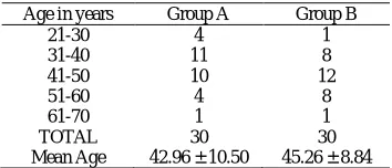
The mean age in Group A was 42.96 years while group B was 45.26 years.(Table 1) In the former group, there were 14 (46.66%) males and 16 (53.33%) females, while in the lattergroup, there were 16 (53.33%) males and 14 (46.66%) females.(Table 2)
Mean time required for extraction in Group A patients was 122.13 seconds with SD of 87.78 and in group B it was 40.46 seconds with SD of 24.10. Effect size was 0.5357, confidence interval was 95% and p value was <0.0001 and the result was highly significant.(Figure 5).
Table No 1 Age wise distribution of patients
Age in years Group A Group B
21-30 4 1
31-40 11 8
41-50 10 12
51-60 4 8
61-70 1 1
TOTAL 30 30
Mean Age 42.96 ± 10.50 45.26 ± 8.84
Table No 2 Sex wise distribution of patients
Sex Group A Group B
Male 14 16
Female 16 14
TOTAL 30 30
Fig 4 Extraction of Maxillary Molar (Left) using Conventional
Randomised Controlled Trial
There were cases of incomplete root removal. 6 in group A and 2 in group B. Absolute and relative effect size were 0.33 and 0.285 respectively, confidence interval was 95% and p value was 0.129, and the result was statistically non-significant (Table 3). 4 cases of fractured alveolar bone were reported, 3 in group A and 1 in group B and the result was statistically non-significant. Absolute and relative effect size were 0.33 and 0.3103 respectively, confidence interval was 95% and p value was 0.612 (Table 4).
The mean post-operative pain as measured on VAS scale in group A was 3.5 ±1.40 on 1st POD and 0.56±0.72 on 3rd POD. In group B it was 2.23±1.33 on 1st POD and 0.06±0.23 on 3rd POD. None of the patient had pain on 7th POD. Effect size for 1st and 3rdPOD were 0.421 and 0.4236 respectively, confidence interval was 95% and p value for 1st POD, 3rd POD were 0.0007 and 0.0008 respectively, both of which were highly significant statistically (Figure 6).
Pain as measured on VRS was less than expected in 14 patients of group A and 22 patients of group B, as expected in 14 patients of group A and 8 patients of group B and more than expected in 2 patients of group A. None of the patient
had more than expected pain in group B. p value was 0.0206 and was highly significant (Figure 7). The acceptance of procedure was 100% in both the groups (Table 5)
Dry socket was noted in 1 patient of group A, (absolute and relative effect size-0.33 and 0.3224 respectively, confidence interval was 95%) post-operative infection was noted in 2 patients of group A, (absolute and relative effect size-0.2 and 0.1869 respectively, confidence interval was 95%) and delayed healing was not present in any patient and the results were not significant (Table 6, 7, 8 respectively).
DISCUSSION
Conventional forceps have been the mainstay of exodontia since long, however there is a risk of mutilation in the existing pattern of forceps.
The after effects of extraction with routine forceps may vary from Paltry of gingival trauma to extensive damage to interdental or alveolar bone crest.10 This may lead to dry socket, delayed healing, post-operative pain and infection. All this may result in post-operative inconvenience and may also cause future prosthetic rehabilitation difficult.11
To overcome these drawbacks various techniques have been evolved over the period of time. Physics forceps is one such Table No 3 Incomplete removal of root
Incomplete removal of root Group A Group B
Yes62 No2428 p-value0.129,NS*
*Not Significant
Table No 4 Fractured alveolus
Fractured alveolus Group A Group B
Present 31
Absent 2729
p-value0.612,NS*
*Not Significant
Fig 6 Post-operative pain on VAS after extraction of tooth
*POD- Postoperative Day
Physics Forceps 0
1 2 3 4
1st
POD POD3rd 7th POD
Physics Forceps Conventional Forceps
Fig 7 Pain as measured on VRS after extraction
Table No 5 Acceptance of Procedure
Acceptance of
Procedure Group A Group B
Acceptable3030 Not Acceptable00
p-value--
More than Expected As Expected
Less than Expected 0
5 10 15 20 25
More than Expected As Expected Less than Expected
Fig 5 Mean time (seconds) required for tooth extraction
Physics Forceps Conventional Forceps 0
50 100 150
MEAN SD
Physics Forceps
Conventional Forceps
Table No 7 Post-operative infection
Post-operative infection Group A Group B
Present 20
Absent 2830
p-value0.150, NS*
*Not Significant
Table No.6 Dry socket
Dry socket Group A Group B
Present 10
Absent 2930
p-value1.000,NS*
* Not Significant
Table No.8 Delayed healing
Delayed healing Group A Group B
instrument that has shown promising results in terms of less trauma, greater speed of extraction and more convenience.
The advantage of physics forceps lies in its bio mechanics with 1st class lever mechanism which allows stress distribution without any wrenching or compressing force.12
The bumper which is placed over the gingiva allows the force to be spread over a larger area thus preserving the cortical plates; the tooth gets luxated out easily without crumbling and with minimal trauma to the investing tissues.13 Mechanical advantage offered by Physics forceps leads to lesser incidence of root and alveolar bone fracture.5
In our study,the mean time required for extraction in group A was 122.13 seconds and group B was 40.46 seconds (p<0.0001) this result was highly significant. This can be attributed to the mechanics of Physics forceps, once mastered it is easy to carry out repeat procedure.
The pain as measured on 1st and 3rd post-operative day (p= 0.0007, and 0.0008 respectively) was more in group A than group B and the result was highly significant.
Our study was in consensus that the atraumatic removal of tooth with a constant force could be one of the reasons for less pain at the surgical site especially in the early post-operative period.14
The Physics forceps extraction is an easy to master technique with a shallow learning curve. This can be attributed to the movement of the wrist, which is more ergonomic and leads to less traumatic experience of the patient physically and psychologically whilst it reduces the chair side time and increases the confidence of the operator. However it has a limited use in extraction of grossly carious tooth and root pieces.
Physics forceps thus offers the clinician an easy method for tackling difficult extractions as well as offering the benefits of minimally traumatic procedure and maintaining alveolar height facilitating early prosthetic rehabilitation.
Physics forceps thus constitute a significant inclusion in the armamentarium for extraction.
Acknowledgement
Conflicts of interest: No conflicts of interest. No external funds were required.
The ethical clearance was taken from Institutional Ethics Committee VSPM’S DCRC, Nagpur and the research has been conducted in full accordance with the World Medical Association Declaration of Helsinki.
Consent: Signed, written and printed informed consent was taken from all the patients participating in the study.
References
1. Misch C, Perez H. Dentistry Today. August 2008; 27(8): 1-3.
2. Dym H, Weiss A. Exodontia: tips and techniques for
better outcomes. Dental Clinics of North America. 2012 Jan 31; 56(1):245-66.
3. White J, Holtzclaw D, Toscano N. Powertome assisted
atraumatic tooth extraction. J Implant AdvClin Dent.
2009; 1:6.
4. Tavarez RR, Dos Reis WL, Rocha AT, Firoozmand
LM, Bandéca MC, Tonetto MR, Malheiros AS.
Atraumatic extraction and immediate implant
installation: The importance of maintaining the contour
gingival tissues. Journal of international oral health:
JIOH. 2013 Dec; 5(6):113.
5. El-Kenawy MH, Ahmed WM. Comparison Between
Physics and Conventional Forceps in Simple Dental
Extraction. Journal of maxillofacial and oral surgery.
2015 Dec 1; 14(4):949-55.
6. Perkins NJ, Perez HM, Misch CE, Golden R. P35 The
physics forceps-a breakthrough in dental extraction
technology. British Journal of Oral and Maxillofacial
Surgery. 2010 May 31; 48:S34.
7. Weiss A, Stern A, Dym H. Technological advances in
extraction techniques and outpatient oral surgery.
Dental Clinics of North America. 2011 Jul 31;
55(3):501-13.
8. Nazarian A. An efficient approach to full-mouth
extractions. Dentistry today. 2011 Aug; 30(8):94.
9. Mandal S, Gupta SK, Mittal A, Garg R. Collate On the
Ability of Physics Forceps V/S Conventional Forceps in Multirooted Mandibular Tooth Extractions. IOSR
Journal of Dental and Medical Sciences
(IOSR-JDMS).; 1(14):63-6.
10. Al-Khateeb TH, Alnahar A. Pain experience after
simple tooth extraction. Journal of oral and
maxillofacial surgery. 2008 May 31; 66(5):911-7.
11. Venkateshwar GP, Padhye MN, Khosla AR, Kakkar
ST. Complications of exodontia: a retrospective study.
Indian Journal of dental research. 2011 Sep 1;
22(5):633.
12. Golden RM, inventor; GoldenMischInc, assignee. Dental plier design with offsetting jaw and pad elements for assisting in removing upper and lower teeth utilizing the dental plier design. US patent 6, 910, 89. June 28, 2005.
13. Kosinski T. Use of innovative physics forceps for
extractions in preparation for dental implants. Implant News Views. 2012 Mar; 14:1-2.
14. Hariharan S, Narayanan V, Soh CL. Split-mouth
comparison of Physics forceps and extraction forceps
in orthodontic extraction of upper premolars. British
Journal of Oral and Maxillofacial Surgery. 2014 Dec
31; 52(10):e137-40.
How to cite this article:
Ramakrishna Shenoi et al (2017) 'Comparative Evaluation of Efficacy of Physics Forceps and Conventional Extraction Forceps in Extraction of Upper Molars- A Randomised Controlled Trial', International Journal of Current Advanced Research, 06(08), pp. 5467-5471. DOI: http://dx.doi.org/10.24327/ijcar.2017.5471.0729