J Med Bacteriol. Vol. 5, No. 3, 4 (2016): pp.22-28 jmb.tums.ac.ir
*Corresponding Author: Moslem Papizadeh, Microorganisms Bank, Iranian Biological Resource Center (IBRC), (ACECR), Tehran, Iran.
Tel: +98-26-92108983, E-mail:[email protected]
A Rapid and Reproducible Genomic DNA Extraction Protocol for
Sequence-Based Identification of Archaea, Bacteria, Cyanobacteria,
Diatoms, Fungi, and Green Algae
Farkhondeh Saba
1, Moslem Papizadeh
1*,
Javad Khansha
1,
Mahshid Sedghi
1,
Mehrnoosh Rasooli
1,
Mohammad Ali Amoozegar
2,
Mohammad Reza Soudi
3,
Seyed Abolhassan Shahzadeh Fazeli
1, 41
Microorganisms Bank, Iranian Biological Resource Center (IBRC), (ACECR), Tehran, Iran.
2
Extremophiles Laboratory, Department of Microbiology, Faculty of Biology and Center of Excellence in Phylogeny of Living Organisms, College of Science, University of Tehran, Tehran, Iran.
3
National Laboratory of Industrial Microbiology, Department of Microbiology, Faculty of Biological Sciences, Alzahra University, Tehran, Iran.
4
Department of Molecular and Cellular Biology, Faculty of Basic Sciences and Advanced Technologies in biology, University of Science and Culture, Tehran, Iran.
Please cite this paper as:Saba F, Papizadeh M, Khansha J,Sedghi M, Rasooli M, Amoozegar MA, Soudi MR, Shahzadeh Fazeli SA. A Safe, Rapid, and Reproducible Genomic DNA Extraction Protocol for Sequence-Based Identification of Archaea, Bacteria, Cyanobacteria, Diatoms, Fungi, and Green Algae. J Med Bacterial. 2016; 5 (3, 4): pp.22-28.
ARTICLE INFO ABSTRACT
Article type: Original Article
Background: Sequence-based identification of various microorganisms including Archaea, Bacteria, Cyanobacteria, Diatoms, Fungi, and green algae necessitates an efficient and reproducible genome extraction procedure though which a pure template DNA is yielded and it can be used in polymerase chain reactions (PCR). Considering the fact that DNA extraction from these microorganisms is time consuming and laborious, we developed and standardized a safe, rapid and inexpensive miniprep protocol.
Methods: According to our results, amplification of various genomic regions including SSU, LSU, ITS, β-tubulin, actin, RPB2, and EF-1 resulted in a reproducible and efficient DNA extraction from a wide range of microorganisms yielding adequate pure genomic material for reproducible PCR-amplifications.
Results: This method relies on a temporary shock of increased concentrations of detergent which can be applied concomitant with multiple freeze-thaws to yield sufficient amount of DNA for PCR amplification of multiple or single fragments(s) of the genome. As an advantage, the recipe seems very flexible, thus, various optional steps can be included depending on the samples used.
Conclusion: Having the needed flexibility in each step, this protocol is applicable on a very wide range of samples. Hence, various steps can be included depending on the desired quantity and quality. Article history:
Received: 27 Apr 2016 Revised: 22 May 2016 Accepted: 27 Aug 2016 Published: 15 Oct 2016 Keywords:
Eukaryotes, Genome (DNA) extraction, Identification,
Introduction
Although the classification and identification of microorganisms such as Archaea, Bacteria, Fungi (filamentous fungi and yeasts), and microalgae (cyanobacteria, diatoms and green algae) relies mainly on the phenotypic characteristics, DNA-sequence based approaches have also been used for phylogenetic purposes in eukaryotic and prokaryotic microorganisms (1-3). In recent years, studies on fungal and algal diversity have increased rapidly (4-6). Due to the fact that the culture-dependent biodiversity-oriented studies require the identification of a large number of specimens, fulfillment of an identification rate without an efficient and rapid DNA extraction procedure, followed by PCR amplification, seems impossible.
The simplest way to turn the genome extraction is colony-PCR which does not always have desirable results. Especially, this is highly critical in culture collections which demand a more efficient identification rate to satisfy an increasing deal of service senders. Sequence-based identification typically requires PCR, and the initial and the essential step is extraction of a sufficient amount of as pure genomic DNA followed by amplification of a target gene.
Excluding as much as possible of hazardous chemicals from DNA extraction protocols is a major attention. This is important from a safety point of view and also from negative effects of solvents which may have on the PCR reproducibility.
DNA extraction from cell-wall equipped microorganisms, fungi and microalgae, generally involves two major steps including the physical and/or chemical breaking of cell walls, and the extraction and purification of genomic DNA. The first step is usually fulfilled with detergents like Cetyltrimethylammonium Bromide (CTAB) and sodium dodecyl sulfate (SDS) (7, 8) and the genomic DNA can then be purified through various recipes like phenol⁄chloroform. After this step, usually precipitation, using isopropanol or ethanol, is regarded as the final step (9).
Various methods have been used to break down cell walls and in the most commonly used methods the frozen biomass is grounded using glass rods. In addition, many laboratories have also used dry ice, glass or magnetic beads, enzyme digestion, benzyl chloride, microwave exposure, ultra-sonication or a combination of different methods (10-14). Although such protocols usually provide DNA of an acceptable quantity and quality, most of these recipes are laborious and lengthy and importantly most of them involve the use of dangerous chemicals.
Microorganisms bank, normally performs sequenced-based identification on a wide range of microbial specimens (mostly from various universities and research centers of Iran). Hence, a safe, cheap, rapid and reproducible DNA extraction procedure for these microorganisms would be helpful in reducing the work load significantly, and also to decrease the test volt face time. The current recipe is an evolved version of solvent-free DNA extraction protocols which is optimized to be applied on various groups of eukaryotic and prokaryotic microorganisms (15, 16).
Materials and Methods
100-A Safe, Rapid, and Reproducible Genomic DN100-A Extraction Protocol … Saba F, et al.
J Med Bacteriol. Vol. 5, No. 3, 4 (2016): pp.22-28 jmb.tums.ac.ir
24
200 mM, NaCl 100-150 mM, pH ~7.8) and was briefly grounded using a glass rod. Then, 75 µl of 10-15% SDS was added and the microtube and was maintained at room temperature for 15 min. After gently shaking, again, 400 µl of the lysis buffer was added. (ii) The samples were then freeze-thawed. Each vial was incubated at 65 °C for 20 min. Then, the vial was transferred to -20 °C for 20 min. (iii) Purification; after another round of 65 °C and -20 °C incubations, 150 µl of cool potassium acetate buffer (pH 4.8; which is made of 60 ml of 5 M potassium acetate, 11.5 ml of glacial acetic acid, and 28.5 ml of distilled water) was added. The tube was vortexed for 10 seconds and centrifuged at 11000 rpm for a min. The supernatant was carefully transferred to a new microtube and centrifuged again at the above mentioned condition. After transferring the supernatant to another 1.5 ml microtube, 800 µl of -20 °C absolute isopropanol was added and microtube was gently inverted till the reaction mixture becomes homogenized. Microtube was centrifuged, immediately (5 °C, 15000 r.p.m, 15 min) and the supernatant was gently discarded and the precipitate was washed with 300 µl of -20 °C, 70-80% ethanol. The microtube was centrifuged again as described above and the supernatant was again discarded gently. The pellet was air dried at 37-65 °C and re-suspended in 70-100 µl of sterile DDW. Also, five specimens from each group of microorganisms were selected randomly and four commercially available DNA extraction kits from two brands; CinnaGen (DN8115C) and IBRC (MBK0011 and MBK0041) were applied on each sample.
Results
Our efforts to extract DNA from various groups of microorganisms showed that a general salting out procedure (17, 18) may lead to a significant amount of pure DNA, but such protocols showed a low reproducibility when various kinds of microorganisms examined (data not shown). Additionally, the efficiency of two brands of the available DNA extraction kits were studied and the
results showed that such kits may not be reproducible when dealing with such a diverse microbial samples.
Working on our recipe, the whole procedure can be performed in an hour and even freeze-thawing steps could be excluded in case of easy-going specimens such as routine gram negative bacteria and yeasts. However, in case of recalcitrant materials, freeze-thawing step seems to be compulsory. Additionally, with increasing freeze-thaw rounds a higher amount of DNA can be yielded in such cases.
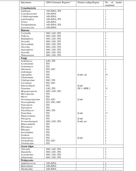
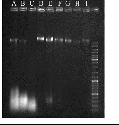
Besides, the higher amount of biomass can affect the extraction efficiency, effectively. Our results showed that in case of non-reproducible PCR amplifications, 10-200 times higher dilutions of the final DNA solution can be applied (Figure 1, A-C). As is shown in figure 1 A and B, the amount of biomass which was described in the recipe is critical and doubling this amount can decrease the yielded DNA effectively (Figure 1 C). The efficiency of this method was examined by amplifying nuclear ribosomal DNA regions; internal transcribed spacer (ITS), large subunit (LSU), and small subunit (SSU) of various fungal and microalgal taxa, and also protein coding regions; actin, calmodulin, β-tubulin, EF-1, RPB2 of a large number of fungal strains. Up to now, we have used this technique to isolate DNA from 131 strains as shown in table 1. Sequence-based identification procedure has been done on all these isolates.
Discussion
Specimens rDNA Genomic Regions a Protein coding Region No. of strains examined
Cyanobacteria
Anabaena 16S rDNA, ITS 1
Cyanobacterium 16S rDNA 1
Cylindrospermum 16S rDNA 1
Leptolyngbya 16S rDNA, ITS 2
Nostoc 16S rDNA 1
Pseudanabaena 16S rDNA, ITS 1
Synechocystis 16S rDNA 1
Diatoms
Cyclotella SSU, LSU, ITS 3
Fallacia SSU, LSU, ITS 1
Halamphora SSU, LSU, ITS 5
Navicula SSU, LSU, ITS 7
Navicymbula SSU, LSU, ITS 1
Nitzschia SSU, LSU, ITS 3
Staurophora SSU, LSU, ITS 1
Surirella SSU, LSU, ITS 1
Thalassiosira SSU, LSU, ITS 2
Fungi
Acidomyces LSU, ITS 1
Acremonium ITS 2
Actinomucor ITS 2
Alternaria ITS, LSU 8
Arthrinium ITS 1
Aspergillus ITS β-tub, cal 19
Chaetomium ITS 2
Cladosporium SSU, ITS 2
Curvularia ITS, LSU 1
Emericellopsis ITS β-tub 3
Fusarium LSU, ITS EF-1, RPB-2 31
Megasporoporia SSU, LSU, ITS 1
Microsporum ITS 1
Mucor ITS 4
Neocamarosporium ITS, LSU β-tub 2
Neosetophoma ITS, LSU, SSU 1
Neurospora ITS 1
Nigrospora ITS 1
Paecilomyces SSU, ITS 1
Peniciilum ITS β-tub 17
Phanerochaete ITS 2
Pleospora ITS β-tub 2
Pyrenochaetopsis SSU, LSU, ITS β-tub, act 2
Rhinocladiella ITS 1
Rhizomucor ITS 1
Rhizopus ITS 2
Sarocladium ITS 2
Sordaria ITS 1
Talaromyces ITS β-tub 4
Trichoderma ITS, LSU 2
Trichotecium ITS 1
Green Algae
Chlorella SSU, LSU, ITS 3
Dunaliella SSU, LSU, ITS 4
Scenedesmus SSU, LSU, ITS 5
Selenastrum SSU, LSU, ITS 2
Prokaryotes
Staphylococcus 16S rDNA 3
Halorubrum 16S rDNA 2
Thiobacillus 16S rDNA 1
A Safe, Rapid, and Reproducible Genomic DNA Extraction Protocol … Saba F, et al.
J Med Bacteriol. Vol. 5, No. 3, 4 (2016): pp.22-28 jmb.tums.ac.ir
26 a
Small subunit of nrRNA gene (SSU), Large subunit of nrRNA gene (LSU), Internal transcribed spacer fragment of nrRNA gene including ITS1-5.8s-ITS2 (ITS), β-Tubulin (tub), Actin (act), Calmodulin (cal).
DNA Concentration (ng/µl) 260/280 Ratio 260/230 Ratio
A 1002 1.789 1.88
B 176 1.811 0.824
C 87 2.253 0.1
However, the developed recipe yielded a reasonable amount of DNA from filamentous fungi, yeasts, diatoms, cyanobacteria and microalgae. A clear DNA band can be frequently seen when 3-5 µl of the extraction product checked in 1% agarose gel. However, the amount of DNA obtained from recalcitrant samples was low, resulting in a faint DNA band. This problem can affect the PCR reproducibility, effectively, and it tends to be a critical bottleneck when trying to amplify the SSU (~1750 bp) fragment (19).
However, addition of a 10 min long
chloroform/isoamyl alcohol purification step before the alcoholic precipitation, or doubling the 70% ethanol washing step followed by an RNase treatment, can increase the harvested DNA, in quality and quantity.
DNA spectrophotometery studies using PicoDrop showed that the procedure has its own limitations to purify the DNA. Firstly, it was assumed that the limitations are related to the lack of the solvent based purification steps which have been omitted from the recipe. But, further investigations indicated that the addition of the solvent based purification steps can lead to a considerable decrease of the DNA yield in expense of an increase of the DNA purity. Our aim was to achieve the highest possible PCR reproducibility which is itself dependent on the DNA purity and quantity. Finally, it was found that the dilution of the final extracted DNA is the best choice to increase the liability of this recipe for downstream procedures. The purity and yield of DNA achieved
Table 1. PicoDrop analysis of the extracted genomes using various versions of this recipe. (A) Performing the
recipe without the solvent based purification and RNase treatment, (B) Performing the recipe with the solvent based purification and RNase treatment (C) Genome DNA extraction from a given microbial specimen using the commercial genomic DNA extraction kit of CinnaGen.
Figure 1. Agarose gel of the extracted genomes
from various versions of this recipe are summarized in table 2 and figure 1. Accordingly, it is obvious that the solvent based purification step followed with an RNase treatment has a detectable effect on the purity. However, DNA extraction from most of the strains, especially the fungal strains, was performed without this step and it didn’t affect the PCR reproducibility.
Conclusion
Our results showed that the present recipe can be used for rapid identification of various groups of microorganisms. Also, having the needed flexibility in each step makes this protocol applicable on a very wide range of samples. Hence, various steps can be included depending on the desired quantity and quality.
Acknowledgements
All the experiments were performed in Microorganisms bank, Iranian Biological Resource Center (IBRC), ACECR, and Iran. Authors would like to thank the research affair of Iranian Biological Resource Center (IBRC).
Conflict of interest
No conflict of interests is declared.
Financial disclosure
This research was financially supported by research affair of Iranian Biological Resource Center (IBRC), Iran.
References
1. Komárek, Modern classification of
cyanobacteria. Cyanobacteria: an economic perspective. John Wiley & Son, West Sussex, UK, 2014.
2. Clerck O, Guiry MD, Leliaert F, et al. Algal taxonomy: a road to nowhere?. J Phycol
2013; 49: 215-225.
3. Hibbett DS, Binder M, Bischoff JF, et al. A higher-level phylogenetic classification of the Fungi. Mycol res 2007; 111: 509-547. 4. Mc Guire KL, Fierer N, Bateman C, et al.
Fungal community composition in
neotropical rain forests: the influence of tree diversity and precipitation. Microb Ecol
2012; 63: 804-812.
5. Dean AJ, Steneck RS, Tager D, et al. Distribution, abundance and diversity of crustose coralline algae on the Great Barrier Reef. Coral Reefs 2015; 34: 581-594. 6. Dighton J, White JF, Oudemans PV. The
fungal community: its organization and role in the ecosystem. CRC Press, New York, USA, 2005.
7. Doyle JJ. A rapid DNA isolation procedure for small quantities of fresh leaf tissue.
Phytochem Bull. 1987; 19: 11-15.
8. Lodhi MA, Ye G, Weeden NF, et al. A simple and efficient method for DNA extraction from grapevine cultivars and Vitis
species. Plant Mol Biol Rep 1994; 12: 6-13. 9. Sachinandan D, Gurpreet K, Amit R, et al. A
simple method for the efficient isolation of genomic DNA from lactobacilli isolated from traditional Indian fermented milk (dahi). Indian J Microbiol 2010; 50: 412-418.
10.Griffin DW, Kellogg C, Peak K, et al. A rapid and efficient assay for extracting DNA from fungi. Lett Appl Microbiol 2002; 34: 210-214.
11.Zhu H, Qu F, Zhu L. Isolation of genomic DNAs from fungi using benzyl chloride.
Acta Mycol. 1994; 13: 34.
12.Müller FMC, Werner KE, Kasai M, et al. Rapid extraction of genomic DNA from medically important yeasts and filamentous fungi by high-speed cell disruption. J Clin Microbiol 1998; 36: 1625-1629.
A Safe, Rapid, and Reproducible Genomic DNA Extraction Protocol … Saba F, et al.
J Med Bacteriol. Vol. 5, No. 3, 4 (2016): pp.22-28 jmb.tums.ac.ir
28
method of genomic DNA extraction suitable for analysis of bulk fungal strains. Lett Appl Microbiol 2010; 51: 114-118.
14.Goodwin D and Lee SB. Microwave
miniprep of total genomic DNA from fungi, plants, protists and animals for PCR.
Biotechniques 1993; 15: 441-432.
15.Liu D, Coloe S, Baird R, et al. Rapid mini-preparation of fungal DNA for PCR. J Clin Microbiol 2000; 38: 471-471.
16.Cenis J. Rapid extraction of fungal DNA for PCR amplification. Nucleic Acids Res 1992;
20(9): 2380.
17.Papizadeh M and Roayaei Ardakani M. Bio
filtration of volatile sulphurous
hydrocarbon-polluted air by hydrocarbon
degrading Pseudomonas NISOC-11. J
Biotechnol 2010: 150; 209-210.
18.Papizadeh M, Roayaei Ardakani M,
Ebrahimipour G, et al. Utilization of dibenzothiophene as sulfur source by
Microbacterium sp. NISOC-06. World J
Microbiol Biotechnol 2010: 26; 1195-1200. 19.Saba F, Noroozi M, Ghahremaninejad F, et
al. Isolation, Purification, and Identification of three Diatom species (Bacillariophyceae) from Gomishan wetland using Phylogeny and Silica-wall Ultra-Structure Analysis.