REVIEW
B waves: a systematic review of terminology,
characteristics, and analysis methods
Isabel Martinez‑Tejada
1,2*, Alexander Arum
1, Jens E. Wilhjelm
2, Marianne Juhler
1and Morten Andresen
1Abstract
Background
:
Although B waves were introduced as a concept in the analysis of intracranial pressure (ICP) recordings
nearly 60 years ago, there is still a lack consensus on precise definitions, terminology, amplitude, frequency or origin.
Several competing terms exist, addressing either their probable physiological origin or their physical characteristics. To
better understand B wave characteristics and ease their detection, a literature review was carried out.
Methods
:
A systematic review protocol including search strategy and eligibility criteria was prepared in advance. A
literature search was carried out using PubMed/MEDLINE, with the following search terms:
B waves
+
review filter
,
slow
waves
+
review filter
,
ICP B waves
,
slow ICP waves
,
slow vasogenic waves
,
Lundberg B waves
,
MOCAIP
.
Results
:
In total, 19 different terms were found,
B waves
being the most common. These terminologies appear to be
interchangeable and seem to be used indiscriminately, with some papers using more than five different terms. Defini‑
tions and etiologies are still unclear, which makes systematic and standardized detection difficult.
Conclusions
:
Two future lines of action are available for automating macro‑pattern identification in ICP signals:
achieving strict agreement on morphological characteristics of “traditional” B waveforms, or starting a new with a fresh
computerized approach for recognition of new clinically relevant patterns.
Keywords:
Intracranial pressure, B waves, Slow waves, Vasogenic waves
© The Author(s) 2019. This article is distributed under the terms of the Creative Commons Attribution 4.0 International License (http://creat iveco mmons .org/licen ses/by/4.0/), which permits unrestricted use, distribution, and reproduction in any medium, provided you give appropriate credit to the original author(s) and the source, provide a link to the Creative Commons license, and indicate if changes were made. The Creative Commons Public Domain Dedication waiver (http://creativecommons.org/ publicdomain/zero/1.0/) applies to the data made available in this article, unless otherwise stated.
Background
Intracranial pressure (ICP) monitoring plays an
impor-tant role in the management of patients with many
neurological and neurosurgical disorders. In the 1960s,
Lundberg described typical macro-patterns: A, B and C
waves [
1
]. B waves were defined as short repeating
eleva-tions in ICP (10–20 mmHg) with a frequency of 0.5–2
waves/min. These classic B wave patterns may be seen
in ICP monitoring in intensive care unit settings (ICU),
but ICP is also monitored in a large number of brain
diseases covering a spectrum from acute and subacute
ICU settings to elective outpatient follow-up. Today a
large proportion of patients undergo ICP monitoring
for milder degrees of disease where pathological
terns are not as prominent. In such scenarios, wave
pat-terns are still called B waves but differ in amplitude and
visual appearance from those defined by Lundberg. Such
‘uncharacteristic’ B waves are often smaller in amplitude
and appear as an irregular pattern, but they have not yet
been formally classified. The current paper uses B waves
as an encompassing umbrella for all variations.
The source of B waves is unknown and although they
are mostly associated with cerebral dysfunction, their
clinical significance is unclear, as they may also appear as
normal physiological phenomena [
2
,
3
]. Their source is
most commonly related to vasogenic activity, but an
ori-gin from a neuro-pacemaker system has also been
sug-gested [
4
]. This diverging information poses a challenge
to a consensus for a general description of B waves and
their quantification, hindering their identification during
diagnosis and treatment of different diseases categories.
Because of these difficulties, clinical practice outside
spe-cialized centers with a focus on ICP-related research is
currently largely restricted to readings of mean ICP.
Identification of waveform abnormalities by simple
vis-ual inspection is still a common clinical practice. This has
Open Access
*Correspondence: [email protected]
1 Clinic of Neurosurgery, Copenhagen University Hospital, Rigshospitalet,
Copenhagen, Denmark
an obvious bias from reliance on personal empiric
expe-rience and raises questions of interobserver
reproduc-ibility. Automated and standardized detection of B wave
patterns would increase the usefulness in both clinical
and research settings. This automated detection is only
possible if the waveform morphological characteristics
are clearly defined; preferably by consensus in the
sci-entific community. A systematic quantitative detection
system could allow for identification of B wave variations
and other ‘non-Lundberg’ patterns, replacing traditional
visual inspection.
The aim of this study was to assess the various terms
and definitions used to describe classical B waves in
order to highlight the lack of consensus in terms of
ter-minology and morphological characteristics, frequency
and amplitude. Therefore, a systematic review was
car-ried out to summarize the different terminologies and
definitions regarding B waves and the methods used for B
wave identification.
Methods
Relevant studies were identified by a single reviewer
using the online database PubMed/Medline. The
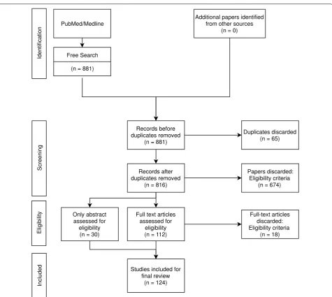
dia-gram in Fig.
1
gives an overview of the literature search
based on the PRISMA systematic review methodology
[
5
]. Studies were selected if they included the key terms
slow vasogenic waves
,
Lundberg B waves
,
slow ICP waves
,
ICP B waves
,
MOCAIP
,
B waves
+
review filter
, and
slow
waves
+
review filter
. A total sum of 816 paper abstracts
were screened initially for content relevance and 124
papers were included in the search review.
Results
Terminologies
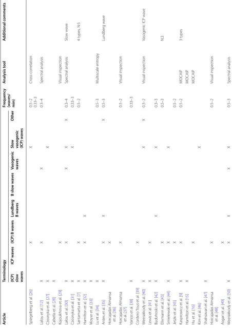
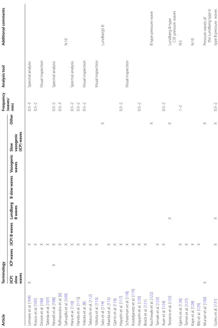
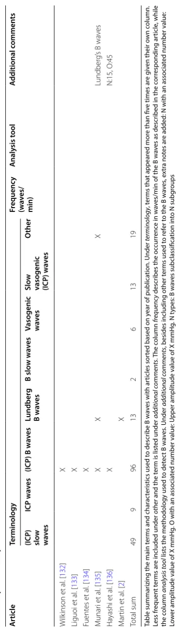
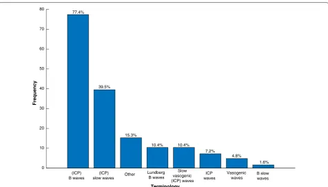
A total of 19 terminologies were found to describe B
waves in the reviewed papers (Table
1
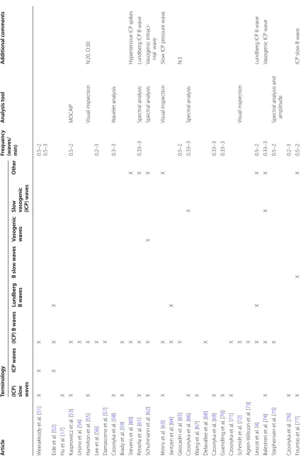
). The most
com-mon terms being
B waves
and
(ICP) slow waves
(Fig.
2
).
Nine articles used four or more terms to refer to B waves.
The choice of terminology is often related to the ongoing
etiology discussion: 22 articles include the word
vasogenic
thereby implying cerebrovascular changes as the origin of
the waves. Raftopoulos [
6
], Santamarta [
7
], Yokota [
8
],
and Kasprowicz [
9
], defined further subgroups in order
to clarify the sources underlying the presence of B waves
(Table
2
).
Characteristics
B waves were identified based on two major wave
param-eters: frequency and amplitude. Frequency is the
num-ber of waves that fit into a certain time period, usually
measured as waves per minute and 27% of the papers
defined a frequency of 0.5–2 waves/min, as originally
defined by Lundberg [
1
]. To accommodate B waves of a
lower frequency, the term
slow
was introduced [
10
]. The
term
slow waves
was then used to define waves with a
fre-quency window of 0.33 to 3 waves/min [
11
]. Two other
papers extended the frequency upper limit to 4 waves/
min [
12
,
13
].
As mentioned, B waves can also be characterized by
their amplitude. Lundberg defined a maximum amplitude
of 50 mmHg back in the 1960s. Under pathological
con-ditions, this level of elevation is less often seen to such
an extent today, and B waves with lower amplitudes are
more likely to be present. As an example, lower
ampli-tude B waves are present in cases of normal pressure
hydrocephalus, where the occurrence of B waves is not
related to high ICP [
14
].
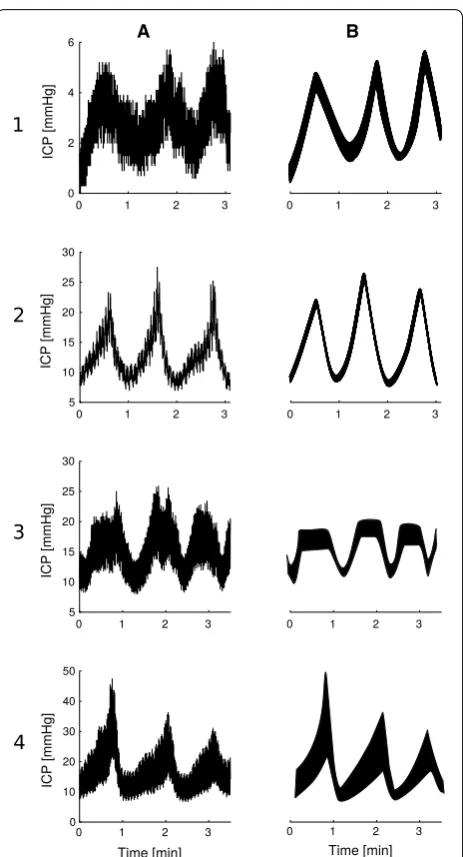
Sub‑classification
In addition to frequency and amplitude, two other
parameters are generally defined for the analysis of B
waves. B waves can also be characterized by their shape
and whether a plateau phase is present or not. The shape
is considered symmetrical if the duration of
ing and descending phases is the same. If the
ascend-ing phase is longer, then the shape is asymmetrical. The
use of these parameters gives rise to different subclasses
within B waves (Table
2
). All subclasses fit into the
tradi-tional definition of B waves with an extended frequency
spectrum, but mainly differ in their morphological
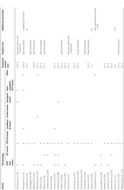
char-acteristics (Fig.
3
).
Besides these four parameters, Raftopolous et al. and
Santamarta et al. also use the duration of the ICP wave
to characterize B waves. They distinguish between three
morphological subclasses: (1) small symmetrical waves
with an amplitude below 10 mmHg, (2) great
symmetri-cal waves with an amplitude above 10 mmHg, and (3)
intermediate waves, with the same frequency as
symmet-rical waves but an amplitude similar to plateau waves [
6
,
7
].
Kasprowicz et al. describe three subcategories of B
waves based on the investigation of their unique shape:
(1) symmetrical ICP B waves, (2) asymmetrical ICP B
waves, and (3) slow ICP B waves with plateau phase. They
show how the different subtypes of B waves are related
to changes in the ICP pulse shape, which indicate that
each has a unique origin [
9
]. Similarly, Yokota et al. also
suggest the existence of three subgroups but from the
analysis of ICP amplitude and occurrence: (1) episodic
B-waves, (2) persistent, high pressure B-waves, and (3)
continuous, regular B-waves, and that these patterns may
better distinguish between different origins of ICP waves
[
8
].
with a plateau phase, and Yokota et al. [
8
] describe
per-sistent high pressure B waves. It is noteworthy that all
sub-classification attempts contain a B wave subtype with
plateau-like features. This raises the question whether
there is a continuous transition from B waves to plateau
waves or whether they have different etiology.
To summarize, B waves are categorized into
differ-ent subclasses if they have distinct shapes
and/or
if their
amplitude is different. These sub-classification attempts
may be used as supplementary evidence that the
classi-cal waveform categories do not adequately address
wave-forms identified in clinical practice today.
Fig. 1 Modified PRISMA 2009 flow diagram. Systematic literature search and selection process overview. Given that the goal of this literature review
was to give an insight into the different terminologies and definitions of B waves, only articles specifically mentioning B waves or related terms were included in the study selection. As an example: slow waves of ABP was not included. Papers simultaneously published by different journals were considered as duplicates and also excluded. The remaining articles (n = 124) were thoroughly examined and included in this study following the PRISMA flow‑diagram. Terminologies, definitions, and methods were identified individually by two independent reviewers and categorized according to a predefined protocol. Disagreements were resolved by consensus. No importance was given to the order of words, ICP B waves was treated equally to B ICP waves. Hyphens were removed, B-waves were grouped together with B waves. Of and in were disregarded, slow waves of ICP were registered as slow waves ICP. Only terminologies associated with ICP B waves were included (i.e. slow waves of ABP was not included). Terminologies in singular form were registered as plural, B wave was registered as B waves. Terms used less than three times were categorized as
Table
1
Summar
y of r
evie
w
ed B w
av
es t
erminolo
gy and char
ac
teristics
A rticle Terminology Fr equenc y (w av es/ min) A naly sis t ool A dditional c ommen ts (ICP ) slo w w ave s ICP w av es (ICP) B w
av es Lundber g B w av es B slo w w av es
Vasogenic wave
s
Slo
w
vasogenic (ICP
) w av es O ther Spiegelber g et al . [ 26 ] X X 0.5–2 0.33–3 Cr oss ‑cor relation Lalou et al . [ 12 ] X X X 0.3–4 Spec tral analysis Cz osn yk a et al . [ 27 ] X X Cabella et al . [ 28 ] X Kojouk ho va et al . [ 29 ] X Visual inspec tion Lalou et al . [ 30 ] X X X X X 0.3–4 Spec tral analysis Slo w wa ve Cz osn yk a et al . [ 31 ] X X 0.33–3 Santamar ta et al . [ 7 ] X X 0.5–2 4 types , N:5 Hamilt on et al . [ 32 ] X X X M oyse et al . [ 33 ] X Lu et al . [ 34 ] X 0.5–3 M ultiscale entr op y Ant es et al . [ 35 ] X X X X 0.5–3 Lundber g wa ve Hor cajadas Almansa et al . [ 36 ] X Hor cajadas Almansa et al . [ 37 ] X 0.5–2 Visual inspec tion Varsos et al . [ 38 ] X 0.33–3 Cor der o Tous et al . [ 39 ] X W eerak kody et al . [ 40 ] X X X 0.3–2 Visual inspec tion
Vasogenic ICP wa
Table
1
(c
on
tinued)
A rticle Terminology Fr equenc y (w av es/ min) A naly sis t ool A dditional c ommen ts (ICP ) slo w w ave s ICP w av es (ICP) B w
av es Lundber g B w av es B slo w w av es
Vasogenic wave
s
Slo
w
vasogenic (ICP
) w av es O ther W eerak kody et al . [ 51 ] X X X 0.5–2 0.5–3 Eide et al . [ 52 ] X X X Hu et al . [ 17 ] X Kaspr owicz et al . [ 53 ] X X 0.5–2 MOCAIP Ursino et al . [ 54 ] X Hamilt on et al . [ 55 ] X X Visual inspec tion N:20, O:30 Lee et al . [ 56 ] X 0.2–3 Damasceno et al . [ 57 ] X Cz osn yk a et al . [ 58 ] X 0.3–3 W av elet analysis Brady et al . [ 59 ] X X St ev ens et al . [ 60 ] X X H yper tensiv
e ICP spik
es Petr ella et al . [ 61 ] X X X 0.33–3 Spec tral analysis Lundber
g ICP B wa
ve Schuhmann et al . [ 62 ] X X X X Spec tral analysis Vasogenic intracr ‑ nial wa ve M inns et al . [ 63 ] X X X Visual inspec tion Slo
w ICP pr
essur e wa ve Jantz en et al . [ 64 ] X X G eocadin et al . [ 65 ] X 0.5–2 N:3 Cz osn yk a et al . [ 66 ] X X 0.33–3 Spec tral analysis W ang et al . [ 67 ] X D ela vallee et al . [ 68 ] X Cz osn yk a et al . [ 69 ] X 0.33–3 Guendling et al . [ 70 ] X 0.33–3 Cz osn yk a et al . [ 71 ] X X Schmidt et al . [ 72 ] X Visual inspec tion A gr en ‑W ilsson et al . [ 73 ] X Lescot et al . [ 4 ] X X X 0.5–2 Lundber
g ICP B wa
ve Balestr er i et al . [ 74 ] X X X X 0.33–3
Vasogenic ICP wa
ve St ephensen et al . [ 75 ] X 0.5–2 Spec
tral analysis and
amplitude Cz osn yk a et al . [ 76 ] X 0.2–3 Fountas et al . [ 77 ] X X X 0.5–2 ICP slo
w B wa
Table
1
(c
on
tinued)
A rticle Terminology Fr equenc y (w av es/ min) A naly sis t ool A dditional c ommen ts (ICP ) slo w w ave s ICP w av es (ICP) B w
av es Lundber g B w av es B slo w w av es
Vasogenic wave
s
Slo
w
vasogenic (ICP
) w av es O ther Cz osn yk a et al . [ 78 ] X X X X 0.33–2 Spec
tral analysis and
amplitude M omjian et al . [ 14 ] X X X X X 0.33–3 Spec tral analysis Lundber
g ICP B wa
ve Lenf eldt et al . [ 79 ] X 0.5–2 N:1 Edsbagge et al . [ 80 ] X Spec tral analysis Balestr er i et al . [ 81 ] X X 0.33–3 Spec tral analysis Ragausk as et al . [ 82 ] X X X 0.3–2 Str ik et al . [ 83 ] X X 0.5–3 Spec tral analysis Dale y et al . [ 84 ] X X 0.5–2 Spec tral analysis St ephensen et al . [ 85 ] X X Poca et al . [ 86 ] X Lemair e et al . [ 87 ] X X X X 0.5–2 Cz osn yk a et al . [ 88 ] X X X X 0.5–3 W alt er et al . [ 89 ] X 0.5–2 Spec tral analysis Schmidt et al . [ 90 ] X 0.5–2 Ek lund et al . [ 23 ] X 0.5–2 Spec
tral analysis and
Table
1
(c
on
tinued)
A rticle Terminology Fr equenc y (w av es/ min) A naly sis t ool A dditional c ommen ts (ICP ) slo w w ave s ICP w av es (ICP) B w
av es Lundber g B w av es B slo w w av es
Vasogenic wave
s
Slo
w
vasogenic (ICP
) w av es O ther Lemair e et al . [ 104 ] X X 0.5–3 Spec tral analysis Krauss et al . [ 105 ] X 0.5–2 Dr ost e et al . [ 106 ] X Visual inspec tion Tak eda et al . [ 107 ] X Ne w ell et al . [ 108 ] X X 0.5–2 Spec tral analysis Raf topoulos et al . [ 6 ] X 0.5–3 Sahuqillo et al . [ 109 ] X N:10 Hara et al . [ 110 ] X 0.5–2 Spec tral analysis Handa et al . [ 111 ]] X 0.5–2 Yok ota et al . [ 8 ] X 0.5–2 Visual inspec tion Tak euchi et al . [ 112 ] X Yok ota et al . [ 113 ] X Visual inspec tion Sat o et al . [ 114 ] X Lundber g’ s B M aeda et al . [ 115 ] X Gjer ris et al . [ 116 ] X Ha yashi et al . [ 117 ] X 0.5–2 Schoeman et al . [ 118 ] X Visual inspec tion Kost eljanetz et al . [ 119 ] X Ha yashi et al . [ 120 ] X 0.5–2 Br ock et al . [ 121 ] X Kuchiwak i et al . [ 122 ] X B t ype pr essur e wa ve Tamak i et al . [ 123 ] X A uer et al . [ 124 ] X 0.5–2 Ter zano et al . [ 125 ] X X Lundber g B ‑t ype CSF pr essur e wa ves Gjer ris et al . [ 126 ] X 1–2 N:5 Tomei et al . [ 127 ] X Ka ye et al . [ 128 ] X N:10 Bilz et al . [ 129 ] X M unar i et al . [ 130 ] X X Pr essur e wa ves of the L undber g t ype b Guieu et al . [ 131 ] X X X 0.5–2
type B pr
essur
e
wa
Table
1
(c
on
tinued)
A
rticle
Terminology
Fr
equenc
y
(w
av
es/
min)
A
naly
sis t
ool
A
dditional c
ommen
ts
(ICP
)
slo
w
w
ave
s
ICP w
av
es
(ICP
) B w
av
es
Lundber
g
B w
av
es
B slo
w w
av
es
Vasogenic wave
s
Slo
w
vasogenic (ICP
) w
av
es
O
ther
W
ilk
inson et
al
. [
132
]
X
Liguoi et
al
. [
133
]
X
Fuent
es et
al
. [
134
]
X
M
unar
i et
al
. [
135
]
X
X
X
Lundber
g’
s B wa
ves
Ha
yashi et
al
. [
136
]
X
N:15, O:45
M
ar
tin et
al
. [
2
]
X
Total sum
49
9
96
13
2
6
13
19
Table summar
izing the main t
er
ms and char
ac
ter
istics used t
o descr
ibe B w
av
es with ar
ticles sor
ted based on y
ear of publica
tion. Under
terminolo
gy
, t
er
ms tha
t appear
ed mor
e than fiv
e times ar
e g
iv
en their o
wn c
olumn.
Less fr
equen
t t
er
ms ar
e included under
other
and the t
er
m is list
ed under
additional c
omments
. T
he
column
frequenc
y
descr
ibes the oc
cur
renc
e in w
av
es/min of the B w
av
es as descr
ibed in the c
or
responding ar
ticle
, while
the c
olumn
analysis t
ool
lists the methodology used t
o det
ec
t B w
av
es
. Under
additional c
omments
, besides including other t
er
ms used t
o r
ef
er t
o the B w
av
es
, e
xtr
a not
es ar
e added: N with an associa
ted number v
alue:
Lo
w
er amplitude v
alue of X mmHg
. O with an associa
ted number v
alue: Upper amplitude v
alue of X mmHg
. N t
ypes: B w
av
es subclassifica
tion in
to N subg
Analysis tools
32% of the papers had an explicitly stated analysis
method. While traditionally the most common
analyti-cal method used was either spectral analysis (40%) or
spectral analysis with an amplitude threshold (7%), there
is now an increasing tendency (10%) to detect B waves
using trained machine learning algorithms, as observed
in more recently published papers [
9
,
15
,
16
]. These
algo-rithms use as input morphological features extracted
from the ICP pulse wave via the Morphological
cluster-ing and analysis of ICP pulse (MOCAIP) algorithm. Thus,
instead of defining B waves in terms of amplitude and
frequency, they define them according to different
mor-phological parameters of the pulse wave. These
param-eters are based on the three subpeaks (
P
1,
P
2, and
P
3)
of the pulse wave: systolic peak, tidal peak, and dicrotic
peak, respectively [
9
]. Examples of these ICP pulse
met-rics include the amplitude of the subpeaks, the latency
between subpeaks, and the start of the ICP pulse wave
and the pulse wave period, among others [
17
].
Terminology 0
10 20 30 40 50 60 70 80
Frequency
77.4%
39.5%
15.3%
10.4% 10.4%
7.2%
4.8%
1.6%
ICP waves
Vasogenic waves B slowwaves Lundberg
B waves Other
(ICP) slow waves (ICP)
B waves
Slow vasogenic (ICP) waves
Fig. 2 Frequency of terminology usage in the reviewed papers. The term B waves was used in most articles, followed by slow waves and ICP slow
waves
Table 2 Major morphological B wave subclasses
Term Shape Plateau Frequency
(waves/min) Amplitude (mmHg)
Raftopolous et al. [6]
Santamarta et al. [7] Small symmetrical wave (SSW)Great symmetrical wave (GSW) SymmetricalSymmetrical NoNo 0.36–50.36–5 < 10> 10 Intermediate wave (IW) Asymmetrical No 0.33–1.67 6–34 Kasprowicz et al. [9] Slow symmetrical ICP wave Symmetrical No – –
Slow asymmetrical ICP wave Asymmetrical No – – Slow ICP B with plateau phase Symmetrical Yes – –
Yokota et al. [8] Type II episodic B‑wave – – – 25–75
Discussion
ICP arises from pressure contributions from the brain,
the heart, and the cerebrospinal fluid (CSF) inside the
skull [
18
]. ICP is monitored invasively with a pressure
transducer inserted either intracranially (subdural,
epi-dural, intraparenchymal or intraventricular placements)
or in the spinal compartment (lumbar puncture). As the
brain is enclosed within the skull and its expandability is
restricted, the ability to compensate for pressure-volume
changes (auto-regulation) is also limited (i.e. compliance
is low). Under normal conditions, auto-regulatory
pro-cesses are responsible for keeping the intracranial volume
constant. As brain compliance starts to decrease, the
compensatory capacity is exhausted so that further
vol-ume changes are no longer accommodated; this causes
ICP to increase. Space-occupying lesions are the main
causes for the changes in intracranial volume.
Hydro-cephalus, intracranial haemorrhage, haematoma, and
brain edema are examples of such lesions [
19
].
Under normal compensatory adaptations, the ICP stays
within a narrow pressure range for each assumed body
posture [
20
,
21
]. This is the simplest way of looking at
ICP, as just a number that should remain within certain
boundary values. Going beyond that, the ICP signal can
be analyzed from a different perspective by studying the
presence of macro-patterns. The diversity of B waves is
the most commonly encountered macro-wave in clinical
practice.
This study demonstrates the lack of agreement with
regard to the terminology and characteristics used
to define B waves. Different names are used to refer
to the same phenomena, in order to either describe
characteristics and morphological variations of the
wave or the etiology behind their occurrence. This
makes mathematical modeling of B waves more
dif-ficult, which consequently complicates the selection
or development of an analysis tool that could be used
to automatically interpret them. Automating B wave
identification may be a way to detect and better
under-stand ICP deviations from a normal physiological state
at an earlier stage. But with the focus of current
analy-sis tools on identifying previously defined B waves,
they share a limitation of throwing away data related to
other potentially relevant waveform deviations. Thus,
underlying patterns of ICP that may contain important
information on the interplay of physiological systems
affecting the brain are potentially neglected.
Open-ing up the analysis of ICP signals without beOpen-ing
lim-ited to previously defined patterns and conventions
could enable fruitful new investigative and diagnostic
techniques.
Sub‑classification
B waves were first defined from ICP monitoring
ses-sions recorded in severely ill patients.
Sub-classifica-tions, which have mainly been qualitative, are the only
attempts at modernizing the description of B waves to
fit the clinical situations we see today [
6
–
9
].
The existence of multiple attempts at B wave
sub-grouping suggests that the overarching B wave
A
B
Fig. 3 Presentation of different B waves sub‑classification
category is not satisfactory for classification purposes
today. A future avenue of research may instead be to
direct attention away from classical B wave detection
and instead focus on the identification of new
param-eters to automate the analysis of repeatable patterns in
the ICP signal. Pattern recognition algorithms will be
the fundamental approach used for this purpose.
Analysis tools
ICP signals arise from the interaction of multiple
physi-ological factors (e.g. heart pump, respiration, ...) that vary
over time. Thus, it may be seen as a time series signal
[
22
]. Traditionally, ICP signals have been inspected
visu-ally for B wave identification. In addition to being a time
consuming technique, it is also subject to investigator
bias due to interpretation subjectivity and dependence
on clinical experience. Since the introduction of
com-puterized algorithms, spectral analysis has led the way in
B wave detection. A general agreement on a certain
fre-quency range that this wave occupies may explain why
spectral analysis is the most reported methodology.
How-ever, there is low frequency activity within the B-wave
range that is unrelated to vasomotor activity
(i.e. respira-tory changes associated with sleep), thereby
introduc-ing a severe limitation in the use of spectral analysis. We
might get unwanted contributions from these signals in
the B-wave frequency range when breaking down the
sig-nal into frequency components. Eklund et al. developed
an algorithm that strives to overcome this problem by
also taking into account the wave amplitude [
23
].
Defining B waves in terms of amplitude is, however,
very ambiguous. In particular, the term amplitude can be
approached as the trough to peak pressure difference in
the signal. If the wave has a sinusoidal appearance there
is no problem in the identification of both its maximum
and minimum values, but their identification becomes
a challenge when the waveform is irregular. At the same
time, the term amplitude can also refer to the distance
from the peak of the wave to the baseline.
MOCAIP extracts morphological parameters from
the pulse wave that are then used to characterize B
waves instead of defining them based on their
ampli-tude and frequency [
24
]. With the advantage of no
longer depending on the classical B wave definition,
this algorithm presents other drawbacks that prevents
it from proper implementation in clinical practice. It
rejects ICP pulses if a corresponding matching
tem-plate is not included within the reference library
pro-posed. This library is limited to intraparenchymal ICP
signals from patients with hydrocephalus and does
not comprise any ICP pulses from other pathologies,
so that ICP pulses could be falsely rejected. Another
limitation is the requirement of a simultaneous
acqui-sition of ECG signal to help in the identification of
the ICP pulse wave. Also, identifying B waves using
MOCAIP assumes that the pulse waves are affected
during the B waves, which is not definitively settled.
Another approach proposed by Elixmann also isolates
the pulse waves and classifies them based on predefined
templates [
25
].
Conclusion
To exploit the potential role of macro-patterns in ICP
dynamics and to automate their identification for
diag-nostic or therapeutic purposes, two approaches for future
work may be considered.
There could be efforts to arrive at strict agreement
on morphological characteristics of classical
macro-patterns, which requires consensus-based definitions to
enable the derivation of relevant metrics to characterize
them.
Alternatively, a new approach could be attempted
with-out relying on classical macro-patterns. Instead it could
be based on recognition of new patterns that more
ade-quately describe variations seen in daily clinical
prac-tice today. This de novo pattern recognition approach
requires relating macro-patterns to clinical information
to ensure that they are biologically relevant.
Abbreviations
ICP: intracranial pressure; CSF: cerebrospinal fluid; ICU: intensive care unit; MOCAIP: morphological clustering and analysis of ICP pulse.
Acknowledgements
The authors are thankful for contributions from the Novo Nordisk Fonden Tandem Programme.
Authors’ contributions
Study conception and design: AA, MA, MJ. Analysis and interpretation of the papers for the work: AA and IMT. Drafting of manuscript: IMT with input from the other authors. Critical revision: MJ, MA, JW. All authors read and approved the final manuscript.
Funding
Funding was provided by Novo Nordisk Fonden (Grant Number: NNF17OC0024718).
Availability of data and materials
Data sharing is not applicable to this article as no datasets were generated or analysed during the current study.
Ethics approval and consent to participate Not applicable.
Consent for publication Not applicable.
Competing interests
Author details
1 Clinic of Neurosurgery, Copenhagen University Hospital, Rigshospitalet,
Copenhagen, Denmark. 2 Department of Health Technology, Technical Univer‑
sity of Denmark, Kongens Lyngby, Denmark.
Received: 12 June 2019 Accepted: 15 September 2019
References
1. Lundberg N. Continuous recording and control of ventricular fluid pressure in neurosurgical practice. Acta Psychiatrica Scandinavica Sup‑ plementum. 1960;36:1–193.
2. Martin G. Lundberg’s B waves as a feature of normal intracranial pres‑ sure. Surg Neurol. 1978;9(6):347–8.
3. Droste DW, Krauss JK, Berger W, Schuler E, Brown MM. Rhythmic oscil‑ lations with a wavelength of 0.5–2 min in transcranial Doppler record‑ ings. Acta Neurologica Scandinavica. 2009;90(2):99–104.
4. Lescot T, Naccache L, Bonnet MP, Abdennour L, Coriat P, Puybasset L. The relationship of intracranial pressure Lundberg waves to electroen‑ cephalograph fluctuations in patients with severe head trauma. Acta
Neurochir. 2005;147(2):125–9 (discussion 129).
5. Liberati A, Altman DG, Tetzlaff J, Mulrow C, Gotzsche PC, Ioannidis JPA, et al. The PRISMA statement for reporting systematic reviews and meta‑ analyses of studies that evaluate healthcare interventions: explanation and elaboration. Br Med J. 2009;339(b2700):b2700.
6. Raftopoulos C, Chaskis C, Delecluse F, Cantrainet F, Bidauti L, Brotchi J. Morphological quantitative analysis of intracranial pressure waves in normal pressure hydrocephalus. Neurol Res. 1992;14:389–96. 7. Santamarta D, González‑Martínez E, Fernández J, Mostaza A. The predic‑
tion of shunt response in idiopathic normal‑pressure hydrocephalus based on intracranial pressure monitoring and lumbar infusion. Acta Neurochir Suppl. 2016;122:267–74.
8. Yokota A, Matsuoka S, Ishikawa T, Kohshi K, Kajiwara H. Overnight recordings of intracranial pressure and electroencephalography in neu‑ rosurgical patients. Part I: intracranial pressure waves and their clinical correlations. J UOEH. 1989;11(4):371–81.
9. Kasprowicz M, Bergsneider M, Czosnyka M, Hu X. Association between ICP pulse waveform morphology and ICP B waves. Acta Neurochir Suppl. 2012;114:29–34.
10. Spiegelberg A, Preuß M, Kurtcuoglu V. B‑waves revisited. Interdisciplin
Neurosurgery: Adv Tech Case Manage. 2016;6:13−7.
11. Czosnyka M, Pickard JD. Monitoring and interpretation of intracranial pressure. J Neurol Neurosurg Psychiatry. 2004;75(6):813–21. 12. Lalou DA, Czosnyka M, Donnelly J, Lavinio A, Pickard JD, Garnett M,
et al. Are slow waves of intracranial pressure suppressed by general anaesthesia? Acta Neurochir Suppl. 2018;126:129–32.
13. Hanlo PW, Gooskens RH, Faber JA, Peters RJ, Hermsen AA, Nijhuis IJ, et al. Relationship between anterior fontanelle pressure measure‑ ments and clinical signs in infantile hydrocephalus. Childs Nerv Syst. 1996;12(4):200–9.
14. Momjian S, Czosnyka Z, Czosnyka M, Pickard JD. Link between vasogenic waves of intracranial pressure and cerebrospinal fluid outflow resistance in normal pressure hydrocephalus. Br J Neurosurg. 2004;18(1):56–61.
15. Hamilton R, Baldwin K, Fuller J, Vespa P, Hu X, Bergsneider M. Intracranial pressure pulse waveform correlates with aqueductal cerebrospinal fluid stroke volume. J Appl Physiol. 2012;113(10):1560–6.
16. Hu X, Hamilton R, Baldwin K, Vespa PM, Bergsneider M. Automated extraction of decision rules for predicting lumbar drain outcome by analyzing overnight intracranial pressure. Acta Neurochir Suppl. 2012;114:207–12.
17. Hu X, Xu P, Asgari S, Vespa P, Bergsneider M. Forecasting ICP elevation based on prescient changes of intracranial pressure waveform mor‑ phology. IEEE Trans Biomed Eng. 2010;57(5):1070–8.
18. Raboel PH, Bartek J, Andresen M, Bellander BM, Romner B. Intracranial pressure monitoring: Invasive versus non‑invasive methods—a review. Crit Care Res Pract. 2012;2012:950393.
19. Lavinio A, Menon DK. Intracranial pressure: why we monitor it, how to monitor it, what to do with the number and what’s the future? Curr Opin Anesthesiol. 2011;24:117–23.
20. Andresen M, Hadi A, Petersen LG, Juhler M. Effect of postural changes on ICP in healthy and ill subjects. Acta Neurochir. 2014;157(1):109–13. 21. Petersen LG, Lawley JS, Lilja‑Cyron A, Petersen JCG, Howden EJ,
Sarma S, et al. Lower body negative pressure to safely reduce intrac‑ ranial pressure. J Physiol. 2019;597(1):237–48.
22. Pham H. Springer handbook of engineering statistics. London: Springer; 2006.
23. Eklund A, Agren‑Wilsson A, Andersson N, Bergenheim AT, Koskinen LO, Malm J. Two computerized methods used to analyze intracranial pressure B waves: comparison with traditional visual interpretation. J Neurosurg. 2001;94(3):392–6.
24. Hu X, Xu P, Scalzo F, Vespa P, Bergsneider M. Morphological clustering and analysis of continuous intracranial pressure. IEEE Trans Biomed Eng. 2009;56(4667641):696–705.
25. Elixmann IM, Hansinger J, Goffin C, Antes S, Radermacher K, Leon‑ hardt S. Single pulse analysis of intracranial pressure for a hydroceph‑ alus implant. In: Proceedings of the annual international conference of the IEEE engineering in medicine and biology Society, Embs, vol. 2012, no. 6346828. 2012. p. 3939–42.
26. Spiegelberg A, Krause M, Meixensberger J, Seifert B, Kurtcuo‑ glu V. Significant association of slow vasogenic ICP waves with normal pressure hydrocephalus diagnosis. Acta Neurochir Suppl. 2018;126:243–6.
27. Czosnyka M, Donnelly J, Calviello L, Smielewski P, Menon DK, Pickard JD. Do ICP‑derived parameters differ in vegetative state from other outcome groups after traumatic brain injury? Acta Neurochir Suppl. 2018;126:17–20.
28. Cabella B, Donnelly J, Cardim D, Liu X, Cabeleira M, Smielewski P, et al. An association between ICP‑derived data and outcome in TBI patients: the role of sample size. Neurocrit Care. 2017;27(1):103–7. 29. Kojoukhova M, Vanha KI, Timonen M, Koivisto AM, Nerg O, Rummu‑
kainen J, et al. Associations of intracranial pressure with brain biopsy, radiological findings, and shunt surgery outcome in patients with suspected idiopathic normal pressure hydrocephalus. Acta Neuro‑ chir. 2017;159(1):51–61.
30. Lalou DA, Czosnyka M, Donnelly J, Lavinio A, Pickard JD, Garnett M, et al. Influence of general anaesthesia on slow waves of intracranial pressure. Neurol Res. 2016;38(7):587–92.
31. Czosnyka M, Varsos GV, Czosnyka ZH, Smielewski P, Saadoun S, Jamous A, et al. Waveform analysis of intraspinal pressure after traumatic spinal cord injury: an observational study (O‑64). Acta Neurochir Suppl. 2016;122:335–8.
32. Hamilton R, Fuller J, Baldwin K, Vespa P, Hu X, Bergsneider M. Relative position of the third characteristic peak of the intracranial pres‑ sure pulse waveform morphology differentiates normal‑pressure hydrocephalus shunt responders and nonresponders. Acta Neurochir Suppl. 2016;122:339–45.
33. Moyse E, Ros M, Marhar F, Swider P, Schmidt EA. Characterisation of supra‑ and infratentorial ICP profiles. Acta Neurochir Suppl. 2016;122:37–40.
34. Lu CW, Czosnyka M, Shieh JS, Pickard JD, Smielewski P. Continuous monitoring of the complexity of intracranial pressure after head injury. Acta Neurochir Suppl. 2016;122:33–5.
35. Antes S, Tschan CA, Heckelmann M, Breuskin D, Oertel J. Telemetric intracranial pressure monitoring with the Raumedic Neurovent P‑tel. World Neurosurg. 2016;91:133–48.
36. Horcajadas Almansa A, Cordero Tous N, Roman Cutillas A, Jorques Infante A, Saura Rojas E, Ibanez Velasco B, et al. Usefulness of continu‑ ous intracranial pressure monitoring in long‑standing overt ventricu‑ lomegaly in adults. Neurocirugia. 2015;26(2):64–72.
37. Horcajadas Almansa A, Cordero Tous N, Roman Cutillas A, Jorques Infante A, Saura Rojas E, Ibanez Velasco B, et al. Usefulness of intracranial pressure continuous monitoring in pseudotumor cerebri. Neurocirugia. 2015;26(4):157–66.
39. Cordero Tous N, Roman Cutillas AM, Jorques Infante AM, Olivares Granados G, Saura Rojas JE, Ibanez Velasco B, et al. Adult chronic idi‑ opathic hydrocephalus‑diagnosis, treatment and evolution. Prospec‑ tive study. Neurocirugia. 2013;24(3):93–101.
40. Weerakkody RA, Czosnyka M, Zweifel C, Castellani G, Smielewski P, Brady K, et al. Near infrared spectroscopy as possible non‑invasive monitor of slow vasogenic ICP waves. Acta Neurochir Suppl. 2012;114:181–5.
41. Lewis PM, Smielewski P, Rosenfeld JV, Pickard JD, Czosnyka M. Moni‑ toring of the association between cerebral blood flow velocity and intracranial pressure. Acta Neurochir Suppl. 2012;114:147–51. 42. Budohoski KP, Schmidt B, Smielewski P, Kasprowicz M, Plontke R,
Pickard JD, et al. Non‑invasively estimated ICP pulse amplitude strongly correlates with outcome after TBI. Acta Neurochir Suppl. 2012;114:121–5.
43. Elixmann IM, Walter M, Kiefer M, Leonhardt S. Simulation of existing and future electromechanical shunt valves in combination with a model for brain fluid dynamics. Acta Neurochir Suppl. 2012;113:77–81.
44. Smielewski P, Czosnyka Z, Kasprowicz M, Pickard JD, Czosnyka M. ICM+:
a versatile software for assessment of CSF dynamics. Acta Neurochir Suppl. 2012;114:75–9.
45. Jetzki S, Weinzierl M, Krause I, Hahne S, Rehbaum H, Kiausch M, et al. A multisensor implant for continuous monitoring of intracranial pressure dynamics. IEEE Trans Biomed Circuits Syst. 2012;6(4):356–65.
46. Kim DJ, Czosnyka Z, Kasprowicz M, Smieleweski P, Baledent O, Guergue‑ rian AM, et al. Continuous monitoring of the Monro‑Kellie doctrine: is it possible? J Neurotrauma. 2012;29(7):1354–63.
47. Shahsavari S, McKelvey T, Ritzén CE, Rydenhag B. Cerebrovascular mechanical properties and slow waves of intracranial pressure in TBI patients. IEEE Trans Biomed Eng. 2011;58(7):2072–82.
48. Horcajadas Almansa A, Roman A, Olivares G, Saura E, Jorques A, Cordero N, et al. Usefulness of IPC continuous monitoring in shunt dysfunction. Neurocirugia. 2011;22(4):310–23.
49. Asgari S, Vespa P, Bergsneider M, Hu X. Lack of consistent intracranial pressure pulse morphological changes during episodes of microdialysis lactate/pyruvate ratio increase. Physiol Meas. 2011;32(10):1639–51. 50. Weerakkody RA, Czosnyka M, Schuhmann MU, Schmidt E, Keong N,
Santarius T, et al. Clinical assessment of cerebrospinal fluid dynamics in hydrocephalus. Guide to interpretation based on observational study. Acta Neurol Scand. 2011;124(2):85–98.
51. Weerakkody RA, Czosnyka M, Zweifel C, Castellani G, Smielewski P, Keong N, et al. Slow vasogenic fluctuations of intracranial pressure and cerebral near infrared spectroscopy—an observational study. Acta Neurochir. 2010;152(10):1763–9.
52. Eide PK, Sorteberg W. Simultaneous measurements of intracranial pressure parameters in the epidural space and in brain parenchyma in patients with hydrocephalus. J Neurosurg. 2010;113(6):1317–25. 53. Kasprowicz M, Asgari S, Bergsneider M, Czosnyka M, Hamilton R, Hu
X. Pattern recognition of overnight intracranial pressure slow waves using morphological features of intracranial pressure pulse. J Neurosci Methods. 2010;190(2):310–8.
54. Ursino M, Giannessi M, Frapparelli M, Magosso E. Effect of cush‑ ing response on systemic arterial pressure. IEEE Eng Med Biol Mag. 2009;28(6):63–71.
55. Hamilton R, Xu P, Asgari S, Kasprowicz M, Vespa P, Bergsneider M, et al. Forecasting intracranial pressure elevation using pulse waveform morphology. Conf Proc IEEE Eng Med Biol Soc. 2009;2009:4331–4. 56. Lee JK, Kibler KK, Benni PB, Easley RB, Czosnyka M, Smielewski P, et al.
Cerebrovascular reactivity measured by near‑infrared spectroscopy. Stroke. 2009;40(5):1820–6.
57. Damasceno BP. Normal pressure hydrocephalus: diagnostic and predic‑ tive evaluation. Dement Neuropsychol. 2009;3(1):8–15.
58. Czosnyka M, Brady K, Reinhard M, Smielewski P, Steiner LA. Monitor‑ ing of cerebrovascular autoregulation: facts, myths, and missing links. Neurocrit Care. 2009;10(3):373–86.
59. Brady KM, Lee JK, Kibler KK, Easley RB, Koehler RC, Shaffner DH. Continu‑ ous measurement of autoregulation by spontaneous fluctuations in cerebral perfusion pressure: comparison of 3 methods. Stroke. 2008;39(9):2531–7.
60. Stevens SA, Stimpson J, Lakin WD, Thakore NJ, Penar PL. A model for idiopathic intracranial hypertension and associated pathological ICP wave‑forms. IEEE Trans Biomed Eng. 2008;55(2 Pt 1):388–98. 61. Petrella G, Czosnyka M, Keong N, Pickard JD, Czosnyka Z. How
does CSF dynamics change after shunting? Acta Neurol Scand. 2008;118(3):182–8.
62. Schuhmann MU, Sood S, McAllister JP, Jaeger M, Ham SD, Czosnyka Z, et al. Value of overnight monitoring of intracranial pressure in hydroce‑ phalic children. Pediatr Neurosurg. 2008;44(4):269–79.
63. Minns RA, Jones PA, Chambers IR. Low frequency pressure waves of possible autonomic origin in severely head‑injured children. Acta Neurochir Suppl. 2008;102:85–8.
64. Jantzen JPAH. Prevention and treatment of intracranial hypertension. Best Pract Res Clin Anaesthesiol. 2007;21(4):517–38.
65. Geocadin RG, Varelas PN, Rigamonti D, Williams MA. Continuous intrac‑ ranial pressure monitoring via the shunt reservoir to assess suspected shunt malfunction in adults with hydrocephalus. Neurosurg Focus. 2007;22(4):E10.
66. Czosnyka M, Smielewski P, Timofeev I, Lavinio A, Guazzo E, Hutchinson P, et al. Intracranial pressure: more than a number. Neurosurg Focus. 2007;22(5):E10.
67. Wang EC, Ang BT, Wong J, Lim J, Ng I. Characterization of cerebrovas‑ cular reactivity after craniectomy for acute brain injury. Br J Neurosurg. 2006;20(1):24–30.
68. Delavallée M, Raftopoulos C. Normal pressure hydrocephalus in a patient with myotonic dystrophy: case report with a 10‑year follow‑up.
Neurosurgery. 2006;58(4):E796 (discussion E796).
69. Czosnyka M, Hutchinson PJ, Balestreri M, Hiler M, Smielewski P, Pickard JD. Monitoring and interpretation of intracranial pressure after head injury. Acta Neurochir Suppl. 2006;96:114–8.
70. Guendling K, Smielewski P, Czosnyka M, Lewis P, Nortje J, Timofeev
I, et al. Use of ICM+ software for on‑line analysis of intracranial and
arterial pressures in head‑injured patients. Acta Neurochir Suppl. 2006;96:108–13.
71. Czosnyka Z, van den Boogaard F, Czosnyka M, Momjian S, Gelling L, Pickard JD. The relationship between CSF circulation and cerebro‑ vascular pressure‑reactivity in normal pressure hydrocephalus. Acta Neurochir Suppl. 2005;95:207–11.
72. Schmidt B, Bocklisch SF, Pässler M, Czosnyka M, Schwarze JJ, Klingel‑ höfer J. Fuzzy pattern classification of hemodynamic data can be used to determine noninvasive intracranial pressure. Acta Neurochir Suppl. 2005;95:345–9.
73. Agren‑Wilsson A, Eklund A, Koskinen LOD, Bergenheim AT, Malm J. Brain energy metabolism and intracranial pressure in idiopathic adult hydro‑ cephalus syndrome. J Neurol Neurosurg Psychiatry. 2005;76(8):1088–93. 74. Balestreri M, Czosnyka M, Steiner LA, Hiler M, Schmidt EA, Matta B, et al. Association between outcome, cerebral pressure reactivity and slow ICP waves following head injury. Acta Neurochir Suppl. 2005;95:25–8. 75. Stephensen H, Andersson N, Eklund A, Malm J, Tisell M, Wikkelsö C.
Objective B wave analysis in 55 patients with non‑communicating and communicating hydrocephalus. J Neurol Neurosurg Psychiatry. 2005;76(7):965–70.
76. Czosnyka M, Balestreri M, Steiner L, Smielewski P, Hutchinson PJ, Matta B, et al. Age, intracranial pressure, autoregulation, and outcome after brain trauma. J Neurosurg. 2005;102(3):450–4.
77. Fountas KN, Sitkauskas A, Feltes CH, Kapsalaki EZ, Dimopoulos VG, Kassam M, et al. Is non‑invasive monitoring of intracranial pressure waveform analysis possible? Preliminary results of a comparative study of non‑invasive vs. invasive intracranial slow‑wave waveform analysis monitoring in patients with traumatic brain injury. Med Sci Monit. 2005;11(2):CR58–63.
78. Czosnyka M, Czosnyka Z, Momjian S, Pickard JD. Cerebrospinal fluid dynamics. Physiol Meas. 2004;25(5):R51–76.
79. Lenfeldt N, Andersson N, Agren‑Wilsson A, Bergenheim AT, Koskinen LOD, Eklund A, et al. Cerebrospinal fluid pulse pressure method: a possible substitute for the examination of B waves. J Neurosurg. 2004;101(6):944–50.
81. Balestreri M, Czosnyka M, Steiner LA, Schmidt E, Smielewski P, Matta B, et al. Intracranial hypertension: what additional information can be derived from ICP waveform after head injury? Acta Neurochir. 2004;146(2):131–41.
82. Ragauskas A, Daubaris G, Ragaisis V, Petkus V. Implementation of non‑invasive brain physiological monitoring concepts. Med Eng Phys. 2003;25(8):667–78.
83. Strik C, Klose U, Kiefer C, Grodd W. Slow rhythmic oscillations in intrac‑ ranial CSF and blood flow: registered by MRI. Acta Neurochir Suppl. 2002;81:139–42.
84. Daley ML, Pasley RL, Connolly M, Timmons SD, Angel J, Stidham G, et al. Spectral characteristics of B‑waves and other low‑frequency activity. Acta Neurochir Suppl. 2002;81:147–50.
85. Stephensen H, Tisell M, Wikkelsö C. There is no transmantle pressure gradient in communicating or noncommunicating hydrocephalus.
Neurosurgery. 2002;50(4):763–71 (discussion 771–773).
86. Poca MA, Sahuquillo J, Ibañez J, Amorós S, Arikan F, Rubio E. Intracranial hypertension after surgery in patients with Chiari I malformation and normal or moderate increase in ventricular size. Acta Neurochir Suppl. 2002;81:35–8.
87. Lemaire JJ, Khalil T, Cervenansky F, Gindre G, Boire JY, Bazin JE, et al. Slow pressure waves in the cranial enclosure. Acta Neurochir. 2002;144(3):243–54.
88. Czosnyka ZH, Czosnyka M, Whitfield PC, Donovan T, Pickard JD. Cerebral autoregulation among patients with symptoms of hydrocephalus.
Neurosurgery. 2002;50(3):526–32 (discussion 532–533).
89. Walter M, Kiefer M, Leonhardt S, Steudel WI, Isermann R. Online analysis of intracranial pressure waves. Acta Neurochir Suppl. 2002;81:161–2. 90. Schmidt EA, Czosnyka M, Smielewski P, Piechnik SK, Pickard JD. Asym‑
metry of cerebral autoregulation following head injury. Acta Neurochir Suppl. 2002;81:133–4.
91. Vanneste JA. Diagnosis and management of normal‑pressure hydro‑ cephalus. J Neurol. 2000;247(1):5–14.
92. Schoeman JF, Laubscher JA, Donald PR. Serial lumbar CSF pressure measurements and cranial computed tomographic findings in child‑ hood tuberculous meningitits. Childs Nerv Syst. 2000;16(4):203–8 (discussion 209).
93. Schuhmann MU, Engel M, Runge L, Samii M, Brinker T. Application of clinically recorded ICP patterns‑an extension of conventional shunt testing. Childs Nerv Syst. 2000;16(12):856–61.
94. Czosnyka M, Smielewski P, Piechnik S, Al‑Rawi PG, Kirkpatrick PJ, Matta BF, et al. Critical closing pressure in cerebrovascular circulation. J Neurol Neurosurg Psychiatry. 1999;66(5):606–11.
95. Droste DW, Krauss JK. Intracranial pressure B‑waves precede corre‑ sponding arterial blood pressure oscillations in patients with suspected normal pressure hydrocephalus. Neurol Res. 1999;21(7):627–30. 96. Qureshi AI, Williams MA, Razumovsky AY, Hanley DF. Magnetic reso‑
nance imaging, unstable intracranial pressure and clinical outcome in patients with normal pressure hydrocephalus. Acta Neurochir Suppl. 1998;71:354–6.
97. Czosnyka M, Smielewski P, Kirkpatrick P, Piechnik S, Laing R, Pickard JD. Continuous monitoring of cerebrovascular pressure‑reactivity in head injury. Acta Neurochir Suppl. 1998;71:74–7.
98. Czosnyka M, Smielewski P, Kirkpatrick P, Laing RJ, Menon D, Pickard JD. Continuous assessment of the cerebral vasomotor reactivity in head
injury. Neurosurgery. 1997;41(1):11–7 (discussion 17–19).
99. Newell DW, Aaslid R, Stooss R, Seiler RW, Reulen HJ. Evaluation of hemo‑ dynamic responses in head injury patients with transcranial Doppler monitoring. Acta Neurochir. 1997;139(9):804–17.
100. Lemaire JJ. Slow pressure waves during intracranial hypertension. Ann Fr Anesth Reanim. 1997;16(4):394–8.
101. Steinmeier R, Bauhuf C, Hübner U, Bauer RD, Fahlbusch R, Laumer R, et al. Slow rhythmic oscillations of blood pressure, intracranial pressure, microcirculation, and cerebral oxygenation. Dynamic interrelation and time course in humans. Stroke. 1996;27(12):2236–43.
102. Krauss JK, Droste DW, Bohus M, Regel JP, Scheremet R, Riemann D, et al. The relation of intracranial pressure B‑waves to different sleep stages in patients with suspected normal pressure hydrocephalus. Acta Neuro‑ chir. 1995;136(3–4):195–203.
103. Wayenberg JL, Hasaerts D, Franco P, Valente F, Massager N. Anterior fontanelle pressure variations during sleep in healthy infants. Sleep. 1995;18(4):223–8.
104. Lemaire JJ, Boire JY, Chazal J, Irthum B. A computer software for fre‑ quential analysis of slow intracranial pressure waves. Comput Methods Programs Biomed. 1994;42(1):1–14.
105. Krauss JK, Droste DW. Predictability of intracranial pressure oscillations in patients with suspected normal pressure hydrocephalus by transcra‑ nial Doppler ultrasound. Neurol Res. 1994;16(5):398–402.
106. Droste DW, Krauss JK, Berger W, Schuler E, Brown MM. Rhythmic oscil‑ lations with a wavelength of 0.5–2 min in transcranial Doppler record‑ ings. Acta Neurol Scand. 1994;90(2):99–104.
107. Takeda Y, Onitsuka T, Haraoka J, Koba T, Ito H, Miwa T. Clinical assess‑ ment of meningeal carcinomatosis; from the viewpoint of the analysis of intracranial pressure. No Shinkei Geka. 1993;21(3):213–20. 108. Newell DW, Aaslid R, Stooss R, Reulen HJ. The relationship of blood
flow velocity fluctuations to intracranial pressure B waves. J Neurosurg. 1992;76(3):415–21.
109. Sahuquillo J, Rubio E, Codina A, Molins A, Guitart JM, Poca MA, et al. Reappraisal of the intracranial pressure and cerebrospinal fluid dynam‑ ics in patients with the so‑called “normal pressure hydrocephalus” syndrome. Acta Neurochir. 1991;112(1–2):50–61.
110. Hara K, Nakatani S, Ozaki K, Ikeda T, Mogami H. Detection of the B waves in the oscillation of intracranial pressure by fast Fourier trans‑ form. Med Inform. 1990;15(2):125–31.
111. Handa Y, Hayashi M, Hirose S, Noguchi Y, Kobayashi H. The effect of increased intracranial pressure during the appearance of pressure waves on the brainstem. Neurol Med Chir. 1990;30(5):301–8. 112. Takeuchi S, Koike T, Sasaki O, Kamada K, Tanaka R, Arai H. Intracranial
extradural pressure monitoring after direct operation on ruptured cerebral aneurysms. Neurosurgery. 1989;24(6):878–83.
113. Yokota A, Matsuoka S, Ishikawa T, Kohshi K, Kajiwara H. Overnight recordings of intracranial pressure and electroencephalography in neurosurgical patients. Part II: changes in intracranial pressure during sleep. J UOEH. 1989;11(4):383–91.
114. Sato H, Sato N, Tamaki N, Matsumoto S. The critical threshold of cerebral perfusion pressure in intracranial pressure circumstance of hydrocepha‑ lus during infancy. No Shinkei Geka. 1988;16(4):385–92.
115. Maeda M, Matsuura S, Tanaka K, Katsuyama J, Nakamura T, Sakamoto H, et al. Effects of electrical stimulation on intracranial pressure and systemic arterial blood pressure in cats. Part I: stimulation of brain stem. Neurol Res. 1988;10(2):87–92.
116. Gjerris F, Børgesen SE, Sørensen PS, Boesen F, Schmidt K, Harmsen A, et al. Resistance to cerebrospinal fluid outflow and intracranial pressure in patients with hydrocephalus after subarachnoid haemorrhage. Acta Neurochir. 1987;88(3–4):79–86.
117. Hayashi M, Kobayashi H, Kawano H, Handa Y, Yamamoto S, Kitano T. ICP patterns and isotope cisternography in patients with communicating hydrocephalus following rupture of intracranial aneurysm. J Neurosurg. 1985;62(2):220–6.
118. Schoeman JF, le Roux D, Bezuidenhout PB, Donald PR. Intracranial pres‑ sure monitoring in tuberculous meningitis: clinical and computerized tomographic correlation. Dev Med Child Neurol. 1985;27(5):644–54. 119. Kosteljanetz M. CSF dynamics in patients with subarachnoid and/or
intraventricular hemorrhage. J Neurosurg. 1984;60(5):940–6. 120. Hayashi M, Kobayashi H, Kawano H, Yamamoto S, Maeda T. Cerebral
blood flow and ICP patterns in patients with communicating hydro‑ cephalus after aneurysm rupture. J Neurosurg. 1984;61(1):30–6. 121. Brock M, Cervos‑Navarro J, Holdorff B. Changes in intracranial pres‑
sure associated with delayed cerebral radionecrosis. Surg Neurol. 1984;22(1):8–16.
122. Kuchiwaki H, Kageyama N, Hirai N, Takada S, Inao S, Terashima M, et al. A biological rhythm in a patient with normal pressure hydrocephalus‑ comparative studies in pre‑ and postoperative patients by a polygra‑ phy. No To Shinkei. 1984;36(9):911–6.
•fast, convenient online submission
•
thorough peer review by experienced researchers in your field
• rapid publication on acceptance
• support for research data, including large and complex data types
•
gold Open Access which fosters wider collaboration and increased citations maximum visibility for your research: over 100M website views per year
•
At BMC, research is always in progress.
Learn more biomedcentral.com/submissions
Ready to submit your research? Choose BMC and benefit from: 124. Auer LM, Sayama I. Intracranial pressure oscillations (B‑waves)
caused by oscillations in cerebrovascular volume. Acta Neurochir. 1983;68(1–2):93–100.
125. Terzano MG, Gatti PL, Manzoni GC, Formentini E, Mancia D. Is the EEG cyclic alternating pattern a true autonomous entity? Analytic study in a case of post‑traumatic coma with good prognosis. Eur Neurol. 1982;21(5):324–34.
126. Gjerris F, Børgesen SE, Hoppe E, Boesen F, Nordenbo AM. The conduct‑ ance to outflow of CSF in adults with high‑pressure hydrocephalus. Acta Neurochir. 1982;64(1–2):59–67.
127. Tomei G, Gaini SM, Giovanelli M, Rampini P, Granata G, Villani R. Intrac‑ ranial pressure in subarachnoid hemorrhage. Preliminary report in 36 cases. J Neurosurg Sci. 1981;25(2):57–66.
128. Kaye AH, Brownbill D. Postoperative intracranial pressure in patients operated on for cerebral aneurysms following subarachnoid hemor‑ rhage. J Neurosurg. 1981;54(6):726–32.
129. Bilz D, Pásztor A, Paraicz E. Rhythmic correlations between regional intracranial blood volume and intracranial pressure within minutes dur‑ ing continuous simultaneous recording in infants. Psychiatr Neurol Med Psychol. 1981;33(8):483–90.
130. Munari C, Calbucci F, Casaroli D, Bonora M. Correlations between E.E.G. findings and intracranial pressure (ICP) during treatment of acute post‑ traumatic intracranial hypertension (preliminary report) (author’s transl). Rev Electroencephalogr Neurophysiol Clin. 1980;10(4):319–30.
131. Guieu JD, Lapierre M, Blond S, Hurtevent JF, Jomin M, Pruvot P. Cor‑ relations between intracranial pressure variations and EEG changes in patients with cranial trauma (author’s transl). Rev Electroencephalogr Neurophysiol Clin. 1979;9(2):194–201.
132. Wilkinson HA, Schuman N, Ruggiero J. Nonvolumetric methods of detecting impaired intracranial compliance or reactivity: pulse width and wave form analysis. J Neurosurg. 1979;50(6):758–67.
133. Liguori G, Fiordirosa M, Cicchiello LR, Ambrosio A. Long‑term monitor‑ ing of intracranial pressure in hydrocephalic children not affected by cerebral tumor. J Neurosurg Sci. 1979;23(4):295–302.
134. Fuentes JM, Bouscarel C, Choucair Y, Roquefeuil B, Vlahovitch B, Blan‑ chet P. Monitoring of intracranial pression in acute neurotrauma by extra‑dural screw (author’s transl). Anesth Analg. 1979;36(9–10):429–33. 135. Munari C, Calbucci F. Correlations between intracranial pressure (I.C.P.)
and EEG changes during traumatic coma (author’s transl). Rev Electro‑ encephalogr Neurophysiol Clin. 1979;9(2):185–93.
136. Hayashi M, Marukawa S, Fujii H, Kitano T, Kobayashi H, Yamamoto S. Intracranial hypertension in patients with ruptured intracranial aneu‑ rysm. J Neurosurg. 1977;46(5):584–90.