Influence of Plasmid Type on the
Replication of
Rhodococcus equi
in Host
Macrophages
Jennifer M. Willingham-Lane,aLonda J. Berghaus,bSteeve Giguère,b Mary K. Hondalusa
Department of Infectious Disease, University of Georgia, Athens, Georgia, USAa; Department of Large Animal Medicine, University of Georgia, Athens, Georgia, USAb
ABSTRACT The soil-dwelling, saprophytic actinomyceteRhodococcus equiis a mul-tihost, facultative intracellular pathogen of macrophages. When inhaled by suscepti-ble foals, it causes severe bronchopneumonia. It is also a pathogen of pigs, which may develop submaxillary lymphadenitis upon exposure. R. equi isolates obtained from foals and pigs possess conjugative plasmids housing a pathogenicity island (PAI) containing a novel family of genes of unknown function called the virulence-associated protein orvap family. The PAI regions of the equine and swine plasmids differ in vap gene composition, with equine isolates possessing six vap genes, in-cluding the major virulence determinant vapA, while the PAIs of swine isolates house vapB and five other unique vap genes. Possession of the pVAPA-type viru-lence plasmid by equine isolates bestows the capacity for intramacrophage replica-tion essential for disease development in vivo. Swine isolates of R. equi are largely unstudied. Here, we show that R. equiisolates from pigs, carrying pVAPB-type plas-mids, are able to replicate in a plasmid-dependent manner in macrophages obtained from a variety of species (murine, swine, and equine) and anatomical locations. Simi-larly, equine isolates carrying pVAPA-type plasmids are capable of replication in swine macrophages. Plasmid swapping between equine and swine strains through conjugation did not alter the intracellular replication capacity of the parental strain, indicating that coevolution of the plasmid and chromosome is not crucial for this at-tribute. These results demonstrate that while distinct plasmid types exist among R. equi isolates obtained from equine and swine sources, this tropism is not deter-mined by host species-specific intramacrophage replication capabilities.
IMPORTANCE This work greatly advances our understanding of the opportunistic pathogen Rhodococcus equi, a disease agent of animals and immunocompromised people. Clinical isolates from diseased foals carry a conjugative virulence plasmid, pVAPA1037, that expresses Vap proteins, including VapA, essential for intramac-rophage replication and virulence in vivo. The understudied R. equi isolates from pigs carry a related but different plasmid, pVAPB, expressing distinct Vap proteins, including VapB. In this work, we document for the first time thatR. equiisolates car-rying pVAPB-type plasmids are capable of intramacrophage replication. Moreover, we show that R. equi isolates carrying either plasmid type can replicate in both equine and swine macrophages, indicating that host species tropism is not due to species-specific intramacrophage replication capabilities defined by plasmid type. Furthermore, plasmid swapping between equine and swine strains did not alter in-tracellular replication capacity, indicating that coevolution of the plasmid and chro-mosome is not essential for intracellular growth.
KEYWORDS: Rhodococcus equi, macrophage, species tropism,vap
Received7 July 2016Accepted31 August
2016 Published12 October 2016
CitationWillingham-Lane JM, Berghaus LJ,
Giguère S, Hondalus MK. 2016. Influence of
plasmid type on the replication ofRhodococcus
equiin host macrophages. mSphere 1(5):
e00186-16. doi:10.1128/mSphere.00186-16.
EditorSarah E. F. D'Orazio, University of
Kentucky
Copyright© 2016 Willingham-Lane et al. This
is an open-access article distributed under the
terms of theCreative Commons Attribution 4.0
International license.
Address correspondence to Mary K. Hondalus, hondalus@uga.edu.
Host-Microbe Biology
crossmark
Volume 1 Issue 5 e00186-16 msphere.asm.org 1
on September 8, 2020 by guest
http://msphere.asm.org/
T
he genusRhodococcus is a diverse taxon that includes numerous environmental bacteria, many of which are utilized for biotechnological applications (1). There are only two identified pathogenic members of theRhodococcusgenus, the plant pathogen Rhodococcus fascians, the causative agent of leafy gall disease (2), and the animal and human pathogen Rhodococcus equi(3–5). The soil saprophytic bacteriumR. equiis a facultative intracellular pathogen of macrophages (6, 7). In foals, R. equi exposure occurs through inhalation, typically resulting in pneumonia characterized by the for-mation of pyogranulomatous lesions within the lungs (8, 9). Pigs and cattle are also susceptible to R. equi infection. However, in these hosts, the clinical appearance is different from that of foals, with disease typically presenting as submaxillary lymphad-enitis and abscessation of the respiratory lymph nodes, respectively (10–12).R. equiis also an important opportunistic pathogen of immunocompromised people. In humans, the most common clinical manifestation is necrotizing pneumonia, which is seen in ~80% of immunocompromised patients infected withR. equi(13, 14).All R. equiisolates from diseased foals and the majority of those obtained from affected swine and humans carry a large circular, conjugative plasmid (15–19). In contrast,R. equiisolates obtained from cattle typically carry a linear plasmid (20). To date, fiveR. equiplasmids have been sequenced and annotated. Two were obtained from strains isolated from pneumonic foals (103S and 33701), one isolate was derived from a person with R. equi pneumonia (1593), and the remaining two strains were acquired from heifers with lymphadenitis (PAM1571 and PAM2012) (20–22). Sequence analysis data for the fiveR. equiplasmids showed that the plasmids can be divided into four distinct regions based on gene homology: conjugation, plasmid replication-and-partitioning, unknown function, and a pathogenicity island (PAI) (20–22). Plasmids from the equine isolate 103S and 33701 strains are considered the same element since they are virtually identical in nucleotide sequence and size (80,609 and 80,610 bp, respec-tively). Both plasmids encode the 17-kDa VapA protein, and these plasmids are referred to as pVAPA1037 (22). The third plasmid, sequenced from the human strain 1593, is 79,251 bp, encodes the 20-kDa antigenic protein VapB, and is known as pVAPB1593 (21). Last, the plasmid sequence of R. equi strain 1571, acquired from cattle, is 119,931 bp in length, possesses VapN, and is termed pVAPN1571 (20).R. equivirulence plasmids, encoding VapA, VapB, or VapN, are referred to colloquially as being of the pVAPA, pVAPB, or pVAPN type. Interestingly, plasmids derived from equine isolates are of the pVAPA type, plasmids from swine strains are typically of the pVAPB type, and plasmids obtained from bovine isolates are primarily of the pVAPN type. HumanR. equi isolates carry either the pVAPA- or pVAPB-type plasmid or no plasmid at all (17, 23).
Approximately 75% of the sequence of pVAPA1037 and pVAPB1593 is highly conserved (showing 95% DNA sequence identity). This conserved sequence is referred to as the plasmid backbone and includes the regions of conjugation, of plasmid replication-and-partitioning, and of unknown function (21). The differences found in this backbone sequence are two additional coding sequences (CDSs) encoding putative membrane proteins, which are specific to pVAPB1593, as well as one gene from each plasmid type that is corrupted (21). While the pVAPN1571 linear plasmid also possesses these categorical regions of the plasmid backbone, the genes within these regions are unrelated to those found in pVAPA1037 or pVAPB1593, bearing more similarity to the linear plasmid from theRhodococcusspecies strain NS1 (20, 24).
In contrast to the high degree of sequence similarity observed in the plasmid backbone of pVAPA1037 and pVAPB1593, the PAI regions share only 43% DNA se-quence identity. Despite the divergent sese-quences, the two PAI regions share certain genetic characteristics, such as the presence of a family of genes known as the virulence-associated protein or vap family. The pVAPA1037 plasmid contains 6 full-lengthvapgenes (vapA,-C,-D,-E, -G, and -H) along with 3vappseudogenes (vapF, -I, and -X), pVAPB1593 includes 6 full-lengthvapgenes (vapB,-J, -K1, -K2, -L, and -M), and pVAPN1571 contains 4 full-length vap genes (vapN, vapO, vapP, and vapQ) and 2 pseudogenes (vapRandvapS) (20–22). Although allvapgenes share homology to one another, each plasmid type has a distinctvap gene composition, which may reflect
Willingham-Lane et al.
on September 8, 2020 by guest
http://msphere.asm.org/
different niche specificities among the bacterial isolates. Comparative analyses ofvap gene DNA sequences suggest that the observed differences are a result of gene duplications, translocations, inversions, and insertion/deletion events (21).
AllR. equiisolates obtained from foals contain the pVAPA-type plasmid, which is required for intracellular replication in in vitro-cultured macrophages of equine and murine origin andin vivoreplication in the equine host and murine infection model (4, 15, 25, 26). The vap genes found on this plasmid have been characterized more thoroughly than those of the pVAPB-type plasmid. Of the six full-length vap genes found in pVAPA1037,vapAhas been shown to be a key virulence factor and encodes VapA, the cell envelope-associated protein previously mentioned (27). Through the analysis of avapAdeletion mutant strain, Jain and colleagues demonstrated VapA to be essential for the intracellular growth and virulence ofR. equi. The loss ofvapAresulted in a fully attenuated mutant, no longer able to replicate in macrophages or establish disease in thein vivochronic disease mouse model (27). The role of VapA in macro-phage infection is not fully elucidated, but VapA appears to interfere with normal phagosome maturation (28). R. equi isolates containing pVAPB-type plasmids have been demonstrated to cause disease in mice inin vivoinfection models, although the virulence factors required for disease development have yet to be identified (18). Interestingly, the majority of R. equistrains isolated from the lymph nodes of swine typically carry plasmids encoding VapB (10, 29, 30). The observation that all R. equi isolates obtained from foals carry a pVAPA-type plasmid and the vast majority ofR. equi isolates from swine possess a pVAPB-type plasmid led to the question of whether the genetic differences between the equine pVAPA- and swine pVAPB-type plasmids dictate host species tropism, potentially at the level of intramacrophage replication. This work examines whether plasmid type carriage determines anR. equiisolate’s ability to replicate within macrophages of distinct host species (murine, equine, and swine).
RESULTS
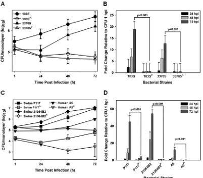
Multiple pVAPB-type plasmid-carryingR. equiisolates are capable of replication within murine macrophages. Currently, there is no published work examining the intramacrophage growth capabilities ofR. equistrains from swine typically containing the pVAPB-type plasmid. In order to assess whether the observed host species-plasmid type carriage dictates species-specific intramacrophage replication, we first assessed whether a swineR. equiisolate (33705) carrying a VapB-type plasmid possessed the ability to replicate within murine macrophages. The murine macrophage model is a well-establishedin vitromodel system of intracellular growth that has been shown to correlate with strain virulence (27, 31, 32). R. equiisolates carrying the pVAPA-type plasmid are known to be capable of replication within murinein vitro-cultured mac-rophages (4), and therefore, a pVAPA-positive isolate (103S) was utilized as a reference to measure the replicative potential of numerous pVAPB-type plasmid-containing strains. Murine bone marrow-derived macrophages (BMDMs) were infected with plasmid-containing and isogenic plasmid-cured derivatives of the indicated R. equi strains, and bacterial intracellular replication was followed by standard lysis and plating of the infected macrophage monolayers over the course of 72 h. As illustrated in Fig. 1A and B, both 103S, a well-characterized equine isolate originally obtained from a foal with R. equipneumonia and carrying pVAPA1037, and the swine isolate 33705, ob-tained from the lymph node of a pig, replicated in murine macrophages, showing an ~20- and an ~15-fold increase in CFU at 72 h postinfection (hpi) compared to 1 hpi, respectively. In contrast, the isogenic strains lacking a virulence plasmid (strains 103SP⫺
and 33705P⫺) failed to replicate intracellularly. To determine if the replicative ability of
33705 was a strain-specific phenotype, three additional pVAPB-type plasmid-containing isolates were similarly analyzed. The examined isolates were strains P117 and 21364B2, both obtained from pigs, as well as strain A5, a humanR. equiclinical isolate from a patient with AIDS (18). Notably, strain A5 has been classified as intermediately virulent in mice and foals relative to an equine isolate carrying pVAPA1037. However, its ability to replicate within macrophages had not been examined (18, 33). Plasmid-cured
Volume 1 Issue 5 e00186-16 msphere.asm.org 3
on September 8, 2020 by guest
http://msphere.asm.org/
isogenic derivatives of the abovementioned strains, created through repeated subcul-ture, were analyzed in parallel. As observed with the 33705 strain, the other swine isolates and the pVAPB-type plasmid-containing human strain A5 were found capable of intramacrophage replication, and plasmid curing was associated with the loss of replicative ability (Fig. 1C and D). Both swine strains, P117 and 21364B2, replicated ~50-fold over 72 h, whereas the human A5 isolate replicated less efficiently, ~15-fold at 72 hpi. These data show that while there is some degree of strain-to-strain variability in the magnitude of intracellular growth, pVAPB-type plasmid-carryingR. equistrains, like equine isolates carrying pVAPA-type plasmids, are able to replicate in murine macro-phages and intracellular replication is plasmid dependent.
The replicative ability of pVAPB-type plasmid-carrying R. equi isolates varies in equine macrophages. Once it was established thatR. equiisolates pos-sessing pVAPB-type plasmids were capable of replication in murine macrophages, the ability of the swine strains to replicate within equine alveolar macrophages was examined. Macrophages were infected with isolates 33705 and P117, along with their isogenic strains lacking a plasmid. As controls, these macrophages were also infected with the equine pVAPA-containing isolate 103S, known to replicate within these cells, and with its intracellular-growth-impaired plasmid-cured derivative, 103SP⫺(32).
Bac-FIG 1 pVAPB-type plasmid-containingR. equiisolates can replicate in murine macrophages. The intracellular growth of
R. equistrains was assessed in murine bone marrow-derived macrophages infected with strains 103S, 103SPⴚ, 33705, and 33705Pⴚ(A and B) and P117, P117Pⴚ, 21364B2, 21364B2Pⴚ, A5, and A5Pⴚ(C and D) at an MOI of 10:1. Following a 1-h incubation allowing for phagocytosis, monolayers were washed and medium supplemented with 20g/ml amikacin was added to the monolayers to prevent extracellular bacterial growth. Triplicate monolayers were lysed at 24 h, 48 h, and 72 h postinfection (hpi). (A and C) Intracellular growth ofR. equistrains following infection of macrophages. (B and D) Fold changes in CFU of intracellular bacteria at 24, 48, and 72 hpi relative to 1 hpi. Error bars represent standard deviations from the means. Data with statistical analysis are a compilation of 3 individual experiments.
Willingham-Lane et al.
on September 8, 2020 by guest
http://msphere.asm.org/
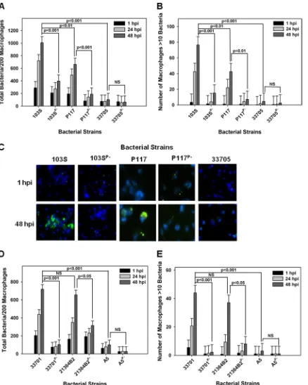
terial replication was examined and quantified by fluorescence microscopy in which the number of bacteria per 200 macrophages was recorded over time in triplicate at each time point. As an additional assessment of intracellular growth, the number of macro-phages containing greater than 10 bacteria per 200 macromacro-phages was also recorded as previously described (32). As expected, the equine isolate 103S carrying pVAPA1037 replicated within equine macrophages, whereas its plasmid-cured derivative 103SP⫺
did not (Fig. 2A to C). The swine isolate 33705 failed to grow within equine macro-phages regardless of plasmid possession. In contrast, strain P117, also originating from swine, exhibited plasmid-dependent replicative ability in these cells (Fig. 2A to C). Given that the two examined swine isolates demonstrated differences in replicative capacity, the intracellular growth of two additional pVAPB-type plasmid-carrying isolates, one from a pig (strain 21364B2) and the other from a person withR. equiinfection (A5), was examined. The replication potential of an additional equine isolate, 33701 carrying pVAPA1037, known to cause disease in mice and foals (15, 34), was used as a positive control for intracellular growth. As expected, strain 33701 displayed virulence plasmid-dependent replication within equine macrophages, as did the swine strain 21364B2 (Fig. 2D and E). In contrast, the pVAPB-type plasmid-containing human isolate A5 was unable to replicate within equine macrophages irrespective of plasmid possession, an outcome similar to that displayed by the swine strain 33705. These data presented in Fig. 2 demonstrate that someR. equiisolates carrying the pVAPB-type plasmid have the capacity to replicate within equine macrophages but that plasmid possession is not the sole criterion conferring replicative ability, since some pVAPB plasmid-containing iso-lates, such as 33705 and A5 specifically, lack this capability.
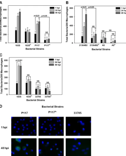
Examination of the intracellular replicative capacity of pVAPA-type and pVAPB-type plasmid-possessing R. equi isolates in swine macrophages. We next evaluated the growth potential of the two equine isolates discussed above, 103S and 33701, in swine monocyte-derived macrophages. Interestingly, as shown in Fig. 3A and B, both equine isolates carrying their pVAPA1037 plasmids were able to replicate in swine macrophages in a virulence plasmid-dependent manner. Then, the replicative capacity of the pVAPB-type plasmid-containingR. equiisolates was assessed in these cells. Similar to the findings with equine macrophages, we observed variability in the replicative ability among these isolates. For example, swine strains P117 and 21364B2 grew in swine macrophages (Fig. 4A, B, and D). Notably, the intracellular replication displayed by these two strains was plasmid dependent, as plasmid-free derivatives (P117P⫺and 21364B2P⫺) did not replicate. Much like the results observed with
infec-tion of equine alveolar macrophages, the swine strain 33705 (Fig. 4C and D) and the human isolate A5 (Fig. 4B) were unable to grow in swine macrophages despite being able to do so in murine macrophages. Once again, these data showed variable intracellular replicative ability among the pVAPB-type plasmid-containing isolates; however, the strains found capable of intracellular replication in equine macrophages were the same ones demonstrating this capacity in swine macrophages.
R. equi transconjugants demonstrate intracellular growth characteristics consistent with those of the strain’s parental chromosomal background.As mentioned, it has been reported thatR. equiplasmid type possession, pVAPA versus pVAPB type, is typically host species specific (10, 11, 15, 16, 26, 29, 34), with pVAPA- and pVAPB-type plasmid carriage characteristic of equine and swine isolates, respectively. A traditional means to determine whether a gene or set of genes is important for host species tropism is to express the gene(s) of interest in a strain of different tropism and evaluate the consequences. Therefore,R. equistrains were constructed via conjugation in which pVAPA- and pVAPB-type plasmids were transferred to plasmid-cured deriva-tive recipient strains originating from a different host species (19). For example, 103SP⫺/A, a pVAPA1037-cured derivative of equine origin, was provided the
pVAPB-type plasmid from the swine isolate 33705, and a pVAPB-free version of the latter (33705P⫺/T) received the pVAPA1037 plasmid of strain 103S. Complete virulence
plasmid transfer was verified through PCR analysis using primer pairs that amplified various regions of the plasmid and also confirmed the presence of eithervapAorvapB
Volume 1 Issue 5 e00186-16 msphere.asm.org 5
on September 8, 2020 by guest
http://msphere.asm.org/
FIG 2 pVAPB-type plasmid-containingR. equiisolates display variable replication ability in equine macrophages. The intracellular growth ofR. equiin equine alveolar macrophages infected with strains 103S, 103SPⴚ, P117, P117Pⴚ, 33705, 33705Pⴚ, 33701, 33701Pⴚ, 21364B2, 21364B2Pⴚ, A5, and A5Pⴚat an MOI of 5:1 was assessed. Following a 1-h incubation allowing for phagocytosis, monolayers were washed and medium supplemented with amikacin was added to the monolayers to kill any extracellular bacteria. Triplicate monolayers were fixed at 1 h, 24 h, and 48 h postinfection (hpi), stained as described in Materials and Methods, and examined under fluorescence microscopy. The number of bacteria per 200 macrophages (A and D) and the number of macrophages with greater than 10 bacteria per 200 macrophages (B and E) were counted. Representative images of the infected macrophage monolayers are shown (ⴛ100 magnification) (C). In these representative microscopy images,R. equi displays green fluorescence and the macrophage nucleus is DAPI stained. Error bars represent standard deviations from the means. Data and statistical analysis are a compilation of 4 individual experiments. NS, not significant.
Willingham-Lane et al.
on September 8, 2020 by guest
http://msphere.asm.org/
(see Fig. S1A to C in the supplemental material) as well as the presence of the chromosomal antibiotic resistance gene used to mark and identify the specific recipient strain (see Fig. S1D). Subsequently, the intracellular growth potential of these transcon-jugant strains was examined initially in murine macrophages. As demonstrated in Fig. 5A and B, the transconjugant strains 103SP⫺/A-p33705 and 33705P⫺/T-p103
car-rying nonnative plasmids were found capable of replication in these cells. However, the transconjugant 103SP⫺/A-p33705 replicated significantly less efficiently than 103S with
its native plasmid, with an ~18-fold increase in bacterial numbers compared to an ~30-fold increase in bacterial numbers over 72 hpi, respectively. To further examine the growth potential ofR. equiisolates in possession of nonnative plasmids, two additional transconjugant strains were created, verified by PCR analysis as previously described, and then similarly examined. The plasmid-cured derivative 103SP⫺/A of equine origin
was provided the swine P117 pVAPB-type plasmid (see Fig. S1B), and the plasmid-free product 21364B2P⫺/Z (see Fig. S1C) of swine origin was given the equine 33701
pVAPA1037 virulence plasmid. Similarly, these transconjugants, 103SP⫺/A-pP117 and
2134B2P⫺/Z-p33701, respectively, were found proficient for intracellular replication in
murine macrophages (Fig. 5C to F). Cumulatively, these data illustrate that within the murine model system of intramacrophage growth, nonnative-plasmid-type carriage does allow anR. equistrain to replicate intracellularly.
We next extended our analysis of the intracellular growth phenotype of the transconjugant strains to equine macrophages. Figure 6A, B, and D illustrates that the equine 103SP⫺strain possessing a pVAPB-type plasmid from either swine isolate 33705
or P117 replicated in equine macrophages. Likewise, the swine 21364B2P⫺ strain
possessing the pVAPA1037 plasmid from the equine strain 33701 (strain 21364B2P⫺/
Z-p33701) demonstrated replicative ability in equine macrophages (Fig. 6C and D). In contrast, the swine 33705P⫺ isolate possessing the equine 103S plasmid did not
replicate in equine macrophages (Fig. 6A and D). The latter finding is particularly interesting in light of the fact that the wild-type parent 33705 swine isolate carrying its endogenous pVAPB-type plasmid was also unable to multiply within equine cells. These results suggest that some component(s) essential for equine macrophage intracellular replication is lacking in or not induced in the genetic background (i.e., chromosome) of this strain.
Last, we assessed the intracellular growth capabilities of the transconjugants in swine macrophages. As shown in Fig. 7A and C, the plasmid-cured strain of equine
FIG 3 EquineR. equiisolates containing the pVAPA1037 plasmids replicate in swine macrophages. The intracellular growth of equine
R. equistrains 103S, 103SPⴚ, 33701, and 33701Pⴚ(A and B) was assessed in swine monocyte-derived macrophages infected at an MOI of 5:1. Triplicate monolayers were fixed and stained at 1 h, 24 h, and 48 h postinfection (hpi), and the number of bacteria per 200 macrophages was determined (A). Representative microscopy images of monolayers infected withR. equistrains 103S and 33701 at 1 h and 48 h postinfection are shown (ⴛ60 magnification) (B). In these images,R. equiexhibits green fluorescence and the macrophage nucleus is blue because of DAPI staining. Statistical analysis was performed on the data compiled from 2 individual experiments.
Volume 1 Issue 5 e00186-16 msphere.asm.org 7
on September 8, 2020 by guest
http://msphere.asm.org/
origin, 103SP⫺/A, in possession of a pVAPB-type plasmid transferred from the swine
isolate 33705 replicated in these macrophages as efficiently as the parent strain (103S) carrying its native pVAPA1037 plasmid. Similarly, the swine 21364B2P⫺/Z isolate
carry-ing the equine 33701 pVAPA1037 plasmid displayed intracellular growth equivalent to that of swine isolate 21364B2 containing its native plasmid (Fig. 7B and C). Like the
FIG 4 pVAPB-type plasmid-containingR. equiisolates display variable replication ability in swine macrophages. Bacterial intracel-lular growth was assessed in swine monocyte-derived macrophages infected withR. equistrains 103S, 103SPⴚ, P117, and P117Pⴚ(A), 21364B2, 21364B2Pⴚ, A5, and A5Pⴚ(B), and 103S, 103SPⴚ, 33705, and 33705Pⴚ(C) at an MOI of 5:1. Triplicate monolayers were fixed at 1 h, 24 h, and 48 h postinfection (hpi) and stained. Then, the number of bacteria per 200 macrophages (A, B, and C) was counted using fluorescence microscopy. Representative microscopy images are shown (ⴛ60 magnification) (D). Statistical analysis was performed on data compiled from 3 individual experiments. NS, not significant.
Willingham-Lane et al.
on September 8, 2020 by guest
http://msphere.asm.org/
FIG 5 R. equicarriage of nonnative plasmids enables replication in murine macrophages. The intracellular growth of R. equi
transconjugant strains was assessed in bone marrow-derived macrophages infected with 103S, 103SPⴚ, 103SPⴚ/A-p33705, 33705,
(Continued)
Volume 1 Issue 5 e00186-16 msphere.asm.org 9
on September 8, 2020 by guest
http://msphere.asm.org/
findings observed in equine macrophages, transconjugant strain 33705P⫺/T-p103,
originally of swine origin carrying the 103S pVAPA1037 equine plasmid, was unable to grow within swine macrophages (Fig. 7A and C). In sum, the findings that the transcon-jugant strains 103SP⫺/A-p33705 and 21364B2P⫺/Z-p103 can replicate in swine
macro-phages confirm once again that nonnative plasmid carriage can promote the intracel-lular growth of some R. equi strains and that coevolution of the plasmid and chromosome is not essential for this trait. The observation that strain 33705P⫺/T-p103
cannot replicate in these cells implies that the chromosomal background also plays a critical role in determining a given R. equi isolate’s ability to survive and grow in macrophages. Cumulatively, these data show that the host species tropism displayed by pVAPA- and pVAPB-type plasmid-containingR. equiisolates cannot be explained by species-specific intramacrophage replication.
DISCUSSION
It has long been acknowledged that clinical isolates of the facultative intracellular bacteriumR. equiobtained from foals with pneumonia and associated disease mani-festations carry an ~80-kb virulence plasmid (pVAPA1037) and express the crucial virulence determinant VapA (15, 16, 34). Also well established is the finding that the majority of R. equi isolates from swine carry a highly related but different plasmid (pVAPB-type) and express Vap proteins distinct from VapA, including VapB (10, 11, 21, 29). We hypothesized that particular plasmid types were associated with specific hosts because plasmid type allowed for intramacrophage replication capabilities in a host-specific manner. Interestingly, the data presented here reveal that plasmid type does not confer species-specific macrophage replication abilities.R. equiisolates possessing either pVAPA-type or pVAPB-type plasmids are capable of replication within macro-phages derived from various host species, including the mouse, horse, and pig.
Since it appears thatR. equihas the capacity to replicate in macrophages regardless of plasmid type or macrophage species, it raises the question why pVAPB-type plasmid-carryingR. equistrains are not isolated from diseased foals. A simple explanation is that foals are not exposed to strains ofR. equithat carry this plasmid type. Currently, there is a paucity of knowledge regarding the epidemiology of R. equi isolates carrying pVAPB-type plasmids. Despite the large number of studies examining the presence of R. equiin the soil and its impact on foal disease, the majority of studies have simply determined plasmid possession through the presence or absence of vapA (35–40). Other plasmid components, including the conserved genetraA, are rarely screened for, but if that were done, it could identifyR. equiisolates carrying various plasmid types, and additional screening could be used to determine specific plasmid type (41). Occasionally, studies have analyzed soil samples for the presence of both pVAPA- and pVAPB-type plasmids, and interestingly, most of the collected R. equi isolates are negative for both types (42, 43). In Hungary, Major and colleagues analyzed 48 soil samples from stud farms forvapAandvapB, where 54.2% of the isolates werevapA positive and the remaining were negative for both genes (44). If these results are generally representative of the R. equi populations found in the soil of horse farms worldwide, the absence of foals infected withR. equistrains possessing pVAPB-type plasmids could be due to a lack of exposure of the foal to bacteria carrying this plasmid type and not because of an inability of such a bacterium to cause disease in the equine host. pVAPB-type plasmid-carryingR. equistrains have been isolated from the soil of pig farms (23); however, very little is known about the frequency at whichR. equiisolates containing pVAPB-type plasmids are found in the environment in general or their environmental stability, especially within the environment of foals.
Figure Legend Continued
33705Pⴚ, and 33705Pⴚ/T-p103 (A and B), 103S, 103SPⴚ, 103SPⴚ/A-pP117, and P117 (C and D), and 21364B2, 21364B2Pⴚ, 21364B2Pⴚ/ Z-p33701, and 33701 (E and F) at an MOI of 10:1. Triplicate monolayers were lysed at 24 h, 48 h, and 72 h postinfection (hpi). (A, C, and E) Intracellular growth ofR. equistrains following infection of macrophages expressed as CFU per monolayer over time. (B, D, and F) Fold changes in CFU of intracellular bacteria at 24, 48, and 72 hpi relative to 1 hpi. Error bars represent standard deviations from the means. Statistical analysis was performed on a compilation of 2 separate experiments. NS, not significant.
Willingham-Lane et al.
on September 8, 2020 by guest
http://msphere.asm.org/
FIG 6 Effect of nonnative plasmid carriage and chromosomal background onR. equiintracellular growth phenotype in equine alveolar macrophages. (A to C) The intracellular growth ofR. equitransconjugant strains was assessed in equine alveolar macro-phages infected with 103S, 103SPⴚ, 103SPⴚ/A-p33705, 33705, 33705Pⴚ, and 33705Pⴚ/T-p103 (A), 103S, 103SPⴚ, 103SPⴚ/A-pP117, and P117 (B), and 21364B2, 21364B2Pⴚ, 21364B2Pⴚ/Z-p33701, and 33701 (C) at an MOI of 5:1. Triplicate monolayers were fixed 1 h, 24 h, and 48 h postinfection (hpi) and stained, and the number of bacteria per 200 macrophages was counted under fluorescence microscopy. (D) Representative microscopy images are shown (ⴛ60 magnification). Statistical analysis was performed on data compiled from 2 individual experiments. NS, not significant.
Volume 1 Issue 5 e00186-16 msphere.asm.org 11
on September 8, 2020 by guest
http://msphere.asm.org/
Given thatR. equistrains carrying pVAPB-type plasmids have the ability to replicate in equine alveolar macrophages, it would be of interest to perform a head-to-head challenge experiment in foals wherein the disease-causing capacity of a pVAPA-type-possessing R. equistrain is directly compared to that of an R. equistrain carrying a pVAPB-type plasmid. Such a study was attempted several years ago (34). In this study, two foals were intratracheally infected with either 105CFU or 106 CFU of the clinical equine isolate 33701, containing pVAPA1037 (33). Two other foals were similarly infected withR. equistrain A5, the human isolate possessing pVAPB, at a challenge dose of 106or 109CFU, respectively. Both foals infected with 33701, at either bacterial dose, developed severe clinical signs, including depression, anorexia, and pyrexia (40.5°C), at days 12 to 14 postchallenge. The foal challenged with 106CFU ofR. equistrain 33701 succumbed to infection on day 22 postinfection. In contrast, the foal infected with 106
FIG 7 Effect of nonnative plasmid carriage and chromosomal background onR. equigrowth capabilities in swine macrophages. The intracellular growth ofR. equistrains carrying native plasmids and transconjugant strains carrying nonnative plasmids was assessed in swine monocyte-derived macrophages infected with 103S, 103SPⴚ, 103SPⴚ/A-p33705, 33705, 33705Pⴚ, and 33705Pⴚ/T-p103 (A) and 21364B2, 21364B2Pⴚ, 21364B2Pⴚ/Z-p33701, and 33701 (B) at an MOI of 5:1. Following a 1-h incubation allowing for phagocy-tosis, monolayers were washed and medium supplemented with amikacin was added to kill any extracellular bacteria. Triplicate monolayers were fixed at 1 h, 24 h, and 48 h postinfection (hpi) and stained, and the number of bacteria per 200 macrophages (A and B) was determined. Representative microscopy images are shown (ⴛ60 magnification) (C). Statistical analysis was performed on data compiled from 2 individual experiments. NS, not significant.
Willingham-Lane et al.
on September 8, 2020 by guest
http://msphere.asm.org/
CFU ofR. equipVAPB-type plasmid-possessing strain A5 displayed no clinical signs at any time, andR. equiwas not isolated from tracheal washes or fecal samples. The foal infected with a 1,000-fold-higher bacterial dose of strain A5, 109 CFU, did develop clinical signs, including pyrexia (40°C) and depression, at day 21 postinfection, but symptoms were far less severe than those of the foals infected with a much lower dose of the pVAPA1037-containingR. equiisolate. These data implied that anR. equiisolate in possession of a pVAPB-type plasmid is less virulent in foals than strains of R. equi carrying a pVAPA-type plasmid. Interestingly, the results of our experiments presented here demonstrate that the pVAPB-containing isolate A5 is less efficient at replicating within murine macrophages than other plasmid-containingR. equiisolates in posses-sion of either a pVAPA- or pVAPB-type plasmid. Moreover, we found it completely unable to replicate within equine alveolar or monocyte-derived swine macrophages. Our data provide an explanation for why the A5 strain was unable to establish an infection in foals unless a high inoculum was used, essentially overpowering the biological or immunological system. We speculate that if another pVAPB-type plasmid-containingR. equistrain (for example, strain 21364B2 or P117) that displayed replicative ability in equine alveolar macrophages were utilized in a similar challenge of foals, then overt disease might develop at an infection dose similar to that observed in foals challenged with a clinicalR. equiisolate possessing the pVAPA-type plasmid.
In the event that R. equiisolates carrying a pVAPB-type plasmid are found in the environment, foals exposed to these isolates may develop characteristicR. equi pneu-monia but are treated without confirmative plasmid type carriage determination by diagnostic microbiology laboratories. Additionally, microbiological identification of R. equican be and often is done without assessment ofvapgene possession; therefore, it is possible that microbiology laboratories are not recognizing pVAPB-type plasmid possession by R. equi isolates in clinical samples from foals. Alternatively, R. equi carriage of a pVAPB-type plasmid may lead to self-resolving subclinical disease in foals. Ultrasonography has been a method utilized to identifyR. equidisease prior to clinical presentation (45–47). During these studies, it was determined that the majority of foals that developed ultrasonographic evidence of pulmonaryR. equidisease resolved the infection prior to the development of clinical disease presentation (47). It is feasible that R. equiisolates in possession of either a pVAPA- or a pVAPB-type plasmid are capable of establishing an infection but that only those strains in possession of a pVAPA-type plasmid progress to clinical disease in foals.
Another potential explanation of whyR. equistrains carrying pVAPB-type plasmids are not isolated from foals is that these strains do not establish infection and produce disease in foals in the same manner as do pVAPA-containing strains. Perhaps,R. equi possessing a pVAPB-type plasmid causes a disease state in foals with a presentation more like that observed in swine. Swine are frequently clinically asymptomatic with lymph node abscessation revealed only upon postmortem examination at slaughter. Since the submandibular lymph nodes of seemingly healthy foals are not routinely examined for the presence ofR. equi, if disease were located there it could be missed altogether. Additionally, as the foal ages and becomes less immunologically naive, presumably the pVAPB-containing isolate, if present, would be cleared.
Little work has been done addressing the pathogenesis ofR. equidisease in pigs. However, one study examined the pathogenicity of the pVAPA1037-containing equine clinicalR. equiisolate 33701 and the human clinicalR. equiisolate, A5, in possession of a pVAPB-type plasmid in pigs. Pigs were challenged in one of two ways. The first was a single intravenous (ear vein) dose of 108or 109CFU ofR. equistrain 33701 or A5 in possession of the pVAPA1037- or pVAPB-type plasmid, respectively, along with the plasmid-cured derivative of 33701 given at an inoculum of 109CFU (48). In the second challenge experiment, pigs were administered two bacterial doses of 109CFU ofR. equi strain 33701, A5, or the plasmid-cured 33701 by intramuscular injection, given 7 days apart (48). Overall, regardless of plasmid type possession or route of infection, none of the pigs developed clinical signs beyond a transient low-grade fever and weight loss. Interestingly, upon examination of the group of intramuscularly challenged pigs,
Volume 1 Issue 5 e00186-16 msphere.asm.org 13
on September 8, 2020 by guest
http://msphere.asm.org/
bacterial recovery from the mandibular lymph nodes of the pig inoculated with the pVAPB plasmid-containingR. equiisolate was higher than that from the pig inoculated with the R. equi strain carrying the pVAPA1037-type plasmid, specifically 6 ⫻ 106 compared to 1⫻105CFU/g of tissue, respectively (48). Additionally, the plasmid-cured derivative of the equine strain 33701 was not isolated from the mandibular lymph nodes of pigs challenged with thisR. equistrain (48). All other organs obtained from these pigs were negative for all three bacterial strains. This may suggest that there is a preference for the establishment of an infection by R. equi isolates carrying a pVAPB-type plasmid in the mandibular lymph nodes of swine. It would be intriguing to know if a pVAPB-containingR. equiisolate shown to replicate in swine macrophages (R. equistrain 21364B2 or P117) would produce more dramatic results.
It has been demonstrated that VirR, VirS, and VapA are the only plasmid-encoded proteins on the pVAPA1037-type plasmid required for intracellular replication in murine macrophages (49). The presence of VapA, the 17-kDa protein product of thevapAgene, is thought to interfere with normal phagosomal acidification, thereby creating a vacuolar environment conducive to bacterial intracellular survival and replication (4, 28, 50, 51). Importantly, VirR and VirS not only regulatevapAexpression but also influence the expression of ~18% of chromosomal genes, having a large impact on the regulatory network of the cell and modulating bacterial physiology to allow adaption to the macrophage environment (49). It has been suggested that acquisition of the horizon-tally acquired PAI region was advantageous for R. equi to survive interactions with predatory protozoa found in abundance in the soil and water (49, 52). While initially VirR and VirS likely minimally influenced the bacterial chromosome, over time and through evolutionary pressure the chromosomal binding efficiency of these proteins improved and their binding was a pivotal event promoting intracellular survival and replication. It is of relevance that the pVAPB-type plasmid carries homologs of the pVAPA-type VirR and VirS proteins, specifically pVAPB1593_0480 and pVAPB1593_0530, which share 92% and 86% amino acid sequence identity, respectively (21). Due to the high degree of sequence similarity, it is likely that the pVAPB-type plasmid-encoded VirR and VirS homologs function similarly and engage in cross talk with the bacterial chromosome to enable intramacrophage survival. However, the differences in protein sequence of these homologs could impart alterations in chromosomal gene regulation between anR. equiisolate possessing the pVAPA-type plasmid and one possessing the pVAPB-type plasmid, and such differences in cross talk could contribute to the host species-specific niche of the equine and swine isolatesin vivo.
Despite the coevolution of the PAI-encoded regulators with a given bacterial chromosome, virulence plasmid swapping via conjugation showed that intramac-rophage growth was possible in a strain carrying a nonnative plasmid of a different host species origin. In addition, it was shown that the replicative ability of an isolate is affected by the chromosomal background, wherein genes essential for this trait are known to reside (53–56). Chromosomal influence was illustrated through the exami-nation of several transconjugant strains, for example, strain 103SP⫺/A-p33705, the
equine 103SP⫺strain housing a pVAPB-type plasmid transferred from the swine isolate
33705. 103SP⫺with its native pVAPA1037-type plasmid, known as 103S, was able to
replicate within all three macrophage species analyzed. In contrast, the swine isolate 33705 demonstrated growth only in murine macrophages, lacking the capacity to replicate in macrophages of both equine and swine origin. Intriguingly, the transcon-jugant 103SP⫺/A-p33705 displayed an intracellular growth phenotype like that of 103S
carrying its native VAPA1037-type plasmid, suggesting that the swine pVAPB-type plasmid from 33705 is fully functional, and the inability of this swine isolate to replicate in equine and swine macrophages aligns with the chromosomal background. This observation was further exemplified by analysis of the reverse transconjugant, 33705P⫺/T-p103, a strain that, like 33705, was unable to replicate in equine and swine
macrophages, once again showing the influence of the bacterial chromosome. To date, only oneR. equichromosome sequence, that of strain 103S of equine origin, has been published. The availability of additional chromosomal sequences of both
Willingham-Lane et al.
on September 8, 2020 by guest
http://msphere.asm.org/
equine and swine isolates would be informative and might reveal features specific to R. equiisolates possessing different plasmid types. While these chromosomal charac-teristics may not directly influence bacterial replication within the macrophage, they may nonetheless play a role in vivo in the host. For example, differences in gene expression amongR. equiisolates could result in variedin vivohost cytokine induction profiles that affect microbial survival, thus contributing to the observed host species tropism.
In conclusion,R. equistrains possessing either a pVAPA- or pVAPB-type plasmid are capable of replication in the three examined macrophage species (murine, equine, and swine). This work suggests that the apparent host species tropism of R. equiis not expressed at the level of the intramacrophage replication and is determined by an as-yet-to-be-defined bacterium-host interaction not observable through the analyses performed here. Additionally, through the examination of various transconjugant strains, it was shown that the chromosomal background influences the degree of replicative ability of the bacterial strain regardless of plasmid type, findings which further support previous work showing the profound impact of cross talk between the virulence plasmid and chromosome (49).
MATERIALS AND METHODS
Bacterial strains and growth conditions.R. equistrains used in this study are listed in Table 1.R. equi
103S was originally isolated from a foal withR. equipneumonia. ATCC 33701 and 33705 were obtained from the American Type Culture Collection (ATCC, Manassas, VA) and were originally isolated from a foal and pig, respectively, withR. equidisease. Strains P117 and 21364B2 were isolated from individual pigs,
TABLE 1 Bacterial strains and plasmids
Strain or plasmid Genotype or characteristic Reference or source
Rhodococcus equistrains
103S Wild-type strain with virulence plasmid pVAP1037, expressingvapA; originally isolated from a pneumonic foal
31
103SP⫺ Plasmid-cured variant of 103S 57
103SP⫺/A 103P⫺containing anaac(3)-IVgene integrated on the chromosome; Aprr 19 33701 Wild-type strain with virulence plasmid pVAP1037, expressingvapA; originally isolated
from a pneumonic foal
ATCC
33701P⫺ Plasmid-cured variant of 33701 19
33705 R. equistrain with pVAPB-type virulence plasmid originally isolated from the lymph node of a pig
ATCC
33705P⫺ Plasmid-cured variant of 33705 19
33705P⫺/T 33705P⫺containing adhfrgene of mouse origin integrated on the chromosome; Trimr This study P117 R. equistrain with pVAPB-type virulence plasmid originally isolated from the lymph
node of a pig
Gift from John Prescott, University of Guelph
P117P⫺ Plasmid-cured variant of P117 This study
21364B2 R. equistrain with VAPB-type virulence plasmid originally isolated from a pig Gift from John Prescott, University of Guelph
21364B2P⫺ Plasmid-cured variant of 21364B2 This study
21364B2P⫺/Z 21364B2P⫺containing theblegene ofStreptoalloteichus hindustanusintegrated on the
chromosome; Zeor
This study
A5 R. equistrain with virulence plasmid pVAPB1593 originally isolated from a person with R. equipneumonia
33
A5P⫺ Plasmid-cured variant of A5 This study
103SP⫺/A-p33705 TC Transconjugant of strain 103P⫺/A and strain 33705, carrying the pVAPB-type plasmid
from 33705; Aprr
This study
103SP⫺/A-pP117 TC Transconjugant of strain 103P⫺/A and strain P117, carrying the pVAPB-type plasmid
from P117; Aprr
This study
33705P⫺/T-p103 TC Transconjugant of strain 33705P⫺/T and 103S, carrying the pVAPA1037 plasmid from
103S; Trimr
This study
21364B2P⫺/Z-p33701 TC Transconjugant of strain 21364B2P⫺/Z and 33701, carrying the pVAPA1037 plasmid from
33701; Zeor
This study
Plasmids
pSET152 aac(3)-IVphiC31 integraseattP; Aprr 58
pVM6 pSET152 withdhfrgene of mouse origin;aac(3)-IVremoved; phiC31 integraseattP; Trimr 19 pSET152.zeo.1 pSET152 withblegene ofStreptoalloteichus hindustanus; AprrZeor This study
pSET152.zeo pSET152.zeo.1 lackingaac(3)-IV; Zeor This study
Volume 1 Issue 5 e00186-16 msphere.asm.org 15
on September 8, 2020 by guest
http://msphere.asm.org/
whereas A5 was obtained from a person withR. equipneumonia, and all were a gift from John Prescott (University of Guelph). Plasmid-cured derivatives of the abovementionedR. equistrains, 103P⫺(57),
33701P⫺, 33705P⫺, P117P⫺, 21364B2P⫺, and A5P⫺, were created by serial subculture at 37°C until the
virulence plasmid was lost. Virulence plasmid loss was confirmed by PCR analysis using primer pairs (Table 2) which anneal to various regions of the plasmid. Bacterial strains were grown at either 30°C or 37°C (shaking, 200 rpm) in brain heart infusion (BHI) broth. When necessary, antibiotics were added at the indicated concentrations: apramycin (Apr), 80g/ml; trimethoprim (Trim), 50g/ml; zeocin (Zeo), 50g/ml.
Plasmid construction.To mark the chromosome of recipient cells, a new version of the previously described integrating vector pSET152 (58) was created, wherein the apramycin resistance gene was replaced by theblegene ofStreptoalloteichus hindustanus, providing resistance to zeocin. To do this, pSET152 was digested with XbaI and EcoRV, resulting in a linearized 5,689-bp DNA fragment. The pEM7/Zeo plasmid (Thermo Scientific) was similarly digested, leading to the isolation of the zeocin resistance cassette under the EM7 bacterial promoter (477 bp). The zeocin resistance cassette was ligated with the linearized pSET152 vector, creating pSET152.zeo.1. For removal of the apramycin resistance cassette, pSET152.zeo.1 was digested with SacI, producing a 5.4-kb and a 751-bp DNA fragment. The larger fragment was then self-ligated, generating pSET152.zeo.
Creation of transconjugants through bacterial conjugation.Mating was performed as described by Tripathi et al. (19); severalR. equistrains were marked via the insertion of an integrating vector carrying a specific and varied antibiotic resistance cassette (Table 1). The virulence plasmid-cured 103P⫺
derivative was chromosomally marked with the geneaac(3)-IVproviding apramycin resistance (desig-nated Aprror A). Similarly, theR. equi strain 33705P⫺ was marked via insertion of a dihydrofolate
reductase (dhfr) gene conferring trimethoprim resistance (designated Trimror T) (19). Strain 21364B2P⫺
was marked with theblegene ofStreptoalloteichus hindustanus, providing resistance to zeocin (desig-nated Zeoror Z). To facilitate virulence plasmid transfer, equal numbers of chromosomally marked
recipient (103P⫺/A, 33705P⫺/T, or 21364B2P⫺/Z) and unmarked donor (103S, 33705, P117, or 33701)
strains were used. The donor and recipient strains were grown overnight at 37°C in BHI broth with the recipient cultures supplemented with the appropriate antibiotic; the next day, the optical density at 600 nm (OD600) was adjusted to 1.0 (~2⫻108CFU/ml). Approximately 107CFU of both donor and recipient
bacteria was centrifuged together, resuspended in a small volume (5 to 10l), and spotted on BHI agar, and the plates were incubated for 72 h at 30°C. Afterward, the cell mixture was scraped from the plates and resuspended in 1 ml phosphate-buffered saline (PBS). Serial dilutions (up to 1:10⫺7) of the
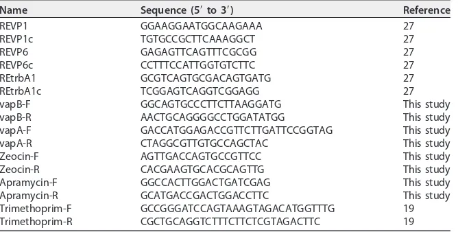
resuspended cells were plated on agar containing the appropriate antibiotics for selection of putative transconjugants as well as recipients. A mixture of colonies from these plates was used to infect murine macrophages as described below in order to amplify and select the lesser-represented transconjugant over the plasmid-cured recipient. At 48 h postinfection, the macrophage monolayer was lysed, the lysate was plated on BHI agar with the appropriate antibiotic, and the plates were incubated for 48 h. Resultant putative transconjugant colonies were screened for the existence of the transferred virulence plasmid using PCR analysis, confirming the presence of the replication/partitioning (primers REVP1/c [Table 2]), conjugation (primers REVP6/c [Table 2]), unknown function (primers REtrbA1/c [Table 2]), and pathoge-nicity (primers vapA-F and vapA-R or vapB-F and vapB-R [Table 2]) regions as well as the appropriate antibiotic resistance marker (Table 2).
Bone marrow-derived macrophages.To obtain macrophage precursors, the marrow of femurs and tibias of female BALB/c mice was flushed into a 50-ml conical tube using cold cation-free phosphate-buffered saline (PBS) supplemented with penicillin G (100 U/ml)-streptomycin (100g/ml) (PGS). The cells were centrifuged at 260⫻gfor 10 min at 4°C. The cell pellet was then resuspended in complete medium consisting of Dulbecco modified Eagle medium (DMEM) containing 10% fetal bovine serum (FBS), 10% colony-stimulating factor 1 (CSF-1) from CSF-1-producing L929 cells, and 2 mM glutamine. Cells were washed by centrifugation at 260⫻gfor 10 min. The final resuspension of cells was done in complete medium with 24 ml per mouse used. Then, the precursor cells were plated in 6-well non-tissue-culture-treated plates (4 ml per well) and incubated at 37°C with 5% CO2. On the third day,
4 ml of complete medium was added to each well. On day 5, the medium was removed and replaced with complete medium without antibiotic; then, on day 6, the medium was aspirated and each well was washed with 4 ml PBS to remove any nonadherent/dead cells. Then, 4 ml of cold, cation-free PBS was added and the plates were refrigerated at 4°C for 15 min. Afterward, the cells were collected, quantified, and seeded into tissue-culture-treated 24-well plates at a concentration of 2⫻105per well.
Equine alveolar macrophages.To acquire alveolar macrophages, a bronchoalveolar lavage (BAL) was performed on adult horses sedated with xylazine hydrochloride (0.5 mg/kg of body weight intravenously [i.v.]) and butorphanol tartrate (0.02 mg/kg i.v.). A sterile BAL catheter was passed via the nasal cavity and wedged within a bronchus. Four aliquots of 60 ml (~240-ml total) of sterile physiologic saline (0.9% NaCl) solution was infused into the horse’s lungs and aspirated immediately. Once collected, the BAL fluid was centrifuged at 260⫻gfor 10 min at 4°C, and the pellet was resuspended in 50 ml PBS followed by another similar centrifugation. This washing step was repeated two additional times. The final cell pellet was resuspended in minimum essential medium alpha (MEM␣) supplemented with 10% donor horse serum (DHS) (Thermo Scientific), 2 mM glutamine, and PGS. Following quantification, 4⫻ 105cells were pipetted into each well of a 24-well tissue culture plate, wherein each well contained a
13-mm glass coverslip. The plates were incubated at 37°C with 5% CO2for 4 h to allow for macrophage
adherence. After 4 h, the wells were washed 3 times with MEM␣to remove nonadherent cells and then the medium was replaced with antibiotic-free MEM␣plus 10% DHS plus 2 mM glutamine and the cells were incubated overnight at 37°C with 5% CO2.
Willingham-Lane et al.
on September 8, 2020 by guest
http://msphere.asm.org/
Porcine monocyte-derived macrophages.From each pig, 50 ml of blood was drawn and placed into a collection vial containing 158 USP units of sodium heparin. The blood was diluted with an equal volume of PBS and layered over Ficoll (15 ml Ficoll per 35 ml of blood-PBS) and then centrifuged at 700⫻
gfor 30 min at 4°C without braking to allow for proper Ficoll gradient formation. After centrifugation, the buffy coat layer, containing leukocytes, was transferred to a new sterile 50-ml conical tube which was then filled to a final volume of 35 ml with PBS and centrifuged at 260⫻gfor 15 min at 4°C. Following centrifugation, the supernatant was discarded and the cells were resuspended in 35 ml PBS supple-mented with PGS and centrifuged two additional times. After the final wash, the cells were quantified and resuspended at a final concentration of 5⫻106per ml in DMEM supplemented with 10% FBS, 10%
CSF-1, 2 mM glutamine, and PGS. The cell suspension was transferred to a 75-cm2tissue-culture-treated
flask (5⫻107cells per flask) and incubated at 37°C with 5% CO
2overnight. The next day, the medium
was gently removed and replaced with fresh medium supplemented with PGS. The cells were incubated in antibiotic-containing medium for 3 days, and then the medium was replaced with non-antibiotic-containing DMEM supplemented with 10% FBS and 2 mM glutamine. The non-antibiotic-non-antibiotic-containing medium was removed and replaced with fresh medium at least once more prior to macrophage harvesting on day 7 of culture. For recovery, cells were washed once with PBS and incubated at 37°C for 5 min in PBS supplemented with 1 mM EDTA. Following collection and centrifugation at 4°C, the cells were resuspended in DMEM supplemented with 10% FBS and 2 mM glutamine. Cells were placed in a 24-well tissue culture plate containing 13-mm glass coverslips at a concentration of 2⫻105cells per well.
Bacterial intracellular growth assay.Overnight bacterial broth cultures were grown to an optical density at 600 nm (OD600) of 1.0 (~2.0⫻108CFU/ml), washed once with PBS, and resuspended to the
original culture volume in PBS. Macrophage monolayers (bone marrow-derived macrophages [BMDMs] and equine and porcine macrophages) were washed once with warm DMEM. The medium was replaced with DMEM supplemented with 10% FBS and 2 mM glutamine (equine macrophages) and 10% FBS, 10% CSF-1, and 2 mM glutamine (BMDMs and porcine macrophages). Bacteria were added at a multiplicity of infection (MOI) of 5 to 10 bacteria per macrophage. Following 60 min of incubation at 37°C to allow for bacterial binding and uptake, the monolayers were washed 3 times with DMEM to remove any unbound bacteria and the appropriate medium containing 20g/ml of amikacin sulfate was added to the monolayers in order to prevent extracellular bacterial replication. At various times postinfection, the macrophage monolayers were washed repeatedly and lysed by the addition of 500l of sterile water, and the lysate was collected and plated onto BHI agar. The number of CFU associated with the macrophage lysate was determined after a 48-h incubation at 37°C (BMDMs). Alternatively, the monolayers were fixed with cold 100% methanol for 30 min at 4°C and the associated bacteria were stained with polyclonal rabbit anti-R. equi
antibody followed by a fluorescein isothiocyanate (FITC)-labeled goat anti-rabbit secondary antibody allowing for the enumeration of the bacteria under fluorescence microscopy.
Fluorescent staining ofR. equi-infected macrophages.Macrophage monolayers on glass cover-slips were fixed with 100% methanol for 30 min at 4°C and then washed once with PBS. Then, primary polyclonal rabbit anti-R. equiantibody, diluted 1:1,000 in PBS containing 5% normal goat serum (NGS), was added to the fixed monolayers and incubated for 60 min at room temperature (RT). Following washing with PBS containing 5% NGS, goat anti-rabbit antibody conjugated with Alexa Fluor 488 (diluted 1:1,000 in PBS with 5% NGS) was added and the monolayers were incubated for 60 min at RT. Following washing with PBS containing 5% NGS, the coverslips were mounted onto microscope slides using ProLong Gold containing 4=,6-diamidino-2-phenylindole (DAPI) stain (Invitrogen).
Statistical analysis.Normality of the data and equality of variances were assessed using the Shapiro-Wilks and Levene tests, respectively. The effects of bacterial strain, time, and interactions between bacterial strain and time on intracellularR. equiwere assessed using two-way repeated-measures analysis of variance (ANOVA) or with mixed-effects linear modeling with experiment modeled as a random effect and bacterial strain, time, and two-way interactions modeled as fixed nominal factors. When indicated, multiple pairwise comparisons were done using the Holm-Sidak method. Significance was set at aPvalue of⬍0.05. Statistical
TABLE 2 Primers utilized forR. equistrain confirmation
Name Sequence (5=to 3=) Reference
REVP1 GGAAGGAATGGCAAGAAA 27
REVP1c TGTGCCGCTTCAAAGGCT 27
REVP6 GAGAGTTCAGTTTCGCGG 27
REVP6c CCTTTCCATTGGTGTCTTC 27
REtrbA1 GCGTCAGTGCGACAGTGATG 27
REtrbA1c TCGGAGTCAGGTCGGAGG 27
vapB-F GGCAGTGCCCTTCTTAAGGATG This study
vapB-R AACTGCAGGGGCCTGGATATGG This study
vapA-F GACCATGGAGACCGTTCTTGATTCCGGTAG This study
vapA-R CTAGGCGTTGTGCCAGCTAC This study
Zeocin-F AGTTGACCAGTGCCGTTCC This study
Zeocin-R CACGAAGTGCACGCAGTTG This study
Apramycin-F GGCCACTTGGACTGATCGAG This study
Apramycin-R GCATGACCGACTGGACCTTC This study
Trimethoprim-F GCCGGGATCCAGTAAAGTAGACATGGTTTG 19 Trimethoprim-R CGCTGCAGGTCTTTCTTCTCGTAGACTTC 19
Volume 1 Issue 5 e00186-16 msphere.asm.org 17
on September 8, 2020 by guest
http://msphere.asm.org/
analyses were performed using the SigmaPlot (Systat Software, San Jose, CA) or SPSS (IBM SPSS Statistics for Windows, Version 23.0; IBM Corp., Armonk, NY) statistical package.
Accession number(s).The pSET152.zeo cloning vector sequence has been deposited into GenBank under accession numberKX709879.
SUPPLEMENTAL MATERIAL
Supplemental material for this article may be found at http://dx.doi.org/10.1128/ mSphere.00186-16.
Figure S1, PDF file, 3.6 MB.
ACKNOWLEDGMENTS
We thank Charles Dove and Andrew Parks for their generous donation of time and help in acquiring porcine blood. We are grateful to Garry Coulson for critical reading of the manuscript and Lindsay Wright for discussions regarding this work.
FUNDING INFORMATION
This work, including the efforts of Mary K. Hondalus, was funded by HHS | National Institutes of Health (NIH) (R01 AI060469).
REFERENCES
1. Larkin MJ, Kulakov LA, Allen CC. 2005. Biodegradation and Rhodococ-cus—masters of catabolic versatility. Curr Opin Biotechnol16:282–290.
http://dx.doi.org/10.1016/j.copbio.2005.04.007.
2. Goethals K, Vereecke D, Jaziri M, Van Montagu M, Holsters M. 2001. Leafy gall formation byRhodococcus fascians. Annu Rev Phytopathol
39:27–52.http://dx.doi.org/10.1146/annurev.phyto.39.1.27.
3. Prescott JF. 1991.Rhodococcus equi: an animal and human pathogen. Clin Microbiol Rev4:20 –34.http://dx.doi.org/10.1128/CMR.4.1.20. 4. Hondalus MK, Mosser DM. 1994. Survival and replication of
Rhodococ-cus equiin macrophages. Infect Immun62:4167– 4175.
5. Johnson JA, Prescott JF, Markham RJ. 1983. The pathology of exper-imental Corynebacterium equi infection in foals following intrabronchial challenge. Vet Pathol 20:440 – 449. http://dx.doi.org/10.1177/ 030098588302000407.
6. Zink MC, Yager JA, Prescott JF, Fernando MA. 1987. Electron micro-scopic investigation of intracellular events after ingestion of Rhodococ-cus equiby foal alveolar macrophages. Vet Microbiol14:295–305.http:// dx.doi.org/10.1016/0378-1135(87)90117-9.
7. Hietala SK, Ardans AA. 1987. Interaction ofRhodococcus equiwith phagocytic cells fromR. equi-exposed and non-exposed foals. Vet Mi-crobiol14:307–320.http://dx.doi.org/10.1016/0378-1135(87)90118-0. 8. Yager JA. 1987. The pathogenesis ofRhodococcus equipneumonia in
foals. Vet Microbiol 14:225–232. http://dx.doi.org/10.1016/0378 -1135(87)90109-X.
9. Giguère S, Prescott JF. 1997. Clinical manifestations, diagnosis, treat-ment, and prevention ofRhodococcus equiinfections in foals. Vet Mi-crobiol56:313–334.http://dx.doi.org/10.1016/S0378-1135(97)00099-0. 10. Takai S, Fukunaga N, Ochiai S, Imai Y, Sasaki Y, Tsubaki S, Sekizaki
T. 1996. Identification of intermediately virulentRhodococcus equi iso-lates from pigs. J Clin Microbiol34:1034 –1037.
11. Katsumi M, Kodama N, Miki Y, Hiramune T, Kikuchi N, Yanagawa R, Nakazawa M. 1991. Typing ofRhodococcus equiisolated from submax-illary lymph nodes of pigs in Japan. Zentralbl Veterinarmed B 38:
299 –302.http://dx.doi.org/10.1111/j.1439-0450.1991.tb00874.x. 12. Flynn O, Quigley F, Costello E, O’Grady D, Gogarty A, Mc Guirk J,
Takai S. 2001. Virulence-associated protein characterisation of Rhodo-coccus equi isolated from bovine lymph nodes. Vet Microbiol 78:
221–228.http://dx.doi.org/10.1016/S0378-1135(00)00297-2.
13. Kedlaya I, Ing MB, Wong SS. 2001. Rhodococcus equiinfections in immunocompetent hosts: case report and review. Clin Infect Dis32:
E39 –E46.http://dx.doi.org/10.1086/318520.
14. Harvey RL, Sunstrum JC. 1991.Rhodococcus equiinfection in patients with and without human immunodeficiency virus infection. Rev Infect Dis13:139 –145.http://dx.doi.org/10.1093/clinids/13.1.139.
15. Takai S, Sekizaki T, Ozawa T, Sugawara T, Watanabe Y, Tsubaki S. 1991. Association between a large plasmid and 15- to 17-kilodalton antigens in virulentRhodococcus equi. Infect Immun59:4056 – 4060. 16. Takai S, Koike K, Ohbushi S, Izumi C, Tsubaki S. 1991. Identification of
15- to 17-kilodalton antigens associated with virulentRhodococcus equi. J Clin Microbiol29:439 – 443.
17. Takai S, Sasaki Y, Ikeda T, Uchida Y, Tsubaki S, Sekizaki T. 1994. Virulence ofRhodococcus equiisolates from patients with and without AIDS. J Clin Microbiol32:457– 460.
18. Takai S, Imai Y, Fukunaga N, Uchida Y, Kamisawa K, Sasaki Y, Tsubaki S, Sekizaki T. 1995. Identification of virulence-associated anti-gens and plasmids inRhodococcus equifrom patients with AIDS. J Infect Dis172:1306 –1311.http://dx.doi.org/10.1093/infdis/172.5.1306. 19. Tripathi VN, Harding WC, Willingham-Lane JM, Hondalus MK. 2012.
Conjugal transfer of a virulence plasmid in the opportunistic intracellular actinomycete Rhodococcus equi. J Bacteriol 194:6790 – 6801. http:// dx.doi.org/10.1128/JB.01210-12.
20. Valero-Rello A, Hapeshi A, Anastasi E, Alvarez S, Scortti M, Meijer WG, MacArthur I, Vázquez-Boland JA. 2015. An invertron-like linear plasmid mediates intracellular survival and virulence in bovine isolates of Rhodococcus equi. Infect Immun 83:2725–2737. http://dx.doi.org/ 10.1128/IAI.00376-15.
21. Letek M, Ocampo-Sosa AA, Sanders M, Fogarty U, Buckley T, Leadon DP, González P, Scortti M, Meijer WG, Parkhill J, Bentley S, Vázquez-Boland JA. 2008. Evolution of theRhodococcus equivap pathogenicity island seen through comparison of host-associated vapA and vapB
virulence plasmids. J Bacteriol 190:5797–5805. http://dx.doi.org/ 10.1128/JB.00468-08.
22. Takai S, Hines SA, Sekizaki T, Nicholson VM, Alperin DA, Osaki M, Takamatsu D, Nakamura M, Suzuki K, Ogino N, Kakuda T, Dan H, Prescott JF. 2000. DNA sequence and comparison of virulence plasmids from Rhodococcus equi ATCC 33701 and 103. Infect Immun 68:
6840 – 6847.http://dx.doi.org/10.1128/IAI.68.12.6840-6847.2000. 23. Takai S, Tharavichitkul P, Sasaki C, Onishi Y, Yamano S, Kakuda T,
Tsubaki S, Trinarong C, Rojanasthien S, Sirimalaisuwan A, Tesapra-teep T, Maneekarn N, Sirisanthana T, Kirikae T. 2002. Identification of virulence-associated antigens and plasmids inRhodococcus equifrom patients with acquired immune deficiency syndrome and prevalence of virulentR. equiin soil collected from domestic animal farms in Chiang Mai, Thailand. Am J Trop Med Hyg66:52–55.
24. Zhu Y, Xu M, Shen M, Chen Z, Qin Z. 2010. Cloning, sequencing and identification of replication origin ofRhodococcus linearplasmid pNSL1. Wei Sheng Wu Xue Bao50:1098 –1103. (In Chinese.)
25. Takai S, Watanabe Y, Ikeda T, Ozawa T, Matsukura S, Tamada Y, Tsubaki S, Sekizaki T. 1993. Virulence-associated plasmids in Rhodo-coccus equi. J Clin Microbiol31:1726 –1729.
26. Tkachuk-Saad O, Prescott J. 1991.Rhodococcus equiplasmids: isolation and partial characterization. J Clin Microbiol29:2696 –2700.
27. Jain S, Bloom BR, Hondalus MK. 2003. Deletion ofvapA encoding virulence associated protein A attenuates the intracellular actinomycete
Rhodococcus equi. Mol Microbiol50:115–128.http://dx.doi.org/10.1046/ j.1365-2958.2003.03689.x.
28. Von Bargen K, Polidori M, Becken U, Huth G, Prescott JF, Haas A.
Willingham-Lane et al.