Iran J Public Health, Vol. 46, No.9, Sep 2017, pp.1167-1175
Review Article
Bulge Region as a Putative Hair Follicle Stem Cells Niche: A
Brief Review
Sanaz JOULAI VEIJOUYE
1, 2, Abazar YARI
3, Fatemeh HEIDARI
4,
Nayereh SAJEDI
5, Fatemeh GHOROGHI MOGHANI
6, *Maliheh NOBAKHT
1, 5, 71. Physiology Research Center, Iran University of Medical Sciences, Tehran, Iran 2. Dept. of Biology, University Campus 2, University of Guilan, Rasht, Iran 3. Dept. of Anatomy, School of Medicine, Alborz University of Medical Sciences, Karaj, Iran
4. Dept. of Anatomy, School of Medicine, Qom University of Medical Sciences, Qom, Iran 5. Dept. of Anatomy, School of Medicine, Isfahan University of Medical Sciences, Isfahan, Iran 6. Dept. of Anatomy, School of Medicine, Tehran University of Medical Sciences, Tehran, Iran 7. Anti-Microbial Resistance Research Center, Iran University of Medical Sciences, Tehran, Iran
*Corresponding Author: Email: nobakht.m@iums.ac.ir
(Received 15 Oct 2016; accepted 07 Mar 2017)
Introduction
In recent years, the identification and characteri-zation of adult stem cells was one of the biologi-cal and biomedibiologi-cal research interests (1). Adult stem cells with slow-cycling nature are capable to differentiate into all cell types, and they are self-renewal in order to refill the stem cell pool. These characteristics make them responsible to regenerate many tissues (2,3). Adult stem cells can be found in various tissues, including
hema-topoietic system, skeletal muscle, nervous system, liver and epidermis (3) as well as integument ap-pendages such as feathers, teeth and hair follicles. Hair follicles stem cells are reserved in niche called bulge (1). Bulge is located between the opening of sebaceous gland and the attachment site of the arrector pili muscle (4). Bulge stem cells are multipotent and have high proliferative potential (5). These cells generate all epithelial
Abstract
Background: Hair follicle stem cells exist in different sites. Most of the hair follicle stem cells are reside in niche called bulge. Bulge region is located between the opening of sebaceous gland and the attachment site of the arrector pili muscle.
Methods: Data were collected using databases and resources of PubMed, Web of Science, Science Direct, Scopus, MEDLINE and their references from the earliest available published to identify English observational studies on hair follicle bulge region.
Results: Bulge stem cells are pluripotent with high proliferative capacity. Specific markers allow the bulge cells to be isolated from mouse or human hair follicle. Stem cells isolated from bulge region are label retaining and slow cycling hence these cells are defined as label-retaining cells. Bulge cell populations, due to their plasticity nature are able to differentiate into distinct linage and could contribute in tissue regeneration.
Conclusion: The current review discuss about bulge stem cells characteristics and biology including their cycle, loca-tion, plasticity, specific markers and regenerative nature. Also the differences between mouse and human hair follicles are investigated.
lineages of the skin, including keratinocytes, se-bocytes and hair (4).
Hair follicles reconstitute during the cycle that initiate with growing phase (anagen), followed by regression phase (catagen) and finally resting phase (telogen) (6).
Since in recent years, adult stem cell therapy plays an important role in clinical application, thus more investigating about hair follicle stem cells may lead to treatments of injuries and diseases, therefor the aim of this study is to discuss about bulge stem cells characteristics and biology.
Methods
Databases and resources of PubMed, Web of Science, Science Direct, Scopus, and MEDLIN from the earliest available online indexing year through 2016 were searched. The search key-words included bulge, hair follicle, stem cell, marker, regeneration, differentiation, pluripotent, and self-renewal. The reference lists of articles were read to obtain additional information. Only the English publications were studied. Lately published cohort and case-control studies on the interesting topic are cited. First, the titles and ab-stracts of all retrieved studies were screened, then the full texts of potentially eligible studies were reviewed before definitive inclusion and relevant data were extracted from studies.
Hair Follicle Anatomy
Hair as one of the skin appendages grows from hair follicle (7). Hair follicle is composed of 3 regions: the lower part (bulb and suprabulb) that begins from the follicle base and continuous to the insertion of the arrector pili muscle, the mid-dle part (isthmus) is a short section that begins from insertion of the arrector pili muscle and continuous to the entrance of the sebaceous gland duct and the upper part (infundibulum) that begins from the entrance of the sebaceous gland duct and continuous to the follicular ori-fice. The bulbous part in the lower hair follicle is hair bulb which includes of the dermal papilla and matrix. Suprabulb segment is composed of hair shaft, root sheath; glassy layer and fibrous
root sheath extends from bulb to the isthmus. Hair shaft is made up of 3 layers: cuticle, cortex and medulla (8). Hair root is consisting of 2 lay-ers: outer root sheath (ORS) and inner root sheath (IRS). IRS is consisting of 3 layers from out to in: Cuticle, Huxley and Henle (9). The IRS extends from matrix cells to entrance of the se-baceous gland duct. Between arrector pili muscle and sebaceous gland duct IRS make bulge region which is the reservoir of the stem cells (2). The outermost layer of follicles is the fibrous root sheath that covers the entire follicle (8).
Hair Follicle Cycle
Hair growth rather than occurring continually is an alternating cycles of growth and quiescence (10). For generating new hair, hair follicles un-dergo cyclical changes consist of anagen (growth), catagen (regression), telogen (rest) and exogen (11).
In each growth cycle, for generating transiently amplified cells, stem cells resident in the niche proliferate, subsequently a differentiation process occurs and the new hair shaft is built (2). Indeed, hair bulge stem cells are responsible for generat-ing the follicle in hair cycle (12).
Follicles generate hair shaft during anagen stage, subsequently within catagen and telogen follicles reset to be able to receive signals in order to on-set the next cycle (13). BMP and WNT signaling supposed to play an important role in onset of anagen (14).
Matrix keratinocytes located in the hair follicle bulb, rapidly proliferate and form the hair during anagen.
The duration of anagen is variable in hairs of dif-ferent sites (5). Accordingly the hair shaft size depends on the anagen period (14). For instant anagen duration in human scalp follicles with 5 mm long hair lasts for many years while in mouse pelage follicles with the length of 1 mm hair it is only 2–4 weeks (5, 14).
move to undermost bulge region, and generate secondary germ (14). Right after telogen, by start-ing a new anagen phase, while the new hair grows the club hair is shed during exogen (5).
LRC population localized at bulge area, it was though that, the hair follicle bulb area harbors stem cell and during anagen phase, the secondary germ ambulate to the follicle bulb in order to produce new hair, and in return during catagen, secondary germ move upward with dermal papil-la (5) (15).
Bulge Stem Cells
Hair follicle stem cells (HFSCs) are major source of pluripotent adult stem cells (16). These cells, like other adult stem cells are slow-cycling cells with the proliferative capacity and ability to gen-erate various tissues (17) HFSCs are reserved in niche called bulge (13). Bulge is located between the opening of sebaceous gland and the attach-ment site of the arrector pili muscle (4). Unna believed that growth of club hair is from bulge region; hence he gave the term “Haarbett” (hair bed) to this region. Stöhr named it “Wulst” (bulge or convexity) (5).
To identify adult stem cells, the main method is using their slow cycling nature. Generally all the cells of a tissue are labeled with a DNA precur-sor, including bromodeoxyuridine (BrdU) or tri-tiated thymidine (3H-T). In this process, only the slow cycling cells retain their labels while rapidly dividing cells lose most of their labels (17). In this way, the labeled slow cycling cells defined as la-bel-retaining cells (LRCs) (6). Lala-bel-retaining cells (LRC) with slow cycling nature are reserved in the bulge region of hair follicles (15). Morris et al. declared that bulge cells are label-retaining for 14 months which is equal to the whole lifespan of mice (18). After it became clear that bulge is reservoir of LRC, many other researchers try to study properties of bulge LRC (1).
Bulge Stem Cell Markers
To identify and isolate stem cells, obtaining spe-cific stem cell marker is essential. Likewis e, de-tecting bulge specific marker helps to identify the existent stem cells. Hence researchers utilized
many experiments for the purification of bulge stem cells (19-21).
Keratin 15 (K15) is an intracellular intermediate filament protein; this marker has high expression in the bulge region. Indeed the LRC reservoir is K15 positive (14).
It is notable that K15 has low level expression in the lower follicle. Thus K15 is not limited to bulge cells and it is not a very specific marker of this region (5, 22). However, K15 promoter in adult transgenic mice carrying LacZ gene, express LacZ confined to hair follicle bulge (6). Morris et al. used K15 promoter to stimulate expression or enhanced green fluorescent protein (EGFP) or LacZ gene in transgenic mice. These expressions are restricted to the LRC of bulge area. These processes allow isolating bulge cells using FACS- based sorting (23).
For the first time Lyle et al. found that human bulge cells are K15 positive (20). Ohyama et al. used K15 marker to isolate stem cells of human hair follicle. Using microarray analysis investi-gated many expressed genes in human bulge cells (22).
CD34 is a surface protein, which is recognize as a specific marker of mouse hair follicle bulge stem cells (24) but not obvious in human bulge area (25,26). Bulge LRC are CD34 positive, Trempus et al. declared CD34 as mouse bulge marker for the first time. Using CD34 antibodies, bulge cells were collected by FACS (21,24). In human bone marrow, CD34 is found as hematopoietic stem cell marker (6).
Ohyama et al. discovered that human bulge LRC expressed CD34 in low level (22). Consequently CD34 is identified as an inappropriate marker for human bulge cells, but a suitable mouse bulge marker. Thus CD34 plays an important role in bulge cells studies (6). Nestin is an intermediate filament. Several groups found that Nestin as a marker of neural stem cells, expresses in bulge cells population (27-29).
the transcriptional factors Gli1, Sox9, Hopx, Nfatc1, Tcf3 and Lhx2 could be markers to iden-tify mouse bulge stem cells (2).
Other parts of hair follicle could be identified by other specific markers (Fig. 1). Isthmus cells are MTS24+, Gli1, Lgr6+ and Lrig1+ (30). Isthmus cells are CD34− and Krt15− (2). In compare, infundibulum cells are Sca-1+ (12) junctional zone contain Lrig1+ cells (12,30) and Blimp1+ cells found in sebaceous gland (1) (Table 1). The bulge area is also reservoir of melanocyte stem cells (MSCs). Melanocytes proliferate to re-populate the melanocytes which raise melanin in order to accomplish hair pigmentation (31).
Human In Compare With Mouse Hair Fol-licle
Major researches for studying hair follicle stem cells are performed on mouse models. Significant differences exist between microanatomy of mouse and human hair follicle stem cells. Hence results from mouse models are necessary to be confirmed in human organism (14). Since the morphology of hair follicle bulge is difference in human and mouse, better understanding of hu-man bulge area is essential.
Fig. 1.Hair follicle stem cells distribution. Different parts of hair follicle could be identified by specific markers. The bulge stem cells (green) are K15+, CD34+, K19+, Sox9+, Lgr5+ and Gli1+. Sebaceous gland (purple) are Blimp1+, Isthmus cells (blue) are MTS24+, Lgr6+ and Lrig1+. Sca-1+ cells (pink) are found in infundibulum.
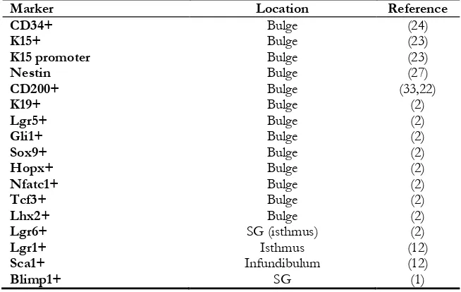
Table 1:Hair follicle stem cells markers. This table contains some of hair follicle putative markers
Marker Location Reference
CD34+ Bulge (24)
K15+ Bulge (23)
K15 promoter Bulge (23)
Nestin Bulge (27)
CD200+ Bulge (33,22)
K19+ Bulge (2)
Lgr5+ Bulge (2)
Gli1+ Bulge (2)
Sox9+ Bulge (2)
Hopx+ Bulge (2)
Nfatc1+ Bulge (2)
Tcf3+ Bulge (2)
Lhx2+ Bulge (2)
Lgr6+ SG (isthmus) (2)
Lgr1+ Isthmus (12)
Sca1+ Infundibulum (12)
In mouse pelage hair follicle, bulge is specified as a distinct swelling section of outer root sheath. Despite human bulge is also well identified in embryonic hair follicle, it is found as a petty dis-tend in adult hair follicle (6).
Human scalp follicles are 5 mm long, while the mouse pelage follicles are 1 mm in length (14). Another difference that can be noted is that the hair cycle has longer period in human hair follicle than in mouse. Each cycle of human hair follicle lasts about a decade, though it last some weeks in mouse follicles (25). Studying of rat whisker (vi-brissa) follicles showed that, these follicles have larger size and unusual structure in compare with pelage follicles. As it is clear, obvious differences could be found between human and mouse fol-licle stem cells characteristics (14). On the other hand, LRC are resident in both mouse and hu-man bulge structure (20,22).
Ohayama et al. found many identical and differ-ent genes that express in human and mouse bulge cells (14), and identified that among these mark-ers CD200 express in human bulge in high level but CD34 has low level expression (6).
CD200, a membrane glycoprotein is a subset of immunoglobulin superfamily (32). Using microar-ray analysis, CD200 was identified as an authentic human bulge cells marker (4), but not mouse bulge stem cells (12). In contrast to human bulge cells K15 (23) and CD34 (21) are specific mark-ers of mouse bulge stem cells which are not iden-tified as human bulge cells (14) (12) (Table 1).
Gene Regulation
Gene expressions control the noncyclic nature and quiescent of hair follicle stem cells, defining gene expression that distinct bulge and non-bulge cell populations provide intuitions of the main-tenance of the stem cell (14). During adult life when the hair follicle is in the rest phase, many different genes are expressed in the bulge region. It becomes complex according to the overlap of markers between different parts of hair follicle niche and the distribution of niche markers dur-ing active hair growth (2).
A combination of bulge cell isolation and mul-tiple gene expression microarray analysis allows
distinguishing bulge stem cells form other diffe-rentiated cells. Mouse bulge cells molecular signa-tures by using different isolation methods were obtained (6).
Bulge cells microarray results declared that genes involved in morphogenesis, organogenesis and development are up regulated, in contrast gene related to cycling and proliferation were down regulated, these results were compatible with quiescent property of bugle stem cells (33). These finding reveal how bulge stem cells of mouse adult hair follicle are quiescent and remain in un-differentiated state. In human and mouse bulge cells, WIF1 and DKK3 as inhibitors of WNT and BMP signaling pathways were down regu-lated (6).
Immunohistological staining declared that K19 and β1-integrin were highly expressed in bulge area (3). CD34 is exclusively express in the bulge, while K15, K19, Lgr5 and Sox9 display in bulge and hair germ. Similarly the expression of Gli1 (the transcription factor) is found in the upper bulge and the hair germ. Integrin 6 is display in bulge cells in different amount according to the vicinity of these cells to the basement membrane (2).
On the other side, human bulge cells surface markers were identified by microarray analyses, such as CD200 and CD59 which are up regulated in bulge and CD24, CD34, CD71 and CD147 are down regulated. Among these markers CD200 is the best human bulge cells marker. In addition, frizzled homolog 1 (FZD1), was up regulated in human bulge cells.
Interestingly, some human bulge up regulated genes including DIO2 and ANGPTL2 were not expressed in mouse bulge cells. The mouse bulge cell marker CD34 were expressed in human bulge in low level (6).
Hair Follicle Signaling Pathways
signaling pathways are required (6). Wnt/β -catenin signaling pathway plays an important role in morphogenesis initiation and onset of hair cycling (anagen) (35). Indeed, cell fate regulation is under control of Wnt/β-catenin signaling. Wnt as secreted glycoprotein, prompt a cascade by binding to Frizzed receptors which results in shifting APC/Axin/3β complex into GSK-3β. When Wnt signaling is absent, β-catenin is phosphorylated, hence could be degraded by APC/Axin/GSK-3β complex. When Wnt is available, Dishevelled is phosphorylated and as result the complex converts to GSK-3β. All these events cause β-catenin stabiles which leads to ac-tivation of transcription. Connection between Wnt signaling and TCF3 transcription factor is require for hair follicle stem cells maintenance. In the case of Wnt suppression, stem cells differen-tiate into sebocytes and keratinocytes. Wnt sig-naling directs mouse hair follicle bulge stem cells to preserve the stem cell circumstance, also the permission to fulfill a differentiation pathway is under control of Wnt signaling pathway (12). In-terestingly Wnt signaling is also needed for ma-trix proliferation (2).
However, Wnt and some other signals induce stem cell proliferation while BMP signals sup-press cell activation. Major BMP signaling role is regulating terminal differentiation of hair shaft and IRS. BMP and FGF18 signals secreted from dermal papilla, the internal K6+ bulge area and bulge stem cells, these signals make bulge stem cells retain their slow-cycling nature. When Wnt signal activates, BMP signal suppresses and re-sults in onset of hair germ proliferation (2). Another signaling pathway that control hair fol-licle stem cell function is sonic hedgehog (Shh) signaling. Shh is a second important signaling pathway that controls hair follicle bulge stem cells (12). Shh/Gli signaling pathway contributes in generating a growing hair follicle by inducing hair progenitor proliferation (35). Shh is a ligand which binds to Patched (Ptc), afterward Ptc binds to Smoothened (Smo) and cause it’s inhibition. At downstream of Smo, the Shh signaling assem-bles. After binding shh to Ptc, Smo is phosphory-lated. Then Ci protein released into nucleus and
stimulates Shh target genes expression. Within embryonic development, Shh suppression cause the follicle remains at bud stage (12). Regulating of mature hair follicle differentiation is under Notch signaling control (36).
Bulge Stem Cell Multipotency
Stem cells due to their mulipotent nature are able to reorganize different lineages in the specific tissue. To study multipotnecy of bulge stem cells, various researches have been performed (6). Bulge stem cells proliferate at the beginning of anagen (5). Taylor trace bulge stem cells of pelage follicle using double labeling, in lower follicle, faint labe-ling was detected in some cells. These results in-dicating that, these cells had originated from bulge region (37). Elsewhere, Tumber et al. used GFP labeling to demonstrate that lower epithelial cells had originated from bulge (19). None of these experiments were able to prove that matrix keratinocytes were originated from bulge, and both were unable to certainly mark bulge cells and their progeny (5).
To study bulge cells multipotency, Oshima et al. focused on transplantation. They worked on ROSA26 (transgenic mice) that express lacZ con-sistently. They dissected the bulge area from RO-SA26 vibrissa follicles. Afterward, labeled bulge were transplanted into the vibrissae of non-ROSA26 mice. After downward migration of bulge cells through vibrissa follicle, expression of lacZ in all epithelial cell layers of some follicles were observed (38).
Moriis et al. to study about bulge cells mulipoten-cy used K15 promoter targeted with CrePR1. K15-CrePR1 transgenic mice were crossed with R26R (reporter mice) which could express lacZ under control of ROSA26 promoter. lacZ ex-pression was observed in bulge cells and all their progeny. This experiment made it clear that bulge progeny could generate all epithelial cell layers located in the lower hair follicle (23).
Interestingly, after culturing isolated bulge cells
invitro, repopulation of hair follicle, epidermis,
Bulge Stem Cells Contribution in Wound Healing
In addition to the production of a new hair fol-licle, bulge stem cells are able to regenerate and contribute in tissue regeneration. During an in-jury, bulge cells migrate into damaged epidermis in order to repair the wound (6, 30). Besides, hair follicle keratinocytes contribute in wounds repo-pulation by emerging from the follicle. Ito et al, labeled bulge cells using K15CrePR;R26R trans-genic mouse, ulceration with trephine lead the bulge cells progeny to migrate into the healing epidermis. The results showed that the origin of the formed epidermis were 25% from bulge cells (5,30).
Bulge Stem Cell Plasticity
Some cell populations, due to their plasticity na-ture are able to differentiate into other linage in-cluding neural cells (6).
Amoh et al. isolated Nestin positive cells from mouse hair follicle bulge, then cultured these cells in vitroandsubjected to classIII beta-tubulin +ve neuronal type cells, then cells were transplanted into nud mice, the result showed that these cells were able to differentiate into several cell lineages including neurons, Schwann cells, glia, keratino-cytes, melanocytes and smooth muscle cells. In injured sciatic nerve, implanted Nestin positive cells were able to differentiate into Schwann cells and repair the severed nerve. Thus bulge cells could contribute in tissue repairing by their plas-ticity nature (27,40).
Nobakht et al experiment on rat vibrissa bulge cells showed that these cells were able to diffe-rentiate into neural and glia lineages (28). Drewa et al. studied on bulge stem cells of rat vibrissa and found that these cells could be used for in vitro restoration of urinary bladder wall grafts (41).
Most of studies were experimented on mouse but not humans (6). However some other findings showed that human follicle cells also demonstrat-ed the plasticity feature (42).
According to these findings, the hair follicle could be considered as a potential source of stem cells in tissue engineering (5).
Conclusion
The hair follicle structure is a remarkable model for studying preserved stem cells within niches. For this purpose, numerous studies have been developed on biology of bulge stem cells. The results demonstrated that label-retaining cells with mulitipotent potential and quiescence fea-ture are reserved in hair follicle bulge region. For isolating bulge cells, specific markers of mouse and human bulge cells such as K15 promoter ac-tivity, CD34 and CD200 are needed. Since signif-icant differences are exist between rodent and human hair follicle stem cells, obtaining results from mouse should be study on human directly. Bulge stem cells are able to differentiate into dif-ferent lineages. In bulge cells difdif-ferentiation process, signals including Wnt and Shh play an important role.
All of these studies about bulge stem cells may lead to finding novel techniques for utilizing stem cells in clinical application.
Ethical considerations
Ethical issues (Including plagiarism, informed consent, misconduct, data fabrication and/or fal-sification, double publication and/or submission, redundancy, etc.) have been completely observed by the authors.
Acknowledgements
The study received no funds form any organiza-tion.
Conflict of Interests
The authors indicate no potential conflicts of in-terest.
References
2. Rompolas P, Greco V (2014). Stem cell dynam-ics in the hair follicle niche. Semin Cell Dev Biol, 25-26:34-42.
3. Zhang Y, Xiang M, Wang Y, et al (2006). Bulge cells of human hair follicles: segregation, cul-tivation and properties. Colloids Surf B Biointer-faces, 47(1): 50-6.
4. Inoue K, Aoi N, Sato T, et al (2009). Differential expression of stem-cell-associated markers in human hair follicle epithelial cells. Lab Invest, 89(8): 844–56.
5. Cotsarelis G (2006). Epithelial stem cells: a folli-culocentric view. J Invest Dermatol, 126(7): 1459–68.
6. Ohyama M (2007). Hair follicle bulge: A fascinat-ing reservoir of epithelial stem cells. J Dermatol Sci, 46(2): 81–9.
7. Harkey MR (1993). Anatomy and physiology of hair. Forensic Sci Int, 63(1-3): 9–18.
8. Ronald Shapiro, Paul Rose, Michael Morgan (2004). Hair anatomy and histology. In: Hair Transplantation. Eds, Unger W, Unger M, Unger WP, Shapiro R . 3rd ed. CRC Press.
New York, pp. 25–33.
9. Botchkareva N, Randall VA (2009). The biology of hair growth. In: Cosmetic applications of laser and light-based system. Eds, Ahluwalia G. 1st Ed.
William Andrew. New York, pp .3-35. 10. Chen CC, Plikus MV, Tang PC et al (2016). The
Modulatable Stem Cell Niche: Tissue Interac-tions during Hair and Feather Follicle Rege-neration J Mol Biol, 428(7):1423-40.
11. Won Oh J, Kloepper J, Langan EA et al (2016). A guide to studying human hair follicle cycl-ing in vivo. J Invest Dermatol, 136(1): 34–44. 12. Eckert RL, Adhikary G, Balasubramanian S et al
(2013). Biochemistry of epidermal stem cells.
Biochim Biophys Acta, 1830(2): 2427–34. 13. Alonso L, Fuchs E (2006). The hair cycle. J Cell
Sci, 119: 391–3.
14. Cotsarelis G (2006). Gene expression profiling gets to the root of human hair follicle stem cells. J Clin Invest, 116(1): 19–22.
15. Cotsarelis G, Sun TT, Lavker RM (1990). Label-retaining cells reside in the bulge area of pilo-sebaceous unit: implications for follicular stem cells, hair cycle, and skin carcinogenesis.
Cell, 61(7): 1329–37.
16. Hoffman RM (2006). The pluripotency of hair follicle stem cells. Cell Cycle, 5(3): 232–233.
17. Ma DR, Yang EN, Lee ST (2004). A review: the location, molecular characterisation and mul-tipotency of hair follicle epidermal stem cells.
Ann Acad Med Singapore, 33(6):784–788. 18. Morris RJ, Potten CS (1999). Highly persistent
label-retaining cells in the hair follicles of mice and their fate following induction of anagen. J Invest Dermatol, 112(4): 470–5.
19. Tumbar T, Guasch G, Greco V et al (2004). De-fining the epithelial stem cell niche in skin.
Science, 303(5656):359-63.
20. Lyle S, Christofidou-Solomidou M, Liu Y et al (1998). The C8/144B monoclonal antibody recognizes cytokeratin 15 and defines the lo-cation of human hair follicle stem cells. J Cell Sci, 111(Pt 21): 3179–88.
21. Trempus C, Morris RJ, Bortner CD et al (2003). Enrichment for living mouse keratinocytes from the hair follicle bulge with the cell sur-face marker CD34. J Invest Dermatol, 120(4):501-11.
22. Ohyama M, Terunuma A, Tock CL et al (2006). Characterization and isolation of stem cell-enriched human hair follicle bulge cells. J Clin Invest, 116(1): 249–260.
23. Morris RJ, Liu YP, Marles L et al (2004). Captur-ing and profilCaptur-ing adult hair follicle stem cells.
Nat Biotechnol, 22(4):411-7.
24. Blanpain C, Lowry WE, Geoghegan A et al (2004). Self-renewal, multipotency, and the existence of two cell populations within an epithelial stem cell niche. Cell, 118(5):635-48. 25. Boehnke K, Falkowska-Hansen B, Stark HJ et al
(2012). Stem cells of the human epidermis and their niche: composition and function in epidermal regeneration and carcinogenesis.
Carcinogenesis,33(7): 1247–58.
26. Gutiérrez-Rivera A, Pavón-Rodríguez A, Jiménez-Acosta F et al (2010). Functional characterization of highly adherent CD34+ keratinocytes isolated from human skin. Exp Dermatol, 19(7): 685–8.
27. Amoh Y, Li L, Katsuoka K et al (2005). Multipo-tent nestin-posetive, keratin-negative hair fol-licle bulge cells can form neurons. Proc Natl Acad Sci U S A, 102(15): 5530–4.
29. Nobakht M, Asalgoo S, Rahbar Roshandel N et al (2011). Effects of Silibinin on Hair Follicle Stem Cells Differentiation to Neural-like Cells. Am J Mol Biol, 1: 212–22.
30. Garcin CL, Ansell DM, Headon, DJ et al (2016). Hair follicle bulge stem cells appear dispensa-ble for the acute phase of wound re-epithelialization. Stem Cells, 34(5):1377-85. 31. Nishimura EK, Jordan SA, Oshima H et al
(2002). Dominant role of the niche in mela-nocyte stem-cell fate determination. Nature, 416: 854–60.
32. Barclay AN, Wright GJ, Brooke G et al (2002). CD200 and membrane protein interactions in the control of myeloid cells. Trends Immunol, 23(6):285-90.
33. Ohyama M, Vogel JC, Amagai M (2007). Gene ontology analysis of human hair follicle bulge molecular signature. J Dermatol Sci, 45(2):147-50.
34. Carrasco E, Calvo MI, Blázquez-Castro A et al (2015). Photoactivation of ROS Production In Situ Transiently Activates Cell Proliferation in Mouse Skin and in the Hair Follicle Stem Cell Niche Promoting Hair Growth and Wound Healing. J Invest Dermatol, 135(11): 2611–22.
35. Huntzicker EG, Oro AE (2008). Controlling hair follicle signaling pathways through polyubi-quitination. J Invest Dermatol, 128(5):1081-7.
36. Demehri S, Kopan R (2009). Notch signaling in bulge stem cells is notrequired for selection of hair follicle fate. Development, 136(6):891-6. 37. Taylor G, Lehrer MS, Jensen PJ et al (2000).
In-volvement of follicular stem cells in forming not only the follicle but also the epidermis.
Cell, 102(4):451-61.
38. Oshima H, Rochat A, Kedzia C et al (2001). Morphogenesis and renewal of hair follicles from adult multipotent stem cells. Cell, 104(2):233-45.
39. Claudinot S, Nicolas M, Oshima H et al (2005). Long-term renewal of hair follicles from clo-nogenic multipotent stemcells. Proc Natl Acad Sci U S A, 102(49):17734-8.
40. Amoh Y, Li LN, Campillo R et al (2005). Im-planted hair follicle stem cells form Schwann cells that support repair of severed peripheral nerves. Proc Natl Acad Sci U S A, 102(41):14677-82.
41. Drewa T, Joachimiak R, Kaznica A et al (2009). Primary cultures from rat vibrissae as a poten-tial cell source for in vitro construction of uri-nary bladder wall grafts. Transplant Proc, 41(5):1932-5.