R E S E A R C H A R T I C L E
Open Access
HPV16 variant lineage, clinical stage, and survival
in women with invasive cervical cancer
Rosemary E Zuna
1*, Erin Tuller
2,4, Nicolas Wentzensen
3, Cara Mathews
2, Richard A Allen
1, Rebecca Shanesmith
1,5,
S Terence Dunn
1, Michael A Gold
1,2,6, Sophia S Wang
3,7, Joan Walker
2and Mark Schiffman
3Abstract
Background:HPV16 variants are associated with different risks for development of CIN3 and invasive cancer, although all are carcinogenic. The relationship of HPV 16 variants to cancer survival has not been studied. Methods:155 HPV16-positive cervical cancers were categorized according to European and non-European variant patterns by DNA sequencing of theE6open reading frame. Clinico-pathologic parameters and clinical outcome were collected by chart review and death registry data.
Results:Of the 155 women (mean age 44.7 years; median follow-up 26.7 months), 85.2% harbored European variants while 14.8% had non-European sequences. HPV16 variants differed by histologic cell type (p = 0.03) and stage (1 vs. 2+; p = 0.03). Overall, 107 women (68.0%) were alive with no evidence of cancer, 42 (27.1%) died from cervical cancer, 2 (1.3%) were alive with cervical cancer, and 4 (2.6%) died of other causes. Death due to cervical cancer was associated with European variant status (p < 0.01). While 31% of women harboring tumors with European variants died from cervical cancer during follow-up, only 1 of 23 (4.4%) non-European cases died of cancer. The better survival for non-European cases was partly mediated by lower stage at diagnosis.
Conclusions:Overall, invasive cervical cancers with non-European variants showed a less aggressive behavior than those with European variants. These findings should be replicated in a population with more non-European cases.
Keywords:cervical neoplasms, human papillomavirus 16, HPV16 variants
Background
While the association of HPV genotypes and cervical carcinoma is well established, the reasons that only a subset of lesions associated with high-risk genotypes progress to invasive cancer remain elusive. Influential variables likely include individual host factors, viral dif-ferences or combinations of both.
One variable that could contribute to differences in biological behavior of HPV- associated lesions is that of HPV DNA sequence variation. HPV16, which is the most prevalent HPV genotype and is associated with approximately half of cervical cancers worldwide [1], has well-documented DNA sequence variants. HPV variants are defined as isolates with primary DNA sequence dif-ferences that total no more than 2% of the L1 open
reading frame (ORF) of the prototype sequence [2]. The sequence variations of the HPV16 E6 ORF have been found to correctly classify the HPV16-variants [3]. Two major categories of HPV16 variants have been defined: European (E) and Non-European (NE) patterns that appear to have evolved principally on a geographic basis [4]. A number of studies have suggested that HPV16 variants differ in risk for progression to high grade intraepithelial lesions [5-7] and in their association with the development of cancer [8-12]. Overall, the 16-NE variants have shown an increased risk for progression to CIN2 or greater when compared with 16-E.
We have previously reported the patterns of HPV16 variants in the spectrum of cervical lesions in our popu-lation [11]. While our study confirmed the increased risk for progression to carcinoma associated with 16-NE variants, 16-E patterns were found in the majority of HPV16 lesions, including cancers. We attributed this to the dominance of the 16-E patterns in our population * Correspondence: rosemary-zuna@ouhsc.edu
1
Department of Pathology, University of Oklahoma Health Sciences Center, 940 SL Young Blvd, Oklahoma City, Oklahoma, 73104-5042, USA Full list of author information is available at the end of the article
and to the fact that all major variant categories can be found in cancers.
In this follow-up study, we address the question of whether HPV16 variant status influences clinical pat-terns and/or prognosis in fully evolved cervical cancers. We performed a cross-sectional analysis of the clinico-pathologic characteristics of an unselected, prospectively accumulated population of women with HPV16-positive invasive cervical cancer from the central United States. The tumors were categorized according to histologic cell type and HPV16 variant category. We also corre-lated the clinico-pathologic characteristics and clinical follow-up with HPV16-variant category in an effort to evaluate the possible association of the HPV16 variants with clinical behavior in cervical cancers.
A similar analysis was performed for the largest sub-population, i.e., HPV16-positive squamous cell cancers harboring European variants, in an effort to identify dif-fering patterns of biologic behavior within sub-lineages of this dominant category.
Results
Patient Demographics
The population of 155 women with HPV16- associated cancers had a mean age of 44.7 years ± 1.0 (SEM), ran-ging from 20-76 years. Median follow-up was 26.7 ± 24.5 months (mean = 31.5 ± 2.0 months). Overall, 107 women (68.0%) were alive with no evidence of cancer, 42 (27.1%) died from cervical cancer, 2 (1.3%) were alive with cervical cancer, and 4 (2.6%) had died of other causes. Of these cases, 132 (85.2%) harbored 16-E var-iants while 23 (14.8%) had 16-NE patterns (AA = 17 cancers, NA-1 = 3, AF1 and AF2 = 3). The patient char-acteristics for this population, sorted according to var-iant status, are summarized in Table 1.
Clinico-pathologic variables
Of 149 women with definite clinical outcome, the overwhelming majority, 91.3%, were diagnosed with SCC. 16-E variants were identified in 119 (87.5%) SCC lesions. These accounted for 93.7% of all 16-E variant cases in this population. Although SCC cancers har-boring 16-E variants dominated in this population, and 16-E cases were more frequent in all other histologic categories as well, the percentage of 16-NE variants ranged from 44.4% (4 of 9) of CAC compared with 25% (1 of 4) for ASC and 12.5% (17 of 136) for SCC. The distribution of individual 16-NE variant categories for histologic cell type is as follows: AA = 12 SCC and 4 CAC; NA-1 = 3 SCC; AF1 = 1 SCC and AF2 = 1 SCC and 1 ASC. Despite the disparity in case numbers, the differences in the distribution of HPV16-variants among the histologic cell types were statistically signif-icant (p = 0.03).
There was no significant difference in the distribution of HPV16 variants according to tumor size, depth of invasion, vascular space invasion, lymph node involve-ment, parametrial involveinvolve-ment, or status of surgical margins. Similarly, none of the clinical parameters showed a significant difference associated with 16-var-iant status including race/ethnicity, age group, FIGO stage and number of pregnancies.
Although significant differences were not identified between the 16-E and 16-NE variant groups for the above prognostic variables affecting survival in cervical cancer patients, there were interesting trends among 16-NE cases for some parameters that may reveal signifi-cance in a larger study with a larger population of 16-NE cases. These variables include younger age, early stage at presentation, three or fewer pregnancies, race/ ethnicity other than white, depth of stromal invasion greater than 5 mm, and the presence of lymphovascular space invasion for 16-NE patients. While not statistically significant (ptrend= 0.13), the modal peak for 16-NE
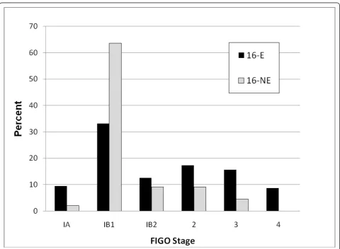
cases was the 31-40 age group (mean = 41.4 ± 2.6 years; median 38 years) compared with the 41-50 group for the 16-E cases (mean = 45.3 ± 1.0; median 45 years). Figure 1 illustrates that the majority of women with 16-NE lesions presented at stage IB1 while those with 16-E lesions presented at a more variable stage (ptrend= 0.02).
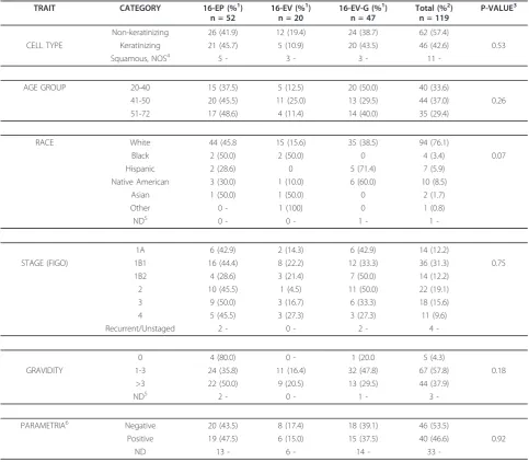
Because of the uneven distribution of cases in this study, a second analysis was performed that was restricted to the 123 cases of SCC associated with Eur-opean variants. This population had a mean age of 45.9 years ± 1.1 (SEM), ranging from 20 to 76 years of age. Median follow-up was 28 months ± 22.4 (mean 33.0 ± 2.0). This population was divided into three categories as follows based on subgroups of European variants: European prototype (16-EP) (N = 53), Eur-opean variants with a small number of nucleotide changes compared to prototype (16-EV) (N = 21), and those European variants harboring the 350 T>G nucleotide change (N = 49).
Table 1 Clinicopathologic Features of 149 Cervical Cancer Cases
TRAIT CATEGORY EUROPEAN (%1) n = 127
NON EUROPEAN (%1) n = 22
TOTAL (%2) P-VALUE3 n = 149
Squamous Carcinoma 119 (87.5) 17 (12.5) 136 (91.3)
CELL TYPE Adenocarcinoma 5 (55.6) 4 (44.4) 9 (6.0) 0.03
Adenosquamous Ca 3 (75.0) 1 (25.0) 4 (2.7)
AGE GROUP 20-40 46 (78.0) 13 (22.0) 59 (39.6)
(YEARS) 41-50 46 (92.0) 4 (8.0) 50 (33.6) 0.13
51-72 35 (87.5) 5 (12.5) 40 (26.8)
White 102 (88.7) 13 (11.3) 115 (77.7)
RACE Black 4 (80.0) 1 (20.0) 5 (3.4) 0.11
Hispanic 7 (63.6) 4 (36.4) 11 (7.4)
Native American 10 (71.4) 4 (28.6) 14 (9.5)
Asian 2 (100) 0 2 (1.4)
Other 1 (100) 0 1 (0.7)
Unknown 1 - - 1
-1A 14 (87.5) 2 (12.5) 16 (11.1)
STAGE (FIGO) 1B1 42 (75.0) 14 (25.0) 56 (38.9) 0.16
1B2 16 (88.9) 2 (11.1) 18 (12.5)
2 22 (91.7) 2 (8.3) 24 (16.7)
3 18 (94.7) 1 (5.3) 19 (13.2)
4 11 (8.9) 0 11 (7.6)
Recurrent/Unstaged 4 - 1 - 5
-0 6 (85.7) 1 (14.3) 7 (4.9)
GRAVIDITY 1-3 73 (82.0) 16 (18.0) 89 (61.8) 0.13
>3 45 (93.8) 3 (6.3) 48 (33.3)
ND4 3 - 2 - 5
-PARAMETRIA5 Negative 53 (77.9) 15 (22.1) 68 (60.7) 0.12
Positive 40 (90.9) 4 (9.1) 44 (39.3)
ND 34 - 3 - 37
-Table 1 (cont’d)
<1 16 (84.2) 3 (15.8) 19 (14.6)
TUMOR SIZE5(cm) 1-4 42 (76.4) 13 (23.6) 55 (42.3) 0.1
>4 51 (91.1) 5 (8.9) 56 (43.1)
ND 18 - 1 - 19
-< 3 15 (93.8) 1 (6.3) 16 (19.3)
INVASION DEPTH6 3.1 - 5.0 10 (83.3) 2 (16.7) 12 (14.5) 0.21
(mm) >5 40 (72.7) 15 (27.3) 55 (66.3)
ND 62 - 4 - 66
-Positive 40 (74.1) 14 (25.9) 54 (62.1)
VSI6 Negative 29 (87.9) 4 (12.1) 33 (37.9) 0.17
-Table 1 Clinicopathologic Features of 149 Cervical Cancer Cases(Continued)
RESECTION Positive 11 (91.7) 1 (8.3) 12 (14.6)
MARGINS6 Negative 53 (75.7) 17 (24.3) 70 (85.4) 0.29
ND 63 - 4 - 67
-LYMPH NODES7 Negative 48 (85.7) 8 (14.3) 56 (50.4)
Positive 44 (80.0) 11 (20.0) 55 (49.6) 0.46
ND 35 - 3 - 38
-NED 86 (80.4) 21 (19.6) 107 (71.8)
STATUS8 DOD 41 (97.6) 1 (2.4) 42 (28.2) 0.008
1
: Percentage calculated across.
2
: Percentage calculated down.
3
: Fisher’s Exact Test
4
: ND: No data available
5
: Parametrial assessment and tumor size were determined by pathologic analysis of a radical hysterectomy or as part of a clinical staging procedure.
6
: Depth of invasion, margin status and status of vascular space invasion were available only for women with early stage tumors who had a conization or hysterectomy.
7
: Lymph node dissections were routinely performed as part of radical hysterectomy. It is not standard for higher stage women although cervical biopsy with lymph node dissection was performed for some higher stage women.
8
: Status: NED = No evidence of cervical cancer at last follow-up; DOD = Dead of cervical cancer
131A>G change in addition to 350T>G. Because of the frequency of the 350T>G pattern, this group of 16-EV tumors was analysed separately in this study in an effort to clarify the clinical significance of this change. The 16-EV-G category specifically excludes the 16-NE variants that contain this same nucleotide 350 alteration. The clinico-pathologic characteristics of this population are shown in Table 2.
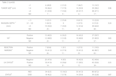
Cross-sectional analysis restricted to the subgroup of 123 women with SCC harboring European variants failed to demonstrate significant differences among the various clinical and pathologic traits for the different variant categories (Table 2). The 16-EP and 16-EV-G categories had similar characteristics. The 16-EV group was somewhat divergent but the numbers were small
and the differences not significant. A comparison of 16-EV-G cases relative to all other 16-E SCC also showed no significant differences (data not shown).
Clinical Follow-up
After a median follow-up of 29.1 months ± 23.3, death due to cervical cancer was highly associated with 16-E variant status (p < 0.01). While 31% (41/127) of women harboring tumors with 16-E variants died from cervical cancer, only 1 of 23 (4.4%) 16-NE cases died of cervical cancer. There were no differences in follow-up status or total follow-up time within the subgroups of 16-E squa-mous cancers.
We attempted to explain the survival differences by adjustment (by stratification or restriction) for possible
Table 2 Clinicopathologic Features of 119 HPV16-Positive Squamous Cell Carcinoma Cases with European Variants
TRAIT CATEGORY 16-EP (%1)
n = 52
16-EV (%1)
n = 20
16-EV-G (%1)
n = 47
Total (%2)
n = 119
P-VALUE3
Non-keratinizing 26 (41.9) 12 (19.4) 24 (38.7) 62 (57.4)
CELL TYPE Keratinizing 21 (45.7) 5 (10.9) 20 (43.5) 46 (42.6) 0.53
Squamous, NOS4 5 - 3 - 3 - 11
-AGE GROUP 20-40 15 (37.5) 5 (12.5) 20 (50.0) 40 (33.6)
41-50 20 (45.5) 11 (25.0) 13 (29.5) 44 (37.0) 0.26
51-72 17 (48.6) 4 (11.4) 14 (40.0) 35 (29.4)
RACE White 44 (45.8 15 (15.6) 35 (38.5) 94 (76.1)
Black 2 (50.0) 2 (50.0) 0 4 (3.4) 0.07
Hispanic 2 (28.6) 0 5 (71.4) 7 (5.9)
Native American 3 (30.0) 1 (10.0) 6 (60.0) 10 (8.5)
Asian 1 (50.0) 1 (50.0) 0 2 (1.7)
Other 0 - 1 (100) 0 1 (0.8)
ND5 0 - 0 - 1 - 1
-1A 6 (42.9) 2 (14.3) 6 (42.9) 14 (12.2)
STAGE (FIGO) 1B1 16 (44.4) 8 (22.2) 12 (33.3) 36 (31.3) 0.75
1B2 4 (28.6) 3 (21.4) 7 (50.0) 14 (12.2)
2 10 (45.5) 1 (4.5) 11 (50.0) 22 (19.1)
3 9 (50.0) 3 (16.7) 6 (33.3) 18 (15.6)
4 5 (45.5) 3 (27.3) 3 (27.3) 11 (9.6)
Recurrent/Unstaged 2 - 0 - 2 - 4
-0 4 (80.0) 0 - 1 (20.0 5 (4.3)
GRAVIDITY 1-3 24 (35.8) 11 (16.4) 32 (47.8) 67 (57.8) 0.18
>3 22 (50.0) 9 (20.5) 13 (29.5) 44 (37.9)
ND5 2 - 0 - 1 - 3
-PARAMETRIA6 Negative 20 (43.5) 8 (17.4) 18 (39.1) 46 (53.5)
Positive 19 (47.5) 6 (15.0) 15 (37.5) 40 (46.6) 0.92
-factors such as stage at diagnosis or age, which showed a relationship both to variant status and outcome. Even within Stage 1 cancers, however, for which survival was favorable, tumors with 16-E variants tended to have somewhat worse outcomes than those with 16-NE var-iants (data not shown).
Discussion
The rationale behind the possibility of biological differ-ences associated with variant status is related to altera-tions in primary HPV DNA sequence that may alter control elements or affect the function of translated pro-teins that interact with the host. For example, the most common E6 sequence variation from prototype described in the 16-E-variants is 350 T> G that results
in a L83V amino acid change in the E6 protein. Such amino acid changes can potentially alter tertiary struc-ture and may influence protein function.In vitrostudies of different HPV16-variants have suggested differences in biological activity [13], but anin vivomechanism of enhanced biological activity has not been described. To date, evidence for different biological effects related to the sequence variants in patients has accumulated on an epidemiologic basis.
The increased risk for progression of 16-NE cervical lesions to high grade CIN and carcinoma has been attributed in part to reports that 16-NE variants are associated with a higher rate of HPV16 persistence and development of high-grade intraepithelial cervical lesions [6,7]. In this report, we explored the question of
Table 2 Clinicopathologic Features of 119 HPV16-Positive Squamous Cell Carcinoma Cases with European Variants (Continued)
Table 2 (cont’d)
<1 6 (40.0) 2 (13.3) 7 (46.7) 15 (14.7)
TUMOR SIZE6(cm) 1-4 18 (46.2) 7 (17.9) 14 (35.9) 39 (38.2) 0.96
>4 21 (43.8) 7 (14.6) 20 (41.7) 48 (47.1)
ND 7 - 4 - 6 - 17
-< 3 3 (23.1) 2 (15.4) 8 (61.5) 13 (22.0)
INVASION DEPTH7 3.1 - 5.0 5 (62.5) 0 3 (37.5) 8 (13.3) 0.24
(mm) >5 19 (50.0) 7 (18.4) 12 (31.6) 38 (64.4)
ND 25 - 11 - 24 - 60
-Positive 15 (40.5) 6 (16.2) 16 (43.2) 37 (59.7)
VSI7 Negative 12 (48.0) 3 (12.0) 10 (40.0) 25 (40.3) 0.83
ND 25 - 11 - 21 - 57
-RESECTION Positive 7 (63.6) 1 (9.1) 3 (27.3) 11 (19.3)
MARGINS7 Negative 19 (41.3) 8 (17.4) 19 (41.3) 46 (80.7) 0.49
ND 26 - 11 - 25 - 65
-Negative 20 (47.6) 4 (9.5) 18 (42.9) 42 (49.4)
LN STATUS8 Positive 18 (41.9) 8 (18.6) 17 (39.5) 43 (50.6) 0.55
ND 14 - 8 - 12 - 34
-NED 34 42.5) 13 (16.3) 33 (41.3) 80 (67.2)
STATUS9 DOD 18 46.2) 7 (17.9) 14 (35.9) 39 (32.8) 0.87
1
: Percentage calculated across.
2
: Percentage calculated down.
3
: Fisher’s exact test
4
: Squamous, NOS: Women with very small tumors or tiny biopsies were difficult categorize as to cell type and were not included in these analyses.
5
: ND: No data available
6
: Tumor size and parametrial assessment were determined by pathologic evaluation of a radical hysterectomy or as part of a clinical staging procedure.
7
: Depth of invasion, margin status and status of vascular space invasion were available only for women with early stage tumors who had a conization or hysterectomy.
8
: Lymph node dissections were routinely performed as part of radical hysterectomy. It is not standard for higher stage women although cervical biopsy with lymph node dissection was performed for some higher stage women.
9
whether this increased risk for progression associated with 16-NE variants also correlates with increased aggressiveness of fully evolved invasive cancers. Our data for 155 HPV16-positive cervical cancer cases fol-lowed for a median of 29.1 months does not support this hypothesis. In fact, the women with tumors harbor-ing 16-E variants had a statistically increased risk of death due to cervical cancer. A major problem in inter-preting these results, however, is the skewed pattern of our cases, with strong predominance of squamous can-cers and 16-E patterns. This imbalance could mask any possible difference in the behavior associated with the 16-NE lesions. For example, 16-NE variants, particularly the AA variant, have been reported to be associated with CAC [9,10] which have a more aggressive course than SCC in some reports [14,15]. In this population, however, only 14 (9.0%) HPV16-positive cases were CAC or ASC.
Survival in cervical cancer patients is related primarily to stage at diagnosis, tumor size, depth of cervical stro-mal invasion, parametrial involvement, lymphovascular space invasion, and lymph node status [16]. Histologic cell type [17] as well as HPV genotype, especially HPV18 [18] have also been reported as prognostic factors.
While survival in this study was correlated with 16-E variant status, there are clues to suggest that this may be an overly simplistic impression given the dispropor-tionate number of cases in the two groups. In this study most of the 16-NE cancer patients presented at a young age and at an early stage (1B1). Additionally, these data showed non-significant trends in which 16-NE lesions had larger size at presentation with increased rate of vascular space invasion and lymph node metastases. It is therefore conceivable that the small number of 16-NE lesions in this population has resulted in a non-repre-sentative distribution of cases. Similarly, it is not clear whether this pattern is representative of all 16-NE cases or if our population is simply anomalous due to small numbers of 16-NE cases. Conceivably, our young 16-NE cases represent a detection bias in which younger women with cancer were identified because of screening while the older 16-E population presented with signs and symptoms of cancer. The relatively small proportion of 16-NE cases could thus result in anomalous results. On the other hand, these results raise the possibility that 16-NE variants have a preferential role in progres-sion to malignancy but do not differentially influence clinical behavior in fully evolved cancers. When we ana-lyzed a subpopulation of cancer cases representing the dominant group of HPV16 European sequence patterns, we were unable to show a difference in clinical behavior based on subcategories of 16-E variants.
Conclusions
While our earlier study showed a significant association of 16-NE variant status with progression to high grade CIN and invasive cancer in cases with a wide spectrum of cervical disease[11], we have not been able demon-strate more aggressive behavior for actual invasive carci-nomas associated with these variants. These results, however, should be interpreted with caution due to the limited number of 16-NE cases in this population. A lar-ger study with a larlar-ger population of 16-NE cases is needed to clarify these issues.
Methods
Patient Population
From 1999-2009, 319 unselected cases of primary, meta-static and recurrent invasive cervical carcinoma were prospectively entered into this study based on the col-lection of a liquid-based cytologic sample that was tested for HPV DNA and a histologic diagnosis of inva-sive cervical cancer. The population seen at our institu-tion is variably screened for cervical cancer so that some cancer patients were identified by routine screening while others presented with clinical signs and symptoms related to cervical cancer. Included in this total were 164 cases from the SUCCEED study [19] and 85 cases that were included in previous reports [20]. These women were staged according to FIGO (International Federation of Gynecology and Obstetrics, 2000) and treated at the OU Medical Center in Oklahoma City using standard therapies of surgery, radiation, and che-motherapy as indicated by individual patient characteris-tics. Of the 167 HPV16-positive cancer cases, 155 were available for variant analysis. Because 80 (51.6%) women did not undergo total hysterectomy and 40 (25.8%) did not have a lymphadenectomy, some pathologic variables were not evaluable in all cases. Follow-up was deter-mined by chart review and death records for the state of Oklahoma. In addition to clinico-pathologic parameters, we compared over-all follow-up taking into account clinical stage at diagnosis, race, age, and censoring due to loss-to-follow-up. Statistical analyses reported below were performed using the population of women (n = 149) with defined outcome (i.e., no evidence of disease or dead of cervical cancer). This study was performed with the approval of the Institutional Review Board for the University of Oklahoma Health Sciences Center.
Pathologic Categorization
criteria [21]. Squamous cell carcinomas (SCC) were designated as “large cell keratinizing” when at least one well-formed keratin pearl was identified [22] and“large cell non-keratinizing” when no pearls were identified. Squamous tumors that were very small or for which only small biopsies were available, were listed as“SCC, not otherwise specified”.
The major histologic categories included SCC (n = 141), both keratinizing and non-keratinizing patterns, adenocarcinoma (CAC) (n = 10), and adenosquamous carcinoma (ASC) (n = 4). There were no HPV16-posi-tive small cell neuroendocrine carcinomas in this population.
HPV Testing Using L1 Consensus Primers
Liquid-based cytologic samples were collected in Pre-servCyt® (Hologic, Malborough, MA) at the time of clinical evaluation and the cellular DNA was extracted using the QIAamp® DNA Blood and Tissue Mini Kit (Qiagen, Valencia, CA). HPV genotyping was performed using the reverse line blot/Linear Array®HPV Genotyp-ing Test (Roche Molecular Systems, Alameda, CA), as previously described [20].
Variant Analysis
Aliquots of cellular DNA from HPV16-positive tumor samples were analyzed for HPV16E6 variant category using PCR amplification and bidirectional PCR-based fluorescent dideoxy chain termination sequencing using the same primers used for initial amplification, as pre-viously described [11].
Sequence alignments were performed using CLUS-TALW [23]. Only nucleotide changes verified as occur-ring on both strands were accepted.
An HPV16-variant category was assigned for each case using the prototype nucleotide sequence for HPV16 [24] (modified as HPV16R [25]) as well as published HPV16 E6sequence patterns that define the different variants [3]. The 16-E category included the prototype sequence and related patterns showing only a small number of nucleotide changes, while the 16-NE categories included Asian-American (AA), Native-American (NA-1), and African (AF-1 and AF-2).
In the secondary analysis restricted to 16-E SCC cases, the variants were subdivided into EP (prototype), 16-EV (European prototype sequence with a small number of additional nucleotide differences), and 16-EV-G (Eur-opean sequence with the common 350T>G substitution; cases with nucleotide changes in addition to 350T>G were also included).
Statistics
Tabular analyses were performed using chi square and Fisher’s exact and Cochran-Mantel-Haenszel trend tests.
Statistical significance was assigned to 2-sided probabil-ity values < .05.
Acknowledgements
This study was supported by grants from the Oklahoma Center for the Advancement of Science and Technology (OCAST HR05-136) and from the National Cancer Institute (03-A117523) as well as from contract N02-CP-31102 from the National Cancer Institute. Drs. Wentzensen and Schiffman are supported by the Intramural Research Program of the National Institutes of Health. Special thanks are due to Greg Rydzak, Information Management Services, Silver Spring, MD for his assistance in data analysis as well as to Tracey Young and her associates in the Department of Obstetrics and Gynecology and Susan Nagelhout, CTR of the OU Medical Center Tumor Registry for their efforts in obtaining patient follow-up information.
Author details
1Department of Pathology, University of Oklahoma Health Sciences Center,
940 SL Young Blvd, Oklahoma City, Oklahoma, 73104-5042, USA.
2
Department of Obstetrics and Gynecology, Division of Gynecologic Oncology, University of Oklahoma Health Sciences Center, 800 NE 10th St. Suite 2001-2, Oklahoma City, Oklahoma, 73104-5418, USA.3Division of Cancer Epidemiology and Genetics, National Cancer Institute, 6120 Executive Blvd, Room 5024, Rockville, Maryland 20852-7234, USA.4Department of
Obstetrics and Gynecology, University of Missouri Health System, 115 Business Loop 70W, Columbia, Missouri 56203-3244, USA.5Department of
Pathology and Laboratory Medicine, Tulane University Health Sciences, 1430 Tulane Avenue SL-79, New Orleans, Louisiana 70112-2699, USA.6Obstetrics
and Gynecology Department, Vanderbilt University Medical Center, 1161 21st Ave S, Nashville, Tennessee 37232-0011, USA.7Department of Cancer
Etiology, City of Hope and the Beckman Institute, 1500 East Duarte Rd, Duarte, CA 91010-3012, USA.
Authors’contributions
REZ: overall leader for this effort including data generation, data analysis and writing of the manuscript; ET: performed chart reviews and patient follow-up; NW: current team leader for SUCCEED who contributed a subset of patients, participated in data interpretation and in writing of manuscript; CM: performed chart reviews and patient follow-up; RAA: technical supervisor responsible for laboratory analyses; RS: developed HPV16 variant methodology for this study; STD: oversaw genotyping and HPV16 variant methodology; MAG: contributed patients including SUCCEED patients for this study; SSW: original team leader for SUCCEED at NCI, contributed cases for this study; JW: contributed patients including SUCCEED cases; MS: original co-team leader for SUCCEED, contributed cases to this study, provided statistical analysis and data interpretation. All co-authors participated in the preparation of this manuscript and approved the final version.
Competing interests
The authors declare that they have no competing interests.
Received: 25 August 2011 Accepted: 28 October 2011 Published: 28 October 2011
References
1. Li N, Franceschi S, Howell-Jones R, Snijders PJ, Clifford GM:Human papillomavirus type distribution in 30,848 invasive cervical cancers worldwide: Variation by geographical region, histological type and year of publication.Int J Cancer2010,128:927-935.
2. de Villiers E-M, Fauquet C, Broker TR, Bernard H-U, zur Hausen H:
Classification of papillomaviruses.Virology2004,324:17-27. 3. Yamada T, Wheeler C, Halpern A, Stewart A, Hildesheim A, Jenison S:
Human papillomavirus type 16 variant lineages in United States populations characterized by nucleotide sequence analysis of the E6, L2, and L1 coding segments.J Virol1995,69:7743-7753.
4. Chan SY, Bernard HU, Ong CK, Chan SP, Hofmann B, Delius H:Phylogenetic analysis of 48 papillomavirus types and 28 subtypes and variants: a showcase for the molecular evolution of DNA viruses.J Virol1992,
5. Hildesheim A, Schiffman M, Bromley C, Wacholder S, Herrero R, Rodriguez A, Bratti MC, Sherman ME, Scarpidis U, Lin QQ, Terai M, Bromley RL, Buetow K, Apple RJ, Burk RD:Human papillomavirus type 16 variants and risk of cervical cancer.J Natl Cancer Inst2001,93:315-318. 6. Xi LF, Koutsky LA, Hildesheim A, Galloway DA, Wheeler CM, Winer RL, Ho J,
Kiviat NB:Risk for High-Grade Cervical Intraepithelial Neoplasia Associated with Variants of Human Papillomavirus Types 16 and 18.
Cancer Epidemiol Biomarkers Prev2007,16:4-10.
7. Villa LL, Sichero L, Rahal P, Caballero O, Ferenczy A, Rohan T, Franco EL:
Molecular variants of human papillomavirus types 16 and 18 preferentially associated with cervical neoplasia.J Gen Virol2000,
81:2959-2968.
8. Berumen J, Ordonez RM, Lazcano E, Salmeron J, Galvan SC, Estrada RA, Yunes E, Garcia-Carranca A, Gonzalez-Lira G, Madrigal-de la Campa A: Asian-American Variants of Human Papillomavirus 16 and Risk for Cervical Cancer: a Case-Control Study.J Natl Cancer Inst2001,93:1325-1330. 9. Burk RD, Terai M, Gravitt PE, Brinton LA, Kurman RJ, Barnes WA,
Greenberg MD, Hadjimichael OC, Fu L, McGowan L, Mortel R, Schwartz PE, Hildesheim A:Distribution of human papillomavirus types 16 and 18 variants in squamous cell carcinomas and adenocarcinomas of the cervix.Cancer Res2003,63:7215-7220.
10. Quint KD, de Koning MNC, van Doorn L-J, Quint WGV, Pirog EC:HPV genotyping and HPV16 variant analysis in glandular and squamous neoplastic lesions of the uterine cervix.Gynecol Oncol2010,117:297-301. 11. Zuna RE, Moore WE, Shanesmith RP, Dunn ST, Wang SS, Schiffman M,
Blakey GL, Teel T:Association of HPV16 E6 variants with diagnostic severity in cervical cytology samples of 354 women in a US population.
Int J Cancer2009,125:2609-2613.
12. Schiffman M, Rodriguez AC, Chen Z, Wacholder S, Herrero R, Hildesheim A, Desalle R, Befano B, Yu K, Safaeian M, Sherman ME, Morales J, Guillen D, Alfaro M, Hutchinson M, Solomon D, Castle PE, Burk RD:A Population-Based Prospective Study of Carcinogenic Human Papillomavirus Variant Lineages, Viral Persistence, and Cervical Neoplasia.Cancer Res2010,
70:3159-3169.
13. Zehbe I, Richard C, DeCarlo CA, Shai A, Lambert PF, Lichtig H,
Tommasino M, Sherman L:Human papillomavirus 16 E6 variants differ in their dysregulation of human keratinocyte differentiation and apoptosis.
Virology2009,383:69-77.
14. Takeda N, Sakuragi N, Takeda M, Okamoto K, Kuwabara M, Negishi H, Oikawa M, Yamamoto R, Yamada H, Fujimoto S:Multivariate analysis of histopathologic prognostic factors for invasive cervical cancer treated with radical hysterectomy and systematic retroperitoneal
lymphadenectomy.Acta Obstet Gynecol Scand2002,81:1144-1151. 15. Macdonald OK, Chen J, Dodson M, Lee CM, Gaffney DK:Prognostic
Significance of Histology and Positive Lymph Node Involvement Following Radical Hysterectomy in Carcinoma of the Cervix.Am J Clin Oncol2009,32:411-416.
16. Hacker N:Cervical Cancer.InPractical Gynecologic Oncology..4 edition. Edited by: Berek JS. Philadelphia PA: Lippincott Williams and Wilkins; 2005:337-395.
17. Look KY, Brunetto VL, Clarke-Pearson DL, Averette HE, Major FJ, Alvarez RD, Homesley HD, Zaino RJ:An Analysis of Cell Type in Patients with Surgically Staged Stage IB Carcinoma of the Cervix: A Gynecologic Oncology Group Study.Gynecol Oncol1996,63:304-311.
18. Im SS, Wilczynski SP, Burger RA, Monk BJ:Early Stage Cervical Cancers Containing Human Papillomavirus Type 18 DNA Have More Nodal Metastasis and Deeper Stromal Invasion.Clin Cancer Res2003,
9:4145-4150.
19. Wang SS, Zuna RE, Wentzensen N, Dunn ST, Sherman ME, Gold MA, Schiffman M, Wacholder S, Allen RA, Block I, Downing K, Jeronimo J, Carreon JD, Safaeian M, Brown D, Walker JL:Human Papillomavirus Cofactors by Disease Progression and Human Papillomavirus Types in the Study to Understand Cervical Cancer Early Endpoints and Determinants.Cancer Epidemiol Biomarkers Prev2009,18:113-120. 20. Zuna RE, Allen RA, Moore WE, Lu Y, Mattu R, Dunn ST:Distribution of HPV
genotypes in 282 women with cervical lesions: evidence for three categories of intraepithelial lesions based on morphology and HPV type.
Mod Pathol2007,20:167-174.
21. Tavassoli FA, Devilee P, Ed:Pathology and Genetics of Tumors of the Breast and Female Genital OrgansLyon: IARC Press; 2003.
22. Wentz WB, Reagan JW:Survival in cervical cancer with respect to cell type.Cancer1959,12:384-388.
23. Thompson JD, Higgins DG, Gibson TJ:CLUSTAL W: improving the sensitivity of progressive multiple sequence alignment through sequence weighting, position-specific gap penalties and weight matrix choice.Nucl Acids Res1994,22:4673-4680.
24. Seedorf K, Krämmer G, Dürst M, Suhai S, Röwekamp WG:Human papillomavirus type 16 DNA sequence.Virology1985,145:181-185. 25. Myers G DH, Icenogle J, Bernard H-U, Baker A, Halpern A, Wheeler C:
Human papillomaviruses: a compilation and analysis of nucleic acid and amino acid sequencesLos Alamos, NM: Los Alamos National Laboratory; 1995.
doi:10.1186/1750-9378-6-19
Cite this article as:Zunaet al.:HPV16 variant lineage, clinical stage, and survival in women with invasive cervical cancer.Infectious Agents and Cancer20116:19.
Submit your next manuscript to BioMed Central and take full advantage of:
• Convenient online submission
• Thorough peer review
• No space constraints or color figure charges
• Immediate publication on acceptance
• Inclusion in PubMed, CAS, Scopus and Google Scholar
• Research which is freely available for redistribution