Pulse pressure findings following upper cervical
care: a practice-based observational study
Robert Kessinger, DC
1Trevor Qualls, DC, MD
1John Hart, DC
1Henri Dallies, DC
1Michael Anderson, DC
1Jered Wayland, DC
1Leldon Bradshaw, DC
11 Private practice
Corresponding author: Robert Kessinger
411 Highway 72 East, Rolla Mo. 65401 USA Tel: 573-382-5724
e-mail: [email protected] © JCCA 2019
The authors have no disclaimers, competing interests, or sources of support or funding to report in the preparation of this manuscript.
Introduction: Pulse pressure is an indicator of
cardiovascular health and is the difference between systolic and diastolic blood pressures. An important etiologic consideration is autonomic nervous system balance. The purpose of this study is to observe pulse pressure changes following a six-week course of care utilizing an upper cervical technique.
Methods: One hundred and thirty patients presenting in five different clinics were separated into three groups based on initial pulse pressure groups with 40 mmHg considered as normal: low (< 40 mmHg), medium (40-49 mmHg), and high (> (40-49 mmHg).
Results: Pulse pressure reduced by 8.9 mmHg in the high group which was statistically significant (p < 0.01) with a large effects size of 0.8. Changes in the low and
Introduction : La tension différentielle est un indicateur
de la santé cardiovasculaire et représente la différence entre la tension artérielle systolique et diastolique. L’équilibre du système nerveux autonome est une considération éthologique importante. L’objectif de cette étude est d’observer les changements de la tension différentielle à la suite d’un traitement utilisant une technique des hautes cervicales d’une durée de six semaines.
Méthodologie : Cent trente patients se sont présentés dans cinq cliniques différentes et ont été répartis par groupes de trois en fonction des groupes de tension différentielle initiaux avec une pression de 40 mmHg considérée comme normale : faible (< 40 mmHg), moyenne (40-49 mmHg) et élevée (> 49 mmHg). Résultats : La tension différentielle réduite de 8,9 mmHg dans le groupe élevé, ce qui était
Introduction
Pulse pressure is an established clinical measurement that may be useful for health professionals that seek to monitor cardiovascular health. Autonomic dysfunction can lead to essential hypertension and arterial stiffness reflected in elevated pulse pressure.1,2 Pulse pressure is the difference between systolic and diastolic blood pres-sures. Elevated pulse pressure is a recognized cardio-vascular risk factor and some studies have suggested it to be a marker for pre-clinical cardiovascular disease.3-5 A meta-analysis involving 8000 patients found that a mod-est increase of 10 mm Hg in pulse pressure increased the risk of cardiovascular events and mortality by almost 20%.6 This study also found that pulse pressure was a better predictor of cardiovascular endpoints than systolic pressure alone.
Pulse pressure is an interplay between the cardiac stroke volume and arterial wall compliance. Compliance is predominantly determined by the arterial wall behavior of the aorta and its large vessels. Increased stiffness of the large vessels decreases their compliance and increases pulse pressure.5 In the case of arteriosclerosis a vicious cycle ensues: stiff arteries increase pulse pressure and an increase in pulse pressure causes mechanical damage to the endothelial lining promoting arteriosclerosis. It can be difficult to determine which is the cause and which is the effect. Regardless, once the cycle begins it can create havoc on the circulatory system.5
The literature indicates a pulse pressure of 40 mm Hg is normal.According to Sherazi S, et al.,7 a pulse pressure of >40 mm Hg has a greater magnitude of left ventricular remodeling due to reduction in left ventricular
end-sys-tolic volume. The main variable of interest in the current study was the pulse pressure difference in pre- versus post (at six weeks, following the initial upper cervical adjust-ment). Low pulse pressure has been defined as a pulse pressure lower than 25% of the systolic pressure.8 Others have defined low pulse pressure as <35 mm Hg.9 Addi-tionally, increased mortality has been attributed to low pulse pressure in patients with advanced heart failure.9 With low pulse pressure it is the loss of stroke volume in an already compromised patient that is detrimental. Elevated pulse pressure is a measure of arterial stiff-ness.10-12 It has been linked to cardiovascular end-points4-7, left ventricular hypertrophy13, cognitive decline14-17, dia-betes18, and brain atrophy15.
There is some evidence that patients with cardiovascu-lar problems such as hypertension sometimes improve following chiropractic adjustments.19-23 The present study explores the potential usefulness of pulse pressure for monitoring cardiovascular progress (or lack thereof) in a chiropractic setting. The purpose of the study was to observe pulse pressure changes following application of an upper cervical chiropractic technique. Improved pulse pressure is defined as a decrease in pulse pressure fol-lowing care in these cases with previously elevated pulse pressure. It was theorized that pulse pressure changes may depend on initial values, e.g., normal pulse pressure may not change while lower and higher pulse pressures would show a change toward normal.
Methods
The practice-based study design was approved by the In-stitutional Review Board at Sherman College of
Chiro-medium groups were not statistically significant (p < 0.05).
Conclusion: In this observational study the group displaying the highest pulse pressure demonstrated statistically significant reduction in pulse pressure.
(JCCA. 2019;63(1):51-58)
k e y w o r d s: chiropractic, pulse pressure, cervical
spine, autonomic nervous system
groupes faibles et moyens n’étaient pas statistiquement importants (p < 0,05).
Conclusion : Dans cette étude par observation, le groupe présentant la tension différentielle la plus élevée a démontré une réduction statistiquement importante de la tension différentielle.
(JCCA. 2019;63(1):51-58)
m o t s c l é s : chiropratique, tension différentielle,
practic. All participants signed informed consent forms prior to participation.
The study included all new patients entering into any of the five participating clinics and who fit the criteria of consenting to the participation within the dates of March 15, 2014 to December 31, 2014. The five attending chiro-practors (one per office) were trained in the knee chest upper cervical specific (KCUCS) chiropractic technique. The technique utilizes the Tytron infra-red scanning tech-nology to assess upper cervical subluxation status on a visit to visit basis along with prone leg check analysis. A three view cervical x-ray series was performed on each case consisting of Anterior-Posterior open mouth full cer-vical, neutral lateral and base posterior views for the pur-pose of assessing alignment of the cranio-cervical junc-tion. A knee chest solid head-piece table set at 14-degree angle was employed for the adjusting technique. All of these procedures were consistent with training through KCUCS chiropractic technique.
Prior to the administration of KCUCS chiropractic technique each participant had their brachial blood pres-sure meapres-sured in the supine position using an automatic Omron model BP760. This instrument has been accepted as valid by the European Society of Hypertension Inter-national Protocol and Association for the Advancement of Medical Instrumentation.24-26 The patient was instructed to rest in the supine position in a quiet rest area for 10 minutes prior to blood pressure examination. Following the initial blood pressure examination, the patient’s initial upper cervical chiropractic adjustment was administered. Following the post adjustment rest period of between 20 to 60 minutes, the blood pressure was measured again in the same position. Knee chest upper cervical care was provided for a six-week period of time.
The knee chest upper cervical care provided for each subject followed the same protocol in each of the five clinics regarding Tytron infra-red scanning, prone leg check analysis, x-ray analysis and the knee chest adjust-ments administered. The exception in patient care be-tween the five clinics was in the rest time of bebe-tween 20 to 60 minutes immediately following each upper cervical adjustment. Early pioneering researchers studying upper cervical chiropractic technique were of the opinion that a rest period immediately following an upper cervical adjustment was essential for improved outcomes.27 It is thought longer rest periods facilitate stability of the
cran-io-cervical junction. There are no current data to support the preference of rest period length other than clinical ex-perience.
The specific vectors of adjustment were assessed via x-ray analysis of the upper cervical misalignment. Fre-quency of upper cervical chiropractic adjustments admin-istered was determined by analysis of Tytron infra-red thermography as well as leg length equality examination. On the six-week follow-up office visit, blood pressure measurements were obtained prior to any care on those visits utilizing the procedure for blood pressure assess-ment used in the initial blood pressure reading.
Statistical Methods
Data were sorted into three groups, according to initial pulse pressure, based on the assumption that a pressure of 40 is normal7: low (< 40), medium (40-49), and high (> 49). The range of 10 mmHg was arbitrarily selected as it was thought to be wide enough to be clinically sig-nificant. Paired testing for differences were used in each category, comparing pulse pressures for pre-adjustment versus at six weeks (three tests; one per each of three pulse pressure categories). Probability plots indicated acceptable normality for all six variables (pre versus six week pulse pressure for low, medium, and high cat-egories). Thus, the paired t-test was used, in Stata 12.1 (StataCorp, College Station, TX). Two-tailed p-values less than or equal to the conventional alpha level of 0.05 were considered statistically significant. Effect size, using a pooled standard deviation, was also used to de-termine the magnitude of pre-post differences that were statistically significant.
Results
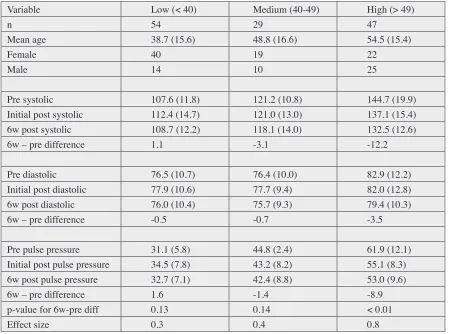
and medium pulse pressure groups (Table 1). The mean change for the high pulse pressure group was relatively large, where a reduction by 8.9 points was observed, and this change was statistically significant (p < 0.01) with a large effect size of 0.8. In the high pulse pressure group similar treatment numbers were observed: the average number of visits were 11.9 and the average number of adjustments was 2.6.
Discussion
Pulse pressure represents the relationship between left ventricle ejection and the properties of the arterial wall. Attributes of the arterial wall determine arterial
compli-ance and the transmission characteristics of the arterial system.5 The Windkessel model of circulation describes the relationship between stroke volume and arterial com-pliance of the aorta and large vessels.28 Windkessel is loosely translated from German to English as ‘air cham-ber’.29 In regard to circulation it is generally accepted as ‘elastic reservoir’. The aorta has remaining blood volume at the end-point of diastole. Upon systole, the left ventri-cular ejection pumps more blood into the aorta. Compli-ance is determined by the aorta’s ability to accommodate further increases in volume. Although compliance occurs throughout the vascular tree, total systemic arterial com-pliance is primarily determined by the aorta and its large Table 1.
Summary data, by pre pulse pressure group, reported as mean (SD). ES = effect size, reported for the main variable of interest in the study, pulse pressure. Initial post is on same visit as initial adjustment. Initial post systolic and diastolic
missing for one patient in low pulse pressure group.
Variable Low (< 40) Medium (40-49) High (> 49)
n 54 29 47
Mean age 38.7 (15.6) 48.8 (16.6) 54.5 (15.4)
Female 40 19 22
Male 14 10 25
Pre systolic 107.6 (11.8) 121.2 (10.8) 144.7 (19.9)
Initial post systolic 112.4 (14.7) 121.0 (13.0) 137.1 (15.4)
6w post systolic 108.7 (12.2) 118.1 (14.0) 132.5 (12.6)
6w – pre difference 1.1 -3.1 -12.2
Pre diastolic 76.5 (10.7) 76.4 (10.0) 82.9 (12.2)
Initial post diastolic 77.9 (10.6) 77.7 (9.4) 82.0 (12.8)
6w post diastolic 76.0 (10.4) 75.7 (9.3) 79.4 (10.3)
6w – pre difference -0.5 -0.7 -3.5
Pre pulse pressure 31.1 (5.8) 44.8 (2.4) 61.9 (12.1)
Initial post pulse pressure 34.5 (7.8) 43.2 (8.2) 55.1 (8.3)
6w post pulse pressure 32.7 (7.1) 42.4 (8.8) 53.0 (9.6)
6w – pre difference 1.6 -1.4 -8.9
p-value for 6w-pre diff 0.13 0.14 < 0.01
Effect size 0.3 0.4 0.8
vessels. Aortic compliance is largely dependent upon the vessels elasticity. As the vessel becomes less elastic, it loses ability to adequately take on more volume upon sys-tole and pulse pressure increases.
There are transmission characteristics of the arterial tree, beyond the Windkessel model, to be considered when evaluating pulse pressure. Circulation depends on a pulse wave initiated by cardiac contraction. Pulse waves travel from the heart to the capillary bed. The forward wave (systolic P1) runs into multiple branch junctions as arteries split into smaller vessels. Each time a wave hits a branch junction there is a wave reflection. A small amount of the wave bounces back toward the heart. The waves reflecting back and having impact on pulse pressure are predominantly from the major branches of the aorta. The reflected wave going back toward the heart can increase aortic systolic pressure, thus pulse pressure, above what is generated via left ventricle ejection. The reflected wave (systolic P2) has an amplifying effect on central pulse pressure. Pulse pressure can be clinically assessed via brachial measurements. Izzo found that brachial systolic blood pressure closely related to centrally measured pres-sure.30
According to Dart and Kingwell 5, there is a common thread between arterial compliance described by the Windkessel model and wave reflection. Both are similar-ly affected by arterial wall properties. Arteries with com-promised elasticity are less compliant and magnify the wave reflection, increasing pulse pressure.
Pulse pressure has a bidirectional effect on cardio-vascular disease.31 Elevated pulse pressure increases mechanical pressure upon the endothelial lining and in-creases the risk of a CVD event. Mechanical pressure, over time, has a damaging effect on the endothelial lining creating a vicious cycle of further increased arterial stiff-ness.
Pulse pressure is subject to any factor that affects aortic elasticity. Aging12 is associated with loss of elasticity in the aorta as is atherosclerosis 32-33. High homocysteine 34-35 and insulin resistance 36-38 have been found to increase pulse pressure. Pulse pressure improvements have been reported with exercise 39-40 as well as diet modifications 41-42 including n-3 fatty acids.
Vascular smooth muscle tone has a direct impact on ar-terial elasticity. Vascular smooth muscle is largely deter-mined by the sympathetic nervous system and is subject
to autonomic balance. The sympathetic nervous system has significant influence on mean arterial pressure and systemic vascular resistance as it modulates the mechan-ical properties of muscular arteries.43 Increased smooth muscle tone can be an explanation for the arterial wall effects seen in elevated pulse pressure.44
A number of studies on upper cervical chiropractic techniques have reported improvements in hypertensive patients’ systolic pressures following care.19-23,45-46 One study that included 42 subjects found that hypertensive subjects’ blood pressure lowered and hypotensive pa-tients’ blood pressure elevated following careindicated a broader normotensive effect.47
Other studies have suggested upper cervical chiro-practic care as being beneficial for autonomic balance. These studies19, 48-51 have focused on heart rate variability (HRV) as an indicator of autonomic modulation.52-53 HRV has been found low in survivors of myocardial infarc-tion (MI) and abnormal HRV patterns precede episodes of life-threatening arrhythmias. Furthermore, HRV is a strong predictor of mortality in patients with MI.54 Our study adds to the body of literature on a possible associ-ation between upper cervical chiropractic care and cardio-vascular assessments.
Various studies have confirmed positive post changes with the application of an upper cervical technique.55-56 One study also reported on positive subjective outcomes and safety of several upper cervical techniques.57
Further research (e.g., a randomized clinical trial) seems warranted as a next step toward possibly intro-ducing a novel approach for a safe, natural approach to cardiovascular health. The present study is limited in its generalizability to other patients since it had an obser-vational study design (case series). In addition, the high pulse pressure group’s readings may simply be temporary outliers. As this is the first study of its kind, further re-search is needed before more definite conclusions can be drawn. In further research, our sample size calculations indicate that 20 patients would be needed in a high pulse pressure group such as ours, based on our data in Table 1 for that group (using 80% power and a two-tailed alpha level of 0.05).
Conclusion
pres-sure in patients determined to previously have had elevat-ed pulse pressure. It is plausible that these observations are related to improved autonomic function. Further re-search with improved designs is a reasonable next step in this line of investigation.
References
1. Giuseppe Mancia, Guido Grassi. The autonomic nervous system and hypertension. Circulation Res. 2014;114: 1804-1814.
2. Eleftheriadou I, Drosos GC, Tentolouris A, Konstantonis G, Sfikakis PP, Protogerou AD, Tentolouris N. Pulse pressure amplification and cardiac autonomic dysfunction in patients with type 2 diabetes mellitus. Journal of Human Hypertension 2018 vol 32, pages 531–539.
3. Vaccarino V, Holford TR, Krumholz HM. Pulse pressure and risk for myocardial infarction and heart failure in the elderly. J Am Coll Cardiol. 2000; 36:130–138.
4. Simone G, Roman MJ, Alderman MH, Galderisi M, Divitiis O, Devereux RB: Is high pulse pressure a marker of preclinical cardiovascular disease? Hypertension. 2005;45: 575-579.
5. Dart AM, Kingwell BA. Pulse pressure – a review of mechanisms and clinical relevance. J Am Coll Cardiol. 2001;37: 975–984.
6. Blacher J, Staessen JA, Girerd X, Gasowski J, Thijs L, Liu L, Wang JG, Fagard RH, Safar ME. Pulse pressure not mean pressure determines cardiovascular risk in older hypertensive patients. Arch Intern Med. 2000; 160 (8): 1085–1089.
7. Sherazi S, Goldenberg I, Moss AJ, Solomon S, Kutyifa V, McNitt S, Shah AH, Huang DT, Aktas MK, Zareba W, Barsheshet A. Comparison of low versus high (>40 mm Hg) pulse pressure to predict the benefit of cardiac resynchronization therapy for heart failure (from the Multicenter Automatic Defibrillator Implantation Trial-Cardiac Resynchronization Therapy Trial). Am J Cardiol. 2014;114(7):1053-1058.
8. American College of Surgeons (2008). Atls, Advanced Trauma Life Support Program for Doctors. Amer College of Surgeons. p. 58. ISBN 978-1-880696-31-6.
9. Yildiran T, Koc M, A, Sahin DY, I, Acarturk E: Low pulse pressure as a predictor of death in patients with mild to advanced heart failure. Tex Heart Inst J. 2010; 37(3): 284– 290.
10. Franklin SS, Gustin W, IV, Wong ND, et al. Hemodynamic patterns of age-related changes in blood pressure: the Framingham heart study. Circulation. 1997;96(1):308–315. 11. Mitchell GF, Wang N, Palmisano JN, et al. Hemodynamic
correlates of blood pressure across the adult age spectrum: noninvasive evaluation in the Framingham heart study. Circulation. 2010;122(14):1379–1386.
12. Steppan J, Barodka V, Berkowitz DE, Nyhan D. Vascular
stiffness and increased pulse pressure in the aging cardiovascular system. Cardiol Res Pract. 2011; 263585. 13. Rizzo V, di Maio F, Petretto F, Marziali M, Bianco G,
Barilla F, Paravati V, Pignata D, Campbell SV, Donato G, Bernardo V, Tallarico D. Ambulatory pulse pressure, left ventricular hypertrophy and function in arterial hypertension. Echocardiography. 2004;21(1): 11-16. 14. Waldstein SR, Rice SC, Thayer JF, Najjar SS, Scuteri
A, Zonderman AB. Pulse pressure and pulse wave velocity are related to cognitive decline in the Baltimore longitudinal study of aging. Hypertension. 2008;51: 99-104.
15. Nation DA, Preis SR, Beiser A, Bangen KJ, Delano-Wood L, Lamar M, Libon DJ, DeCarli C, Seshadri S, Wolf PA. Pulse pressure is associated with early brain atrophy and cognitive decline: modifying effects of APOE4. Alz Dis Assoc Disord. 2016; 30(3): 210-215.
16. Yaneva-Sirakova T, Tarnovska-Kadreva R, Traykov L. Pulse pressure and mild cognitive impairment. J Cardiovasc Med. 2012;13(11):735-740.
17. Yasar S, Ko JY, Nothelle S, Mielke MM, Carlson MC. Evaluation of the effect of systolic blood pressure and pulse pressure on cognitive function: the women’s health and aging study II. PLoS ONE. 2011; 6(12): e27976. 18. Yasuno S, Ueshima K, Oba K, Fujimoto A, Hirata M,
Ogihara T, Saruta T, Nakao K. Is pulse pressure a predictor of new-onset diabetes in high-risk hypertensive patients? A subanalysis of the Candesartan Antihypertensive Survival Evaluation in Japan (CASE-J) trial. Diabetes Care. 2010; 33(5): 1122–1127.
19. Qualls T, Lester C. Resolution of atrial fibrillation and hypertension in a patient undergoing upper cervical Chiropractic Care. J Upper Cerv Chiropr Res. 2012; 1: 9-15.
20. Prater-Manor S, Clifton E, York A. Resolution of anxiety and hypertension in a 60-year old male following subluxation based chiropractic care: a case study and review of the literature. Annals Vert Subl Res. 2015: 146-153.
21. Chung J, Brown J, Busa J: Resolution of hypertension following reduction of upper cervical vertebral subluxation: a case study. J Upper Cerv Chiropr Res. 2014:1-6.
22. Kessinger RC, Moe C. Improvement in chronic
hypertension following a single upper cervical adjustment: a case report. J Upper Cerv Chiropr Res. 2015: 1-5. 23. Bakris G, Dickholtz Sr M, Meyer PM, Kravitz G, Avery E,
Miller M, Brown J, Woldfield C, Bell B. Atlas vertebra realignment and achievement of arterial pressure goal in hypertensive patients: a pilot study. J Human Hypertens. 2007. 21: 347–352.
International Protocol (ESH-IP) for the validation of blood pressure measuring devices and future perspectives. J Hypertens. 2018; 36(3):479-487.
25. Topouchian J, Agnoletti D, Blacher J, Youssef A, Chahine MN, Ibanez I, Assemani N, Asmar R. Validation of four devices: Omron M6 Comfort, Omron HEM-7420, Withings BP-800, and Polygreen KP-7670 for home blood pressure measurement according to the European Society of Hypertension International Protocol. Vasc Health Risk Manag 2014; 10: 33–44.
26. Omron Healthcare Validity for Omron BP 760. Available at https://omronhealthcare.com/service-and-support/ clinical-validation/
27. Palmer BJ. Chiropractic clinical controlled research, Vol. XXV. 1951 W.B. Conkey Company, Hammond, Indiana. 28. Frank O. The basic shape of the arterial pulse. First
treatise: mathematical analysis. 1899. J Mol Cell Cardiol. 1990; 22(3): 255-277.
29. Sagawa K, Lie RK, Schaefer J. Translation of Otto Frank’s paper “Die Grundform des Arteriellen Pulses” Zeitschrift für Biologie 37: 483-526 (1899). J Mol Cell Cardiol. 1990;22(3):253-254.
30. Izzo Jr JL. Brachial vs. central systolic pressure and pulse wave transmission indicators: a critical analysis. Am J Hypert. 2014; 27(12): 1433-1442.
31. Ryan SM, Waack BJ, Weno BL, Heistad DD. Increases in pulse pressure impair acetylcholine-induced vascular relaxation. Am J Physiol. 1995;268(1 Pt 2):H359-363. 32. Ikonomidis I, Kadoglou N, Tsiotra PC, Kollias A, Palios I,
Fountoulaki K, Halvatsiotis I, Maratou E, Dimitriadis G, Kremastinos DT, Lekakis J, Raptis SA. Arterial stiffness is associated with increased monocyte expression of adiponectin receptor mRNA and protein in patients with coronary artery disease. Am J Hypertens. 2012; 25:746– 755.
33. Casalnuovo G, Gerdts E, de Simone G, Izzo R,
De Marco M, Giudice R, Trimarco B, De Luca N. Arterial stiffness is associated with carotid atherosclerosis in hypertensive patients (the campania salute network). Am J Hypertens. 2012; 25: 739–745.
34. Momin M, Fan F, Li J, Qin X, Jia J, Qi L, Zhang Y, Huo Y. Associations of plasma homocysteine levels with peripheral systolic blood pressure and noninvasive central systolic blood pressure in a community-based Chinese population. Sci Rep. 2017; 7: 6316.
35. Williams C1, Kingwell BA, Burke K, McPherson J, Dart AM. Folic acid supplementation for 3 wk reduces pulse pressure and large artery stiffness independent of MTHFR genotype. Am J Clin Nutr. 2005; 82(1): 26-31. 36. Henry RMA, Kostense PJ, Spijkerman AMW, et al.
Arterial stiffness increases with deteriorating glucose tolerance status: the Hoorn study. Circulation. 2003;107(16): 2089–2095.
37. Sengstock DM, Vaitkevicius PV, Supiano MA. Arterial
stiffness is related to insulin resistance in nondiabetic hypertensive older adults. J Clin Endocrinol Metabol. 2005;90(5): 2823–2827.
38. Yamada E, Takeuchi M, Kurata M, Kazumi T, Fukuo K. Association of Pulse Pressure with Serum TNF-α and Neutrophil Count in the Elderly. J Diabetes Res. 2014; 2014: 972431.
39. Cameron JD, Dart AM. Exercise training increases total systemic arterial compliance in humans. Am J Physiol. 1994; 266: H693-H701.
40. 91. Kingwell BA, Cameron JD, Gillies KJ, Jennings GL, Dart AM. Arterial compliance may influence baroflex function in athletes and hypertensives. Am J Physiol. 1995; 268: H411-H418.
41. McVeigh GE, Brennan GM, Cohn JN, Finkelstein SM, Hayes RJ, Johnston GD. Fish oil improves arterial compliance in noninsulin-dependent diabetes mellitis. Arterioscler Thromb. 1994; 14:1425-1429.
42. Nestel PJ, Pomeroy SE, Sasahara T et al. Arterial compliance in obese subjects is improved with dietary plant n-3 fatty acid from flaxseed oil despite increased LDL oxidizability. Arterioscler Thromb Vasc Biol. 1997; 17:1163-1170.
43. Parati G., Salvi P. Arterial stiffness and the sympathetic nervous system. In: Safar M., O’Rourke M., Frohlich E. (eds) Blood Pressure and Arterial Wall Mechanics in Cardiovascular Diseases. Springer, London. 2014. 44. Dobrin P, Rovick A. Influence of vascular smooth muscle
on contractile mechanics and elasticity of arteries. Am J Physiol. 1969; 217:1644–1651.
45. Knutson, G. Significant changes in systolic blood pressure post vectored upper cervical adjustment vs resting control groups: a possible effect of the cervicosympathetic and/or pressor reflex. J Manipulative Physiol Ther, 2001; 24:101-109.
46. Goodman R. Hypertension and the atlas subluxation complex. J Chiropr Res Clin Investigation, 1992; 8(2):30-32.
47. Torns S. Atlas vertebra realignment and arterial blood pressure regulation in 42 subjects. J Upper Cerv Chiropr Res. 2012; 1: 40-45.
48. Burcon MT. Upper cervical specific pattern analysis utilizing paraspinal thermography, leg length inequality and heart rate variability in two patients with tachycardia. J Upper Cervical Chiropr Res. 2016; 1: 6-12.
49. Win NN, et al. Effects of upper and lower cervical spinal manipulative therapy on blood pressure and heart rate variability in volunteers and patients with neck pain: a randomized controlled, cross-over, preliminary study. J Chiropr Med. 2015;1-9.
in pattern analysis, heart rate variability and symptoms following upper cervical chiropractic care. J Upper Cervical Chiropr Res. 2013;2: 32-42.
52. Porges SW. The polyvagal perspective. Biological Psychology. 2007; 74 (2): 116–143.
53. Robinson BF, Epstein SE, Beiser DG, Braunwald E. Control of heart rate by the autonomic nervous system - studies in man on the interrelation between baroreceptor mechanisms and exercise. Circulation Research. 1966;19: 400-411.
54. Kudaiberdieva G, Gorenek B, Timuralp B. Heart rate variability as a predictor of sudden cardiac death. Anatol J Cardiol 2007: 7(Suppl 1); 68-70.
55. Woodfield CH, Dickholtz, Sr. M. Quality of life
improvement in patients with chronic fatigue syndrome following upper cervical chiropractic care. J Upper Cerv Chiropr Res. 2012; 1: 92-99.
56. Fedorchuk C, Lightstone D, Moser III JH. Improvement in symptoms, cervical alignment and quality of life in a 40-Year-old female with fibromyalgia following chiropractic BioPhysics® Technique: A case study and selective review of literature. Ann Vertebr Sublux Res. 2017: 34-46.