R E S E A R C H
Open Access
Changes in protein profile and
encapsulation avoiding responses of
entomopathogenic nematode in the
American bollworm
Helicoverpa armigera
(Hübner) (Lepidoptera: Noctuidae)
Istkhar
1*and Ashok Kumar Chaubey
2Abstract
The American bollwormHelicoverpa armigera(Hübner) (Lepidoptera: Noctuidae) is a highly destructive agriculture pest
of worldwide importance. The aim of the present study was to infect H. armigera larvae with entomopathogenic
nematode juveniles for hematological study of proteins and encapsulation responses to evaluate using this nematode for the management of this pest. Total protein estimation and the electrophoretic profiling carried out in the
hemolymph showed a high pathogenicity of Steinernema abbasi to H. armigera. The control group survived and
succeeded to develop to adults, while the infected ones died within 24 h. An increase in the protein contents in the total and plasma hemolymph was observed just after 3 h of infection with an increase at 6 h and 9 h as symptoms of early defence of the insect. SDS-PAGE profile also showed an evolvement of a protein band of 46 kDa. No self-association or aggregation and binding of other proteins were found in the hemolymph as revealed by Native-PAGE. The encapsulation avoidance rate of nematode juvenile gave good results with (> 33%) in 2 IJs/larva to 5% in 20 IJs/ larva doses at24 h post infection. Loss of hemolymph proteins continued for more than 24 h with a very low recognition of nematode rate in hemolymph, followed by the death of larvae within 48 h, which proved the high pathogenicity ofS. abbasiand suppression of host immune system ofH. armigera.
Keywords:Helicoverpa armigera, Entomopathogenic nematode,Steinernema abbasi, Protein estimation, Encapsulation
Background
Insect immunology is a rapidly growing discipline with ex-citing and innovative application, especially in control of agricultural pests by understanding the host parasite inter-actions such as entomopathogenic nematodes (EPNs). When insect pests of agricultural crops are targeted by microorganisms, they rely on innate immunity for quick response comprises cellular and humoral activity.
The American bollworm,Helicoverpa armigera(Hübner) (Lepidoptera: Noctuidae), is a polyphagous worldwide distributed pest infests more than 181 plant species belonging to 45 families (Singh2005). Defensive strategies
ofH. armigeraare poorly understood, and this insect has developed a strong resistance against several chemical pes-ticides (Bass et al.2015). Studying the immune system of pests helps better understanding of their defenses against invading parasites (EPNs), which are nowadays been used in insect pest management. In this regard, less information is available on immunological defenses in hemolymph of H. armigeracreating its management difficult. Results of pathogenicity of EPNs againstH. armigeralarvae provided a good picture and better management in a comparatively environmentally safer way (Istkhar and Chaubey2017).
The EPNs, steinernematids and heterorhabditids have be-come a new tool as they have a wide variety of host range, high virulence, less killing time, host finding ability, fast acting generations, and safe to vertebrates and non-target invertebrates (Lacey and Georgis 2012). Steinernematids
© The Author(s). 2019Open AccessThis article is distributed under the terms of the Creative Commons Attribution 4.0 International License (http://creativecommons.org/licenses/by/4.0/), which permits unrestricted use, distribution, and reproduction in any medium, provided you give appropriate credit to the original author(s) and the source, provide a link to the Creative Commons license, and indicate if changes were made.
* Correspondence:[email protected]
1Department of Bioscience and Biotechnology, Banasthali Vidyapith, Banasthali, Rajasthan 304022, India
are naturally associated with symbiotic bacteria of the genus Xenorhabdus (Gammaproteobacteria: Enterobacteriaceae). Researchers showed that not only the nematodes, but also the bacterial symbionts are highly pathogenic to many in-sect pests once they reached the hemocoel of the inin-sect (Forst and Nealson1996). During their complex life cycle in a common insect host, bacteria carried by the nematodes in their gut are regurgitated in the hemocoel of insect, where they multiply along with nematodes and produced several toxins, leading to the death of insect. Hemolymph plays a vital role for insects as the place where they com-pete with invading microorganisms. Production of anti-microbial peptides (AMPs), activation of melanization, phagocytosis, and encapsulation are the main events against EPN infection in the insect hemolymph (Castillo et al. 2011). When the nematode enters in the hemocoel of in-sect, soluble proteins in hemolymph and proteins released by hemocytes and hematopoietic organs participate in their encapsulation (Lemaitre and Hoffmann2007). To avoid the targeting, nematode and/or nematode-bacterial complex also attempt to evade the poorly understood immune re-sponse of insect immune system (Brivio et al.2010). Little information is available about the nematode infection and hematological changes inH. armigera.
The present study was planned to study the hematological responses ofH. armigeralarvae infected with 3rd stage in-fective juvenile (IJs) ofSteinernema abbasifor protein and nematode encapsulation.
Materials and methods
Insect’s maintenance
Eggs ofH. armigerawere obtained from the National Bur-eau of Agriculturally Important Resources (NBAIR), ICAR, Bangalore, India (National Accession No. NBAII-MP-NOC-01), and the culture was maintained in the laboratory. Larvae were fed on a semisynthetic diet (Nagarkatti and Prakash1974), while the adults were fed on 10% honey solution containing vitamin E for better egg laying. A culture of the greater wax moth,Galleria mello-nella(L.) (Lepidoptera: Pyralidae), was also maintained in the laboratory for in vivo culturing of the EPNs.
In vivo nematode culture
Steinernema abbasi (Rhabditida: Steinerneamtidae) was found to be the most dominating and pathogenic species during the survey conducted in studied area and showed a strong pathogenicity toH. armigera(Istkhar and Chaubey 2017).Isolate CS2ofS. abbasioriginally isolated from the
soil of District Meerut (U.P.), India, was used to infect the larvae ofH. armigerafor hematological study. Population ofS. abbasiwas maintained in vivo on larvae ofG. mello-nella, which considered as the universal host of EPNs. Freshly emerged 3rd stage infective juvenile (IJs) were col-lected from White trap (Stock and Goodrich-Blair 2012)
and stored in BOD at 15 ± 1 °C and used to infect the lar-vae ofH. armigerafor experimentation within a week of emergence.
Experimental design for protein estimation
For studying the changes in protein quantity and protein profiling through electrophoresis in hemolymph of H. armigera, the hemolymph was collected at different post infection periods (PIP) after infection with 100 IJs of S. abbasi (isolate CS2) in six-well Petri plates (Himedia)
lined with a double layer of Whatman filter Paper (No. 1) by maintaining the adequate moisture. Larvae were then surface sterilized and 50 μl of hemolymph from each larva was drawn directly in pre-chilled 1.5 ml centrifuge tubes containing 450 μl of phosphate buffer saline (PBS, pH 7.2) by cutting their proleg at 0, 3, 6, 9, 12, 18, and 24 h post infection period (PIP). Control group received 450 μl of distilled water and used for hour to hour protein comparison. Control and treated group were placed in BOD at 27°±2 °C. For each hour interval, 10 out of 20 infected larvae were randomly selected for hemolymph collection, and the whole ex-periment was repeated twice. Hemolymph of larvae in the control group was also collected in a similar way. Hemolymph was collected singly from each larva, and mean values of 10 replicates were taken as mean quan-tity of protein in milligram/milliliter of hemolymph.
Protein estimation
For the assessment of combined effect of soluble proteins and hemocytic protein (proteins secreted by hemocytes) changes due to nematode infection in hemolymph and soluble proteins only, the hemolymph samples were tested as total hemolymph and plasma fractions, respectively. Total protein contents in plasma fraction of hemolymph samples, collected from the infected and control larvae of H. armigera in PBS solution (pH 7.2), were estimated according to Bradford’s method (Bradford 1976). Two-hundred microliters of the hemolymph-PBS mixture men-tioned above was retained as a whole hemolymph, and the rest was centrifuge at 10,000 g for 20 min at 4 °C to separ-ate out plasma contents. Protein was estimsepar-ated as total protein in hemolymph and in plasma fraction in terms of soluble proteins. Both samples were used for protein esti-mation and were expressed in milligram/milliliter of hemolymph. Dilutions of bovine serum albumin (BSA) were used to prepare a standard curve for estimation of protein.
Electrophoresis of hemolymph samples
Reducing electrophoresis of cell-free hemolymph was performed using 12% resolving and 4% stacking
poly-acrylamide gel (SDS-PAGE) (Laemmli 1970) on Genei
power supply set at 15 mA for 10 min and at 30 mA for
120 min. A broad range (10–250 kDa) multicolour
ladder (Thermo ScientificTM) was run along with
hemolymph samples for estimating and comparing changes in protein samples. Lanes of gel were loaded with 5-μl plasma sample side by side collected from par-asitized and unparpar-asitized larvae ofH. armigera, respect-ively. After electrophoresis, the gels were stained, using coomasie brilliant blue R250 stain. The analyses of gels were performed through My Image Analysing Software Version 2.0 (Thermo Scientific). For non-reducing electrophoresis, Native-PAGE was performed for all the samples to study self-association or aggregation and the binding of other proteins if any.
Experimental design for encapsulation responses
The encapsulation responses of host insect H. armigera were investigated by injecting IJs/larva (Peters and Ehlers 1997) directly into the hemocoel. The encapsulations were observed microscopically after 24 h as after 48 h all the larvae were found dead. To ensure the entry of nematode IJs, an injection apparatus modified from Ehlers et al. (1997) (Fig. 1) was constructed, using a ca-pillary tube inserted and sealed in a microtip of 20 μl capacity. The tip of the capillary tube was heated, drawn out, and broken into fine pointed tip. The other end of the capillary tube was fixed into the microtip and placed over the micropipette ensuring the fixed amount of nematode suspension to be injected into the body cavity of insect larvae. Eighty larvae were injected by 4 different doses of IJs having 2, 5, 10, and 20 IJs of nematode in 8 μl of PBS solution (Himedia) and dived into 2 batches. The first batch was infected with IJs was dissected to ob-serve the total number of IJs encapsulated and studied under stereomicroscope (NIKON SMZ645). The second batch was placed to observe the mortality rates at the tested doses of nematode IJs applied at the same time of dissection. Ten replicates were maintained for each dose of injection. Ten larvae ofH. armigerareceived only PBS solution and placed as control. Larvae were dissected after each 24 h of infection, and numbers of encapsu-lated nematode juveniles were counted. The larvae were disinfected by 70% alcohol before injecting.
Statistical analyses
Descriptive analysis was performed for quantified pro-tein at each hour interval and presented as mean ± standard error of mean (SEM) of 10 replicates randomly selected from infected with EPNs and control larvae of H. armigera. Two-way ANOVA was performed to find out the effect of time and treatment on total protein contents. One-way ANOVA was executed to compare protein differences in infected and control larvae. Fish-er’s least significance difference (Fisher’s LSD) was used
as post hoc test for comparison within infected and con-trol at each hour interval.
Results and discussion
In the present study, the estimation of protein was per-formed, followed by their electrophoretic profiling. The total protein contents were measured in total and plasma fraction of larval hemolymph ofH. armigera in-fected with S. abbasi-X. Indica separately. Analysis of variance (two-way) showed that total protein contents in total hemolymph challenged withS. abbasiwas affected by time (df = 6;F= 14.584; p= 0.00) and not by treat-ment (df = 1; F= 1.388; p= 0.241). Effect of time and treatment interaction was significant (df = 5;F= 17.784;
p = 0.00). While applying one-way ANOVA, the mean
protein contents varied significantly between the in-fected and control group ofH. armigeralarvae (df = 12; F= 14.825;p= 0.00) (Fig.1a). Insignificant changes were recorded in the control group and the amount of protein was almost steady with very minute fluctuations from 129.23 to 129.62 mg/ml. The increment in protein con-tents was reported over 3 h of infection. This significant increment was 12% as compared to 0 h from 129.49 to 144.81 mg/ml (p< 0.05). With the significant increments of 24.69 and 28.27% protein contents at 6 h and 9 h, re-spectively, an increment in protein profile was detected in the total protein amount reaching the peak amount of 165.77 mg/ml. From 9 h PIP onwards, a dramatic de-cline was recorded continuously up to 24 h with 116.92 mg/ml, 109.55 mg/ml, and 97.50 mg/ml at 12, 18, and 24 h, respectively. In a similar way, soluble protein con-tent fluctuated significantly in the plasma fraction of hemolymph (Fig.1b).
Chaubey 2018). Increment observed in protein contents by spectrophotometric study, just after 3 h, indicated the activation of H. armigera immune system responses by detecting nematode IJs which may be considered an early defense mechanism. The superior pathogenicity of Stei-nernemaspecies perceived with the entry of the IJs in the insect host’s body confirmed by the elevated profile of protein in the hemolymph samples, which was almost steady in the control group up to 9 h.
During the analysis of separation of plasma protein contents through SDS-PAGE, lanes were loaded alterna-tively by plasma fractions of control and infected larval hemolymph plasma of H. armigera. The electrophoretic
profile revealed the 8 major bands of proteins in hemolymph plasma with molecular weights of ~ 173, ~ 96, ~ 68, ~ 59, ~ 52, ~ 48, ~ 34, and ~ 17 kDa (Fig.2). In nematode-bacterium challenged larvae an additional band of ~ 46 kDa was observed at 9 and 12 h of PIPs. This band vanished at 18 h of infection and a new band appeared at 24 h of infection with molecular weight of ~ 28 kDa. When the plasma fractions were analyzed in non-denatured conditions, there were 5 major bands (Rf values 0.09, 0.15, 0.38. 0.53, and 0.57) in the gel and no changes were observed revealed by Native-PAGE elec-trophoretic profile in control and infected groups of H. armigera (Fig. 3). Electrophoretic study performed on
Rhipicephalus microplus (Boophilus) infected with Het-erorhabditis indicashowed no differences in SDS-PAGE profile of hemolymph proteins (Patrícia Silva Golo et al. 2016). In our study, the electrophoretic patterns of in-fected and control hemolymph was almost similar; how-ever, the appearance and disappearance of proteins were observed in denatured electrophoretic profile at 9 h, 12 h, and 24 h in infected hemolymph. This can be explained as after entry of nematode-bacterium complex within the
body of H. armigera, the host produces some defensive factors to combat with invading pathogens. The proteins are secreted during the initial phase of nematode growth more abundantly (Simões et al. 2000) as observed in the present study, where the amount of proteins in hemolymph increased significantly during initial hours of infection. The increment reported in hemocytes during initial hours of infection in the previous study (Istkhar and
Chaubey 2018) acclaimed the increment in protein
Fig. 2A comparative electrophoretic profile of plasma fractions ofacontrol (0, 3HC, 6HC, 9HC) andSteinernema abbasi-Xenorhabdus indica infected (3HT, 6HT, 9HT)Helicoverpa armigera,bcontrol (12HC, 18HC, 24HC) andSteinernema abbasi-Xenorhabdus indicainfected (12HT, 18HT, 24HT)H. armigeraanalyzed by SDS-PAGE. Gels were analyzed through My Image Analysing Software Version 2.0 (Thermo Scientific)
contents in present study and showed the secretion of some major proteins of hemolymph and some unknown proteins by hemocytes, which may help in initial protec-tion and defend host to parasitic infecprotec-tion. This statement was supported by the presence of extra band at 9 h of in-fection. The disappearance of proteins bands occurred at 9 and 12 h as well as in the total protein contents were ex-plained as the action of hydrolysing of the host proteins. These results agree with the Schmidt and Platzer (1980), who reported protein degradation, when Culex pipiens (L.) was infected byRomanomermis culicivorax. They sug-gested the production of some proteases from the nema-todes leads to the degradation of hemolymph proteins of
C. pipiens. Wee et al. (2000) suggested the production of proteases by bacterial cells, followed by the breakdown of insect’s protein and serving as nutritional resources for nematode-bacterium development. In another study, the reduction in total protein contents of hemolymph was
observed in the hemolymph of Schistocerca gregaria
(Forsskål) during the course of infection with EPF, Mer-tarhizium anisopliaevar acridium(Gillespie et al. 2000). Other workers supported the study, where the produc-tions of proteases and subsequent reduction in proteins by Steinernema carpocapsae and bacteria were reported (Toubarro et al.2013). While studying the electrophoretic band pattern in native-PAGE for the agglutination and/or
association of proteins during infection, no changes were observed and similar pattern banding was found in the control and the infected groups.
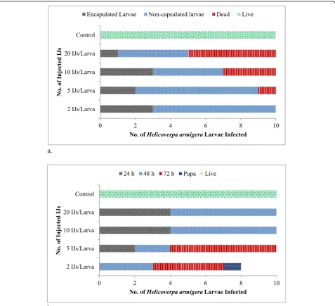
For encapsulation study, the first batch of nematode-infected larvae was dissected after 24 h only and not after 48 h as infected larvae were found dead after 48 h. During the time of dissection, the adhering of hemocytes was observed around the encapsulated nematode. In the first batch, 30, 20, 30, and 10% of larvae injected with 2, 5, 10, and 20 IJs of nematode were found having encapsulated IJs within their hemocoel. No strong melanization was noticed over encapsulated IJs. Mortality was observed in the larvae and 10, 30, and 50% mortality rates were found inH. armigeralarvae infected with 5, 10, and 20 IJs/larva, respectively, at 24 h (Fig.4a). No mortality was observed in the larvae injected with 2 IJs/larva. In a second batch of larvae injected with nematode, all nematodes were found dead in dissected larvae at 48 h in 10 and 20 IJs/larva doses, whereas in 2 and 5 IJs/larva doses, 70 and 80% after 72 h and one of the larvae attained pupation, but could
not achieved emergence (Fig. 4b). The percentage of
encapsulation of IJs was calculated in the larvae found positive for encapsulation, when injected with different doses of IJs. The proportion of encapsulated nematodes declined with increasing dose from 66.67% for 2 IJs/larva to 5% for 20 IJs/larva. For 5 and 10 IJs/larva, the encapsu-lation was 30 and 16.67%, respectively.
However, EPNs were found effective against the insect pests; the insect has also evolved defensive mechanisms against these nematode invaders (Feldhaar and Gross 2008). The innate immune responses included hemocytes, which participated in phagocytosis, nodulation, and encapsulation, and played a significant role and provide a protection to the insect hosts (Schmidt et al. 2001). The
increments in hemocyte number in hemolymph of H.
armigera infected with S. abbasi have already been observed in previous study (Istkhar and Chaubey 2018). The encapsulation responses of different hosts varied according to host species and nematode species. In Manduca sexta(L.),H. bacteriophorawas recognized by (> 99 %) value, whileS. glaserishowed only (28%) recognition (Li et al.2009). The encapsulation study ofS. feltiaeandH. bacteriophora, against the prepupae ofLeptinotarsa decem-lineata (Say) showed a more frequent encapsulation of S. feltiaethan forH. bacteriophora(Ebrahimi et al.2011). On the other hand, in the rose sawflyArge ochropus(Gmelin), the cellular responses were weaker toS. carpocapsaethan inH. bacteriophora (Sheykhnejad et al. 2014). In another study, 6% encapsulation ofS. feltiaewas reported, while it was 24% in H. bacteriophora in Agriotes lineatus (L.) (Rahatkhah et al.2015). Obtained results showed that the S. abbasi isolate CS2 had the strong capability to avoid
encapsulation responses in larval H. armigera leading to the death of larvae within a very short time.
Conclusion
This study revealed that the presence of nematode within the hemolymph significantly decreased the amount of pro-tein contents. The low encapsulation avoiding response led to a high number of nematode in hemolymph and subsequently reduced the amount of defensive proteins. More number of IJs in the hemolymph produced more toxins, which weaken the insect’s immune system because of the quick death of the host larvae. The results showed thatS. abbasi isolate CS2had a high pathogenicity merit
and provided an insight to use it as a better alternative of biological control agent in future insect pest management programs.
Acknowledgements
Authors greatly acknowledge the Department of Zoology, Chaudhary Charan Singh University, Meerut, India, for providing infrastructure.
Authors’contributions
Istkhar and AKC conceived and designed the study and critically revised the manuscript. Istkhar performed the experiments and analyzed the data. Both authors have approved the final manuscript.
Funding
Authors are thankful to Council of Scientific and Industrial Research (CSIR), New Delhi, India, for funding through EMR project (37(1550)/12-EMR-II).
Availability of data and materials
All data generated or analyzed during this study are included in this published article.
Ethics approval and consent to participate
Not applicable.
Consent for publication
Not applicable
Competing interests
The authors declare that they have no competing interests.
Author details
1Department of Bioscience and Biotechnology, Banasthali Vidyapith, Banasthali, Rajasthan 304022, India.2Nematology Laboratory, Department of Zoology, Chaudhary Charan Singh University, Meerut, Uttar Pradesh 250004, India.
Received: 4 July 2019 Accepted: 23 September 2019
References
Bass C, Denholm I, Williamson MS, Nauen R (2015) The global status of insect resistance to neonicotinoid insecticides. Pestic Biochem Physiol 12:78–87 Bradford MM (1976) A rapid and sensitive method for the quantitation of
microgram quantities of protein utilizing the principle of protein-dye binding. Anal Biochem 72:248–254
Brivio MF, Mastore M, Nappi AJ (2010) A pathogenic parasite interferes with phagocytosis of insect immunocompetent cells. Dev Comp Immunol 34: 991–998
Castillo JC, Reynolds SE, Eleftherianos I (2011) Insect immune responses to nematode parasites. Trends Parasitol 27(12):537–547.https://doi.org/10.1016/ j.pt.2011.09.001
Ebrahimi L, Niknam GH, Dunphy GB (2011) Hemocyte responses ofLeptinotarsa decemlineataandGalleria mellonellato the entomopathogenic nematodes, SteinernemafeltiaeandHeterorhabditis bacteriophora. J Insect Sci 11:76.
https://doi.org/10.1673/031.011.7501
oleracea(Diptera) andGalleria mellonella(Lepidoptera). J Invertebr Pathol 69: 212–217
Feldhaar H, Gross R (2008) Immune reactions of insects on bacterial pathogens and mutualists. Microbes Infect. 10:1082–1088
Forst S, Nealson K (1996) Molecular biology of the symbiotic-pathogenic bacteria Xenorhabdusspp. andPhotorhabdusspp. Microbiol Rev 60:21–43
Gillespie JP, Burnettand C, Charnley AK (2000) The immune response of the desert locustSchistocerca gregariaduring mycosis of the fungus,Metarhizium anisopliae. J Physiol 46:429–437
Istkhar, Chaubey AK (2017) Impact of soil dwelling entomopathogenic nematodes, recovered from the soil of Uttar Pradesh (India), on cotton bollwormHelicoverpa armigera(Hübner) (Lepidoptera: Noctuidae). Appl Biol Res 19:63–70
Istkhar, Chaubey AK (2018) Challenging the larvae ofHelicoverpa armigeraand assessing the immune responses to nematode-bacterium complex. Phytoparasitica. 46(1):75–87.https://doi.org/10.1007/s12600-018-0640-3
Lacey LA, Georgis R (2012) Entomopathogenic nematodes for control of insect pests above and below ground with comments on commercial production. J Nematol 44:218–225
Laemmli UK (1970) Cleavage of structural proteins during the assembly of the head of bacteriophage T4. Nature 227:680–685
Lemaitre B, Hoffmann J (2007) The host defence ofDrosophila melanogaster. Ann Rev Immunol 25:97–743
Li XY, Cowles EA, Cowles RS, Gaugler R, Cox-Foster DL (2009) Characterization of immunosuppressive surface coat proteins fromSteinernemaglaserithat selectively kill blood cells in susceptible hosts. Mol Biochem Parasitol 165: 162–169
Nagarkatti S, Prakash A (1974) Rearing ofHeliothis armigera(Hubn) on artificial diet. Technical Bulletin of Commonwealth Institute of Commonwealth Institute of Biological Control, Bangalore, pp 17, pp 169–173 Patrícia Silva Golo PS, de Oliveira dos Santos AS, Monteiro CMO, de Souza
Perinotto WM, Quinelato S, Camargo MG, de Sá FA, da Costa AI, Martins MF, de Azevedo Prata MC, Bittencourt VREP (2016) Lab-on-a-chip and SDS-PAGE analysis of hemolymph protein profile fromRhipicephalus microplus(Acari: Ixodidae) infected with entomopathogenic nematode and fungus. Parasitol Res.https://doi.org/10.1007/s00436-016-5109-z
Peters A, Ehlers RU (1997) Encapsulation of the entomopathogenic nematode Steinernema feltiaeinTipula oleracea. J Invertebr Pathol 69(3):218–222 Rahatkhah Z, Karimi J, Ghadamyari M, Brivio MF (2015) Immune defenses of
Agriotes lineatuslarvae against entomopathogenic nematodes. BioControl 60: 641–653
Schmidt O, Theopold U, Strand M (2001) Innate immunity and its evasion and suppression by hymenopteran endoparasitoids. Bioessays 23:344–351 Schmidt SP, Platzer EG (1980) Hemolymph composition of mosquito larvae
infected with a mermithid nematode. J Nematol 10:299–304
Sheykhnejad H, Ghadamyari M, Ghasemi V, Jamali S, Karimi J (2014) Hemocytes immunity of rose sawfly,Arge ochropus(Hym.: Argidae) against
entomopathogenic nematodes,Steinernema carpocapsaeandHeterorhabditis bacteriophora. J Asia-Pac Entomol 17:879–883
Simões N, Caldas C, Rosa JS, Bonifassi E, Laumond C (2000) Pathogenicity caused by high virulent and low virulent strains ofSteinermena carpocapsaeto Galleria mellonella. J Invertebr Pathol 75:47–54
Singh OP (2005) Consumption pattern of insecticide inHelicoverpa armigera management in India. In: Saxena H, Rai AB, Ahmed R, Gupta S (eds) Recent Advances inHelicoverpa armigeraManagement. Indian Society of Pulses Research and Development, Kanpur, pp 17–24
Stock SP, Goodrich-Blair H (2012) Nematode parasites, pathogens and associates of insects and invertebrates of economic importance. In: Lacey LA (ed) Manual of Techniques in Invertebrate Pathology. Academic Press, London, pp 373–426
Toubarro D, Avila MM, Montiel R, Simões N (2013) A pathogenic nematode targets recognition proteins to avoid insect defenses. PLoS One.https://doi. org/10.1371/journal.pone.0075691
Wee KE, Yonan CR, Chang FN (2000) A new broadspectrum protease inhibitor from the entomopathogenic bacteriumPhotorhabdus luminescens. Microbiol 146:3141–3147
Publisher’s Note