C A S E R E P O R T
Open Access
Adenocarcinoma arising from an ectopic
pancreas in the duodenum: a case report
Tsukasa Kaneko
1*, Masanori Ohara
1, Kunishige Okamura
1, Aki Fujiwara-Kuroda
1, Daisuke Miyasaka
4,
Takumi Yamabuki
1, Ryo Takahashi
1, Kazuteru Komuro
1, Masato Suzuoki
4, Nozomu Iwashiro
1, Mototsugu Kato
2,
Noriko Kimura
3, Hiroshi Kijima
5, Toru Nakamura
6and Satoshi Hirano
6Abstract
Background:The malignant transformation of an ectopic pancreas in the duodenum is extremely rare. Herein, we report a case of an adenocarcinoma that arose from an ectopic pancreas. We also reviewed 14 cases of malignant transformations arising from an ectopic pancreas in the duodenum that were previously published.
Case presentation:An 81-year-old man with a 1-month history of vomiting was admitted to our institution. Esophagogastroduodenoscopy (EGD) and computed tomography (CT) scans revealed an obstruction at the first part of the duodenum. A distal gastrectomy was performed for diagnostic and therapeutic purposes. The histopathological examination of the resected specimen showed adenocarcinoma that arose from an ectopic pancreas (Heinrich type 1). The patient is alive without relapse at 18 months of follow-up.
Conclusions:Adenocarcinoma that arises from an ectopic pancreas should be considered when an obstruction is identified in the duodenum.
Keywords:Ectopic pancreas, Distal gastrectomy, Duodenal adenocarcinoma, Cancer-induced vomiting
Background
An ectopic pancreas, often found during surgery or bi-opsy, is defined as an uncommon pancreatic tissue out-side the normal pancreas, which lacks any connection to the normal pancreas, and has its own vascular and ductal systems [1]. The frequency of the occurrence of ectopic pancreatic tissue is found in 0.25% of abdominal surgeries and 1.2% of gastrectomy operations, and its
frequency at autopsy has been reported to be 0.55–
13.7% [2]. Ectopic pancreatic tissue has been found in both abdominal and extra-abdominal locations, but is
mainly encountered in the duodenum (25–35%) [3] and
stomach (25–60%) [4], though mesocolon [5, 6] and Meckel’s diverticulum [7] are also other rare sites. Malig-nant transformations that arise from ectopic pancreatic tissue are extremely rare, and there are only 14 reported cases in the literature. Here, we report a case of
adenocarcinoma that arose from an ectopic pancreas in the first part of the duodenum.
Case presentation
An 81-year-old Japanese man was admitted to our insti-tution with a 1-month history of vomiting. Although the patient did not complain of any obvious weight loss, he experienced daily persistent vomiting and always felt full. Past medical history was positive for chronic atrial fibrilla-tion, chronic heart failure, Graves’disease, hyperlipidemia, and benign prostatic hyperplasia. The patient had no pre-vious surgical history. Serum tumor markers, such as carbohydrate antigen (CA) 19–9, CA 125, α-fetoprotein (AFP), and carcinoembryonic antigen (CEA), were all within normal ranges. An esophagogastroduodenoscopy (EGD) revealed a submucosal tumor-like lesion with a smooth surface involving the entire circumference of the first part of the duodenum. The demarcation line of the lesion was unclear (Fig.1). We could not pass the endo-scope beyond the first part of the duodenum because of duodenal stenosis. An endoscopic ultrasound (EUS) was not performed; enhanced multi-detector row computed
© The Author(s). 2019Open AccessThis article is distributed under the terms of the Creative Commons Attribution 4.0 International License (http://creativecommons.org/licenses/by/4.0/), which permits unrestricted use, distribution, and reproduction in any medium, provided you give appropriate credit to the original author(s) and the source, provide a link to the Creative Commons license, and indicate if changes were made.
* Correspondence:[email protected]
1Department of Surgery, National Hospital Organization Hakodate National Hospital, Hakodate, Japan
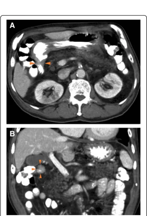
tomography (enhanced MDCT) revealed increased wall thickness in the first part of the duodenum (Fig. 2). No swollen lymph nodes were detected. The forceps biopsy specimen from the submucosal tumor-like lesion did not show evidence of malignancy. As the possibility of a ma-lignant tumor could not be ruled out clinically, a surgical resection was planned for diagnostic and therapeutic purposes.
The patient subsequently underwent an open surgery. A hard mass was palpable in the duodenal bulb, which extended dorsally to the second part of the duodenum. After Kocherization of the duodenum, the area proximal to the pylorus ring to the end of the second part of the duodenum, where the tumor was not palpated, was resected (Fig.3).
A distal gastrectomy was performed. The tumor was 30 × 10 mm and located in the first part of the
duode-num (Fig. 4). It was not continuous with the normal
pancreas as revealed first by imaging and later confirmed during surgery. Microscopically, the tumor was diag-nosed as a moderately differentiated adenocarcinoma that extended from the submucosal layer to the muscu-laris propria of the duodenum. Normal pancreatic tissue was observed adjacent to the tumor, suggesting the
pres-ence of an ectopic pancreas (Fig. 5). Surgical margins
were negative for the presence of tumor cells. Moderate lymphatic invasion, moderate venous invasion, marked neural invasion, and metastases to both superior and in-ferior pyloric lymph nodes were observed. The adjacent ectopic pancreatic tissue had a microscopic appearance consistent with Heinrich’s type 1 [8, 9] and was charac-terized by the presence of ducts, islets, and acini. On im-munohistochemical staining, the islets of the ectopic pancreas and the normal pancreas showed positive stain-ing for chromogranin A, synaptophysin, neural cell adhesion molecule (NCAM), insulin, glucagon, and som-atostatin. Based on these findings, our final diagnosis was of a ductal adenocarcinoma arising from an ectopic pancreas in the first part of the duodenum.
Fig. 1Esophagogastroduodenoscopy showing the obstruction at the first part of the duodenum. Arrow: pylorus ring. Arrow head: obstruction of the first part of the duodenum
Fig. 2Enhanced multi-detector row computed tomography (MDCT) image showing the wall thickness in the duodenum in the transverse (a) and the coronal (b) planes. Arrow heads: wall
The patient was discharged 18 days after surgery, with no complications. Postoperative adjuvant chemotherapy was not administered. We performed a follow-up blood
exam including tumor markers (CEA, CA 19–9) every 3
months and CT images every 6 months. The patient is alive without relapse, at 18 months of follow-up.
Discussion
An ectopic pancreas is defined as an uncommon pan-creatic tissue that exists outside the normal pancreas with no connection to it. The frequency of ectopic pancreatic tissue has been reported to be 0.25% in
abdominal surgery [2], 25–35% of which was found in
the duodenum [3].
Histopathologically, ectopic pancreatic tissue has been classified into four types by Heinrich [8, 9] depending
on the presence or absence of pancreatic ducts, acini, and islet cells. The ectopic pancreas in our patient was located in the first part of the duodenum and contained ducts, acini, and islet cells, making it a Heinrich type 1 ectopic pancreas. The malignant transformation of ec-topic pancreatic tissue is extremely rare, with a fre-quency that ranges from 0.7 to 1.8%, among all cases of ectopic pancreatic tissue [10,11]. These tumors are usu-ally located in the submucosal layer and only occasion-ally expand into the muscularis propria [1]. Jaervi and Lauren [12] have proposed three criteria for diagnosing carcinoma that arises from a heterotopic pancreas:
(1) The tumor must be found within or close to the ectopic pancreatic tissue.
(2) A direct transition must be observed between pancreatic structures and the carcinoma (malignant transformation of an ectopic pancreas must be differentiated from a metastatic deposit or a neoplastic invasion from a neighboring digestive cancer, especially from the stomach, the biliary tract, and the ectopic pancreas).
(3) The non-neoplastic pancreatic tissue must comprise fully developed acini and ductal structures.
In this case, the adenocarcinoma was adjacent to the ectopic pancreas and located in the submucosal layer,
Fig. 4Resected specimen (gastric antrum and duodenum). Arrow heads: tumor. Arrow: pyloric ring
A
B
C
D
away from the normal pancreas. No obvious cancer was seen in other organs. Therefore, we diagnosed the patient with adenocarcinoma of an ectopic pancreas in the duodenum.
To the best of our knowledge, 52 cases of malignant transformation arising from an ectopic pancreas, includ-ing the present case, have been reported in PubMed (keywords: ectopic OR heterotopic OR aberrant pan-creas, carcinoma), 14 of which were malignant trans-formation arising from an ectopic pancreas in the duodenum (Table 1) [13–23]. The mean age of the pa-tients in this group was 70.2 years (range 56–86 years), and eight patients were males and six were females. The
mean tumor size was 27.9 mm (range 12–50 mm).
Eleven of the 14 patients were pathologically diagnosed with adenocarcinoma (tubular adenocarcinoma, poorly differentiated adenocarcinoma, papillary adenocarcin-oma, and mucinous adenocarcinoma), and about half of the carcinomas arose from Heinrich type 1 ectopic pan-creatic tissue. In all except 2 of the 14 cases, the tumors were located in the first or second part of the duodenum.
Except for one patient, no other patient has reportedly been diagnosed with a malignant transformation arising
from an ectopic pancreas prior to surgery. Endo et al. were able to diagnose an ectopic pancreas adenocarcin-oma preoperatively using endoscopic ultrasonography-guided fine-needle aspiration (EUS-FNA) [22]. They sug-gested that EUS-FNA is a useful procedure for
preopera-tive diagnosis in such cases [22] probably because
ectopic pancreatic tissue is usually situated in the sub-mucosal layer [1]. In our case, due to the obstruction of the duodenum, which made curative or palliative surgery necessary, EUS-FNA was not performed. Ordinary pan-creatic cancer is characterized by an ischemic mass on enhanced CT; however, only one in six cases presented with low contrast effects. In the present case, the tumor was not distinctive on enhanced MDCT and the wall of the first part of the duodenum looked similar to the un-involved parts.
Because few reports of malignant transformations aris-ing from ectopic pancreatic tissue are available, no re-ports have compared prognosis between these patients and those with ordinary pancreatic cancer. An analysis of the eight cases of ectopic pancreatic tissue that had reported on patient prognoses after surgery, including the present case, revealed a 5-year survival rate of 64.3%.
Table 1Review of case reports of adenocarcinoma arising from a heterotopic pancreas in the duodenum
Case Year Author Age Sex Part of duodenum involved
Contrast effect on enhanced CT Diagnostic approach Operative method Pathology Outcome
1 1993 Tanaka 72 M ND ND Operation PD Cancer ND
2 1996 Inoue 81 F ND ND Operation DG Adenocarcinoma +
muc
ND
3 2006 Inoue 75 M ND ND Operation PPPD Adenocarcinoma ND
4 2007 Tison 72 M Second portion, vater
ND Operation PD Adenocarcinoma,
CDHP
Death (16 months)
5 2007 Kawakami 65 F Second portion, vater
Heterogeneously enhanced
Operation SSPPD Acinar cell carcinoma
Alive (19 months)
6 2008 Rosok 59 F Proximal Multi-cystic lesion Operation LR IPMC Alive
(36 months)
7 2010 Inoue 75 M Second portion Homogeneously enhanced
Operation PPPD Adenocarcinoma Alive (72 months)
8 2010 Bini 56 M First portion ND Operation PD Adenocarcinoma ND
9 2011 Stock 79 F Fourth portion ND Operation SD Adenocarcinoma ND
10 2012 Kinoshita 62 F First portion Heterogeneously enhanced
Operation PD Adenocarcinoma Alive
(12 months)
11 2013 Ginori 86 F First portion ND Operation STG + DR Adenocarcinoma +
muc
ND
12 2014 Endo 75 M Second portion ND EUS-FNA SSPPD Adenocarcinoma Alive
(60 months)
13 2015 Fukino 62 M Fourth portion Poorly enhanced Operation SD Adenocarcinoma Death (33 months)
14 Present case 81 M First portion Same contrast effect as duodenum
Operation DG Adenocarcinoma Alive
(18 months)
The corresponding survival rate for ordinary pancreatic cancer is about 10% [24]. In malignant transformations that arise from ectopic pancreatic tissue, gastrointestinal symptoms due to stenosis are easier to identify than in ordinary pancreatic cancer, which may result in a better prognosis in the former.
Some patients underwent postoperative adjuvant chemo-therapy similar to patients with ordinary pancreatic cancer, such as S-1 or gemcitabine monotherapy [23]. However, in the present case, no postoperative adjuvant chemotherapy was performed, because no evidence of the efficacy of post-operative adjuvant chemotherapy for adenocarcinoma aris-ing from ectopic pancreatic tissue in an old patient with low ADL exists.
Conclusions
We reported an extremely rare case of an adenocarcin-oma that arose from ectopic pancreatic tissue in the duodenum. Considering the rarity of this disease, gather-ing data from all cases of adenocarcinoma arisgather-ing from ectopic pancreatic tissue in the duodenum will facilitate the development of diagnostic and treatment strategies.
Abbreviations
ADL:Activities of daily living; AFP:α-Fetoprotein; CA: Carbohydrate antigen; CEA: Carcinoembryonic antigen; CT: Computed tomography;
EGD: Esophagogastroduodenoscopy; Enhanced MDCT: Enhanced multi-detector row computed tomography; EUS: Endoscopic ultrasound; EUS-FNA: Endoscopic ultrasonography-guided fine-needle aspiration; NCAM: Neural cell adhesion molecule; PD: Pancreaticoduodenectomy
Acknowledgements
Not applicable
Authors’contributions
TK wrote the manuscript and performed the literature search. MO, KO, AF, TY, RT, KK, MS, and NI treated and observed the patient. NK and HK performed the histological examination. DM, MK, TN, and SH supervised the preparation of this case report. All authors have read and approved the final manuscript.
Funding
This research did not receive any specific grant from any funding agency in the public, commercial, or not-for-profit sectors.
Availability of data and materials
The dataset supporting the conclusions of this article is included within the article.
Ethics approval and consent to participate
The institutional ethics committee approved the publication of this case report.
Consent for publication
Informed consent was obtained from this patient to publish the details of the case, and his identity has been protected.
Competing interests
The authors declare that they have no competing interests.
Author details
1Department of Surgery, National Hospital Organization Hakodate National Hospital, Hakodate, Japan.2Department of Gastroenterology, National Hospital Organization Hakodate National Hospital, Hakodate, Japan. 3Department of Pathology, National Hospital Organization Hakodate National Hospital, Hakodate, Japan.4Department of Surgery, Japanese Red Cross Hakodate Hospital, Hakodate, Japan.5Department of Pathology and
Bioscience, Graduate School of Medicine, Hirosaki University, Hirosaki, Japan. 6Department of Gastroenterological Surgery II, Faculty of Medicine, Hokkaido University, Sapporo, Japan.
Received: 5 June 2019 Accepted: 30 July 2019
References
1. Thoeni RF, Gedgaudas RK. Ectopic pancreas: usual and unusual features. Gastrointest Radiol. 1980;5:37–42.https://doi.org/10.1007/BF01888597. 2. Dolan RV, ReMine WH, Dockerty MB. The fate of heterotopic pancreatic
tissue. A study of 212 cases. Arch Surg. 1974;109:762–5.https://doi.org/10.1 001/archsurg.1974.01360060032010.
3. Mizuno Y, Sumi Y, Nachi S, Ito Y, Marui T, Saji S, et al. Acinar cell carcinoma arising from an ectopic pancreas. Surg Today. 2007;37:704–7.https://doi. org/10.1007/s00595-006-3384-5.
4. Mulholland KC, Wallace WD, Epanomeritakis E, Hall SR. Pseudocyst formation in gastric ectopic pancreas. JOP. 2004;5:498–501.
5. Ishikawa O, Ishiguro S, Ohhigashi H, Sasaki Y, Yasuda T, Imaoka S, et al. Solid and papillary neoplasm arising from an ectopic pancreas in the mesocolon. Am J Gastroenterol. 1990;85:597–601.
6. Tornóczky T, Kálmán E, Jáksó P, Méhes G, Pajor L, Kajtár GG, et al. Solid and papillary epithelial neoplasm arising in heterotopic pancreatic tissue of the mesocolon. J Clin Pathol. 2001;54:241–5.
7. Koh HC, Page B, Black C, Brown I, Ballantyne S, Galloway DJ. Ectopic pancreatic-type malignancy presenting in a Meckel’s diverticulum: a case report and review of the literature. World J Surg Oncol. 2009;7:54.https:// doi.org/10.1186/1477-7819-7-54.
8. Von Heinrich H. Ein Beitrag zur Histologie des sogen, Akzessorischen Pankreas. Virchows Arch A. Pathol Anat Histopathol. 1909;198:392–401.
https://doi.org/10.1007/BF02085327.
9. Gaspar Fuentes A, Campos Tarrech JM, Fernández Burgui JL, Castells Tejón E, Ruíz Rossello J, Gómez Pérez J, et al. Pancreatic ectopias. Rev Esp Enferm Apar Dig. 1973;39:255–68 [Article in Spanish].
10. Makhlouf HR, Almeida JL, Sobin LH. Carcinoma in jejunal pancreatic heterotopia. Arch Pathol Lab Med. 1999;123:707–11.https://doi.org/10.1043/ 0003-9985(1999)123<0707:CIJPH>2.0.CO:2.
11. Guillou L, Nordback P, Gerber C, Schneider RP. Ductal adenocarcinoma arising in a heterotopic pancreas situated in a hiatal hernia. Arch Pathol Lab Med. 1994;118:568–71.
12. Jaervi O, Lauren P. Gastric glandular tumors provided with excretory ducts, and criticism of the theory of the tumors arising in heterotopic pancreas; observations on the occurrence of atypical glands in the stomach. Acta Pathol Microbiol Scand. 1964;62:1–23.
13. Stock C, Keutgen XM, Pisapia D, Crawford C, Zarnegar R. Heterotopic pancreatic neoplasm presenting as an obstructing mass at the fourth portion of the duodenum. JOP. 2011;12:241–3.
14. Tanaka K, Tsunoda T, Eto T, Yamada M, Tajima Y, Shimogama H, et al. Diagnosis and management of heterotopic pancreas. Int Surg. 1993;78:32–5. 15. Inoue Y, Hayashi M, Arisaka Y, Higuchi K, Egashira Y, Tanigawa N.
Adenocarcinoma arising in a heterotopic pancreas (Heinrich type III): a case report. J Med Case Rep. 2010;4:39.https://doi.org/10.1186/1752-1947-4-39. 16. Tison C, Regenet N, Meurette G, Mirallié E, Cassagnau E, Frampas E, et al.
Cystic dystrophy of the duodenal wall developing in heterotopic pancreas: report of 9 cases. Pancreas. 2007;34:152–6.https://doi.org/10.1097/01.mpa. 0000246669.61246.08.
17. Kawakami H, Kuwatani M, Onodera M, Hirano S, Kondo S, Nakanishi Y, et al. Primary acinar cell carcinoma of the ampulla of Vater. J Gastroenterol. 2007; 42:694–7.https://doi.org/10.1007/s00535-007-2070-8.
18. Røsok BI, Rosseland AR, Grzyb K, Mathisen O, Edwin B. Laparoscopic resection of an intraductal papillary mucinous carcinoma in ectopic pancreatic tissue. J Laparoendosc Adv Surg Tech A. 2008;18:723–5.https:// doi.org/10.1089/lap.2007.0168.
19. Bini R, Voghera P, Tapparo A, Nunziata R, Demarchi A, Capocefalo M, et al. Malignant transformation of ectopic pancreatic cells in the duodenal wall. World J Gastroenterol. 2010;16:1293–5.https://doi.org/10.3748/wjg. v16.i10.1293.
21. Ginori A, Vassallo L, Butorano MA, Bettarini F, Di Mare G, Marrelli D. Pancreatic adenocarcinoma in duodenal ectopic pancreas: a case report and review of the literature. Pathologica. 2013;105:56–8.
22. Endo S, Saito R, Ochi D, Yamada T, Hirose M, Hiroshima Y, et al.
Effectiveness of an endoscopic biopsy procedure using EUS-FNA and EMR-C for diagnosing adenocarcinoma arising from ectopic pancreas: two case reports and a literature review. Intern Med. 2014;53:1055–62.https://doi. org/10.2169/internalmedicine.53.1420.
23. Fukino N, Oida T, Mimatsu K, Kuboi Y, Kida K. Adenocarcinoma arising from heterotopic pancreas at the third part of the duodenum. World J Gastroenterol. 2015;2(13):4082–8.https://doi.org/10.3748/wjg.v21.i13.4082. 24. Doi R, Imamura M, Hosotani R, Imaizumi T, Hatori T, Takasaki K, et al. Surgery
versus radiochemotherapy for resectable locally invasive pancreatic cancer. Surg Today. 2008;38:1021–8.https://doi.org/10.1007/s00595-007-3745-8.
Publisher’s Note