C A S E R E P O R T
Open Access
Spinal cord infarction following epidural
and general anesthesia: a case report
Kaori Kobayashi
1, Noriko Narimatsu
1*, Takafumi Oyoshi
1, Takashi Ikeda
2and Toshimitsu Tohya
3Abstract
Background:Epidural anesthesia is widely used for postoperative analgesia and rarely causes permanent neurological complications. We report a case of paraplegia following abdominal surgery under combined epidural/general anesthesia. Case presentation:A 75-year-old woman underwent a scheduled abdominal total hysterectomy and bilateral salpingo-oophorectomy for suspected endometrial cancer. In the operating room, an epidural catheter was inserted at T11/ 12 while the patient was conscious. The needle entered smoothly, with no observed bleeding, paresthesia, or pain, and general anesthesia was induced. During surgery, 4 mL of 0.25% levobupivacaine and 0.1 mg of fentanyl were administered via the epidural catheter, and a solution of 2.5μg/mL fentanyl and 0.2% levobupivacaine was continuously infused at 4 mL/h for postoperative analgesia. The patient promptly regained consciousness and could move her bilateral lower extremities without difficulty upon leaving the operating room. During the first postoperative night, she complained of an absence of sensation and weakness in the lower extremities. By the morning of the second postoperative day, she had developed paralysis and sensory losses associated with touch, temperature, pinprick, and vibration below T5. The epidural infusion was stopped. Magnetic resonance imaging (MRI) revealed a hyperintense area of the thoracic cord from T8 to T11, and spinal cord infarction was suspected. Ossification of the yellow spinal ligaments between T11 and T12, resulting in thoracic canal stenosis and thoracic spinal cord compression, were observed. Notably, the epidural catheter was inserted at the same site where the thoracic canal stenosis was present.
Conclusions:Permanent neurological complications of epidural anesthesia are rare. Studies of neurological complications after epidural/spinal anesthesia have noted the possibility of spinal anomalies, such as lumbar stenosis, in relation to neurological complications after epidural/spinal anesthesia. In this case, the onset of spinal cord infarction may have occurred coincidentally with catheter insertion into the site of existing spinal stenosis. Therefore, it is important to evaluate lower extremity symptoms and consider spinal disease before administering epidural anesthesia. Spinal cord infarction may be prevented by preoperatively identifying spinal lesions using computed tomography or MRI in cases of suspected spinal disease.
Keywords:Spinal cord infarction, Epidural anesthesia, Magnetic resonance imaging
Background
Although epidural anesthesia is widely used for postop-erative analgesia and rarely causes permanent nerve complications, some patients develop subsequent paraly-sis. Here we report a case of spinal cord infarction in a patient who developed paraplegia and sensory impair-ment after surgery under combined epidural and general anesthesia.
Case presentation
The patient was a 75-year-old woman who had been tak-ing oral medication for underlytak-ing hypertension. Prior to surgery, she could independently perform activities of daily living without experiencing symptoms, such as numbness in the lower extremity. She was scheduled to undergo an abdominal total hysterectomy and bilateral salpingo-oophorectomy for suspected endometrial cancer. After admission to the operating room, an epidural cath-eter (Perifix®, B. Braun, Germany) was inserted at T11/12 with the patient in a lateral position using a para-median approach with an 18-gauge Tuohy needle. A loss of
* Correspondence:narimatsu.noriko@kuh.kumamoto-u.ac.jp
1Department of Anesthesiology, Kumamoto Rosai Hospital, 1670,
Takehara-machi, Yatsushiro, Kumamoto, Japan
Full list of author information is available at the end of the article
resistance with normal saline was used to confirm needle advancement into the epidural space. The distance from the skin to the epidural space was 6 cm, and a catheter was inserted 5 cm toward the epidural space. The needle entered smoothly, with no observed bleeding, paresthesia, or pain. General anesthesia was induced with 0.25μg/kg/ min remifentanil, 50 mg of propofol and 40 mg of rocuro-nium and maintained with sevoflurane and remifentanil. The trachea was intubated with an endotracheal tube.
Vasoconstrictors, such as ephedrine and phenyleph-rine, were required to maintain an intraoperative systolic blood pressure of 80–90 mmHg. During surgery, 4 mL of 0.25% levobupivacaine and 0.1 mg of fentanyl were administered via the epidural catheter. The total opera-tive duration was 80 min, and the total anesthesia dur-ation was 123 min. For postoperative analgesia, the patient received a continuous 4 mL/h infusion of a 2.5μg/mL fentanyl and 0.2% levobupivacaine solution.
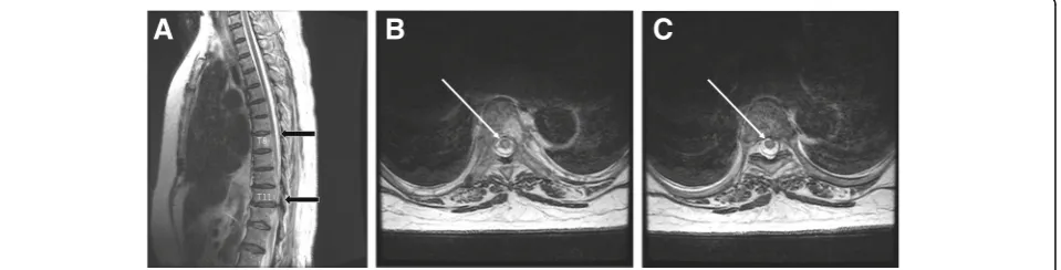
The patient promptly regained consciousness and was transferred from the operating room following extuba-tion. She could move her bilateral lower extremities without difficulty when leaving the operating room. Dur-ing the afternoon of the first postoperative day, she could stand without numbness or weakness in the lower extremities. On the night of the same day, the patient complained of a loss of sensation and weakness in the bilateral lower extremities. Her lower extremity symp-toms failed to resolve by the following morning (second postoperative day), at which point sustained-release epi-dural administration was stopped. However, the patient developed paralysis and a loss of touch, temperature, pinprick, and vibration sensations below the T5 level. In addition, her lower extremities were flaccid. To deter-mine whether an epidural hematoma had occurred in her epidural space, a magnetic resonance imaging (MRI) examination was immediately performed. The MRI re-vealed the following: (1) There were no epidural masses, i.e., no epidural hematomas. (2) The patient exhibited yellow ligament ossification. (3) T2-weighted MRI
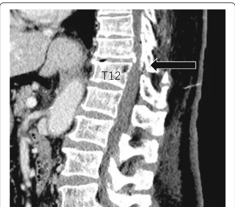
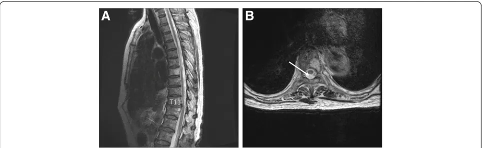
revealed hyperintensity in the T8-T11 spinal cord region (Fig. 1a–c). In addition, a computed tomography (CT) examination revealed that the epidural catheter had been inserted at the same site where the thoracic canal sten-osis was most severe (Fig. 2). Therefore, she was diag-nosed with a spinal cord infarction. Other spinal diseases (e.g., spinal degenerative disease) were excluded because of the rapid onset. Spinal cord infarctions are usu-ally treated conservatively, as surgical interventions for this disease are inadequate. She was managed conserva-tively. She was treated with blood pressure control therapy and rehabilitation consisting of muscle training and walk-ing practice, among other exercises. The epidural catheter was removed on the third postoperative day. One week later, an MRI examination revealed that the hyperintense region had extended superiorly to the T4 level (Fig. 3). The patient underwent rehabilitation and regained the ability to move her lower extremities on postoperative day 30. Although the patient could stand on her left leg, she was unable to stand on her right leg. She received con-tinuous bladder catheterization, and her defecation was controlled by drugs. An MRI examination on postopera-tive day 93 demonstrated that the hyperintense region was localized to T11, where the thoracic canal stenosis was originally present and the epidural catheter was inserted (Fig. 4a, b). On postoperative day 286, the patient was transferred to a rehabilitation hospital.
Discussion
We are reporting a case of spinal cord infarction that oc-curred after epidural anesthesia. There are few similar case reports in the English literature. Spinal cord infarc-tion accounts for only 0.3–1% of stroke cases and there-fore has a low incidence relative to other types of stroke [1]. Some studies have reported that age, hypertension, diabetes, obesity, a history of cerebral infarction, and atherosclerotic lesions are risk factors for spinal cord in-farction [1, 2]. Other studies have reported the roles of aortic disease and related surgical treatments, as well as
spinal disease and related surgical treatments, as risk factors for spinal cord infarction [3, 4]. Although epi-dural anesthesia is widely used for intraoperative and postoperative analgesia, it can lead to several complica-tions, of which permanent nerve damage is the most se-vere. Epidural hematoma after epidural anesthesia is a typical nerve complication. While spinal cord infarction as observed in this case is rare, it should still be taken into consideration.
Concerning the cause of spinal cord infarction after anesthesia and surgery, there are few reports in the litera-ture. Some reports have assumed that spinal cord ische-mia following epidural anesthesia was associated with intraoperative hypotension [5, 6], toxicity from local anesthesia [5], or vasospasm related to the administration of a local epinephrine-containing anesthetic [5–7]. How-ever, no clear associations have been demonstrated in ei-ther case. Anoei-ther study noted that in elderly patients and those with abnormal spine anatomy, an excessive anesthetic agent volume may cause spinal cord compres-sion [8]. Furthermore, a study of neurological complica-tions after epidural and spinal anesthesia estimated that spinal cord infarction occurred in approximately five per million patients [9]. Four of these five patients were re-ported to have anatomical abnormalities, such as lumbar spine stenosis and subarachnoid cysts. Therefore, the authors suggested that these spinal abnormalities are asso-ciated with neurological complications after epidural/ spinal anesthesia [9]. One prior case report showed that spinal cord infarction developed due to severe disc hernia-tion without anesthesia or surgery [10]. Thus, compres-sion to the spinal cord may be an important factor. In our case, the spinal cord infarction occurred at the narrowest area of the spinal cord. This area was narrowed due to the ossification of the yellow ligament and became narrower by the tip of the epidural puncture tube. The small vessels around the narrow spinal cord might have been com-pressed, causing the infarction. Therefore, patients who complain of numbness and/or weakness of the lower limbs, intermittent claudication, etc. should undergo pre-operative MRI examinations of the spinal cord. Some studies have examined paralysis after aortic surgery [11]. The patient in our case underwent soft tissue sur-gery in the lower abdomen, and the relationship be-tween this abdominal surgery and the patient’s spinal cord infarction may have been negative. In our patient, the causal relationship between epidural anesthesia and spinal cord infarction is still not clear. However, it is unique that her clinical symptoms occurred after anesthesia and surgery. Based on this presentation, an-esthesiologists and surgeons must recognize that spinal cord infarction may occur after combined general and epidural anesthesia or surgery.
Concerning the treatment of spinal cord infarction, conservative treatments are usually chosen. In this pa-tient, the motor and sensory deficits were clinically dis-tinct, indicating the infarction of three spinal arteries (one anterior and two posterior). A surgical intervention would have been difficult, and the standard drug therapy is aspirin, based on the consensus recommendation for the acute treatment of ischemic stroke at any site. How-ever, no direct studies have examined the efficacy of drug therapy in spinal cord infarction.
Yellow ligament ossification frequently occurs in middle-aged people and is thought to particularly affect the lower thoracic spine because it often occurs at T10/ 11 and T11/12 [12]. Numbness and pain in the lower ex-tremities are common symptoms of this condition. In this case, MRI revealed ossification of the yellow liga-ments at the T11/12 spinal level, with the accompanying findings of thoracic spinal cord compression. The site of spinal cord infarction was also consistent with the loca-tion of epidural catheter inserloca-tion. Although the patient in this case did not present with clinical symptoms of thoracic canal stenosis, such as numbness or weakness of the lower extremities, prior to surgery, the onset of spinal cord infarction may have occurred simultaneously with the insertion of the catheter into a site where spinal stenosis was already present.
A preoperative examination with a focus on yellow ligament ossification may be necessary in middle-aged patients undergoing lower abdominal surgery with epi-dural anesthesia. A preoperative plain chest radiography evaluation of spinal ossification and an evaluation of lower extremity symptoms may be important for redu-cing the incidence of spinal cord infarction related to epidural anesthesia. When administering epidural anesthesia to patients with a spinal disease, lower thor-acic spine catheterization might have to be avoided. Spinal cord infarction in cases of suspected spinal dis-ease may be prevented by preoperatively identifying spinal lesions via a CT or MRI evaluation.
Conclusions
We report a case of paraplegia following post-epidural analgesia in a patient diagnosed with spinal cord infarc-tion. MRI revealed spinal cord compression resulting from yellow ligament ossification; notably, the site of spinal cord infarction was consistent with the location of epidural catheter insertion. Spinal cord infarction may be prevented by preoperatively identifying spinal lesions
using CT or MRI prior to epidural anesthesia when spinal disease is suspected.
Abbreviations
CT:Computed tomography; MRI: Magnetic resonance imaging
Acknowledgments
The authors thank Crimson Interactive and American Journal Experts for the editorial assistance.
Availability of data and materials
Not applicable.
Authors’contributions
KK and TT collected the patient’s data and drafted the manuscript. NN and TO helped to draft the manuscript. NN, TT, and TI checked the manuscript. All authors have read and approved the final manuscript.
Ethics approval and consent to participate
This study was approved by the Ethics Committee of Kumamoto Rosai Hospital.
Consent for publication
We informed and obtained consent from the patient and the patient’s family for the publication of this case report.
Competing interests
The authors declare that they have no competing interests.
Publisher’s Note
Springer Nature remains neutral with regard to jurisdictional claims in published maps and institutional affiliations.
Author details
1Department of Anesthesiology, Kumamoto Rosai Hospital, 1670,
Takehara-machi, Yatsushiro, Kumamoto, Japan.2Department of Orthopedic Surgery, Kumamoto Rosai Hospital, Yatsushiro, Japan.3Department of
Obstetrics and Gynecology, Kumamoto Rosai Hospital, Yatsushiro, Japan.
Received: 14 February 2017 Accepted: 17 July 2017
References
1. Romi F, Naess H. Characteristics of spinal cord stroke in clinical neurology. Eur Neurol. 2011;66(5):305–9.
2. Kei K, Hiroko M, Keisuke Y, Ken Y. A case of postoperative paraplegia caused by idiopathic spinal cord infarction in a young male. Masui. 2008;57(4):450–2. 3. Robertson CE, Brown RD Jr, Wijdicks EF, Rabinstein AA. Recovery after spinal
cord infarcts. Neurology. 2012;78:114–21.
4. Novy J, Carruzzo A, Maeder P, Bogousslavsky J. Spinal cord ischemia clinical and imaging patterns, pathogenesis, and outcomes in 27 patients. Arch Neurol. 2006;63(8):1113–20.
5. Weinberg L, Harvey WR, Marshall RJ. Post-operative paraplegia following spinal cord infarction. Acta Anaesthesial Scand. 2002;46:469–72. 6. Fernandez MF, Dachy B, Demeurisse G. Lower limb monoplegia and
dissociated hypoesthesia after peridural anesthesia. Rev Neurol (Paris). 1999;155(8):598–600.
7. Ackerman WE, Juneja MM, Knapp RK. Maternal paraparesis after epidural anesthesia and cesarean section. South Med J. 1990;83(6):695–7.
8. Jacob AK, Borowiec JC, Long TR, Brown MJ, Rydberg CH, Wass CT. Transient profound neurologic deficit associated with thoracic epidural analgesia in an elderly patient. Anesthesiology. 2004;100:1470–1.
9. Seze MP, Sztark F, Janvier G, Joseph PA. Severe and long-lasting complications of the nerve root and spinal cord after central neuraxial blockade. Anesth Analg. 2007;104(4):975–9.
10. Reynolds JM, Belvadi YS, Kane AG, Poulopoulos M. Thoracic disc herniation leads to anterior spinal artery syndrome demonstrated by diffusion-weighted magnetic resonance imaging (DWI): a case report and literature review. Spine J. 2014 1;14(6): e17-e22.
11. Ito Z, Matsuyama Y, Ando M, Kawabata S, Kanchiku T, Kida K, Fujiwara Y, Yamada K, Yamamoto N, Kobayashi S, Saito T, Wada K, Tadokoro N, Takahashi M, Satomi K, Shinomiya K, Tani T. Postoperative paralysis from thoracic ossification of posterior longitudinal ligament surgery risk factor of neurologic injury: nationwide multiinstitution survey. Spine (Phila Pa 1976). 2016 1; 41(19): E1159-E1163.