Recognizing Normal Heart
Murmurs: A Logic-based
Mnemonic
A logic-based mnemonic for the auscultory diag-nosis of nonpathologic murmurs, which I developed for teaching medical students and house staff, is presented with the hope of improving the ausculta-tory skills of primary care pediatric practitioners. The objective is positive identification of nonpatho-logic murmurs rather than diagnosis by exclusion. The auscultatory features of these murmurs are largely determined by their causative mechanisms. The logic, therefore, is based on an understanding of these mechanisms so that the listener will know why these murmurs have their characteristic sound fea-tures and specifically what to listen for. The logic can also be used in explaining nonpathologic murmurs to parents. The mnemonic device should reduce the need for rote memorization.
Heart murmurs are present in most pediatric pa-tients, and almost all are nonpathologic. Yet, the most common reason for referral to many pediatric cardiologists is for the evaluation of a heart murmur, two thirds of which are deemed normal.1,2I believe that improving the accuracy of auscultation by pri-mary care practitioners and improving their confi-dence in recognizing normal murmurs will decrease referrals to pediatric cardiologists, without underre-ferral of patients with heart disease. This outcome would be of significant psychologic and economic benefit to patients and their families.
In referring to nonpathologic murmurs, the com-monly used terms are innocent, functional, and nor-mal. I prefer the latter. Perhaps, because “normal” is a less technical, more familiar word to parents, to me it seems easier for them to understand and accept the benign nature of these murmurs with this terminol-ogy. The formulation of the logic begins with a con-sideration of causative mechanisms.
GENESIS OF HEART MURMURS
A heart murmur is a continuous series of sounds, with a variable number of different sound wave frequencies present at each instant. The wave fre-quency content of a murmur determines, to a large extent, its quality, pitch, and loudness.
The auditory quality of a murmur, an important element in differential diagnosis, is related to its spectrum or mix of frequencies. The clinical assess-ment of quality, however, is subjective, and there is no consensus on a descriptive terminology. In 1955, McKusick et al,3using spectral phonocardiographic analysis of heart murmurs, linked quality to fre-quency mix and classified murmurs as either “musi-cal” or “noisy.” A musical murmur, similar to a musical tone, has only a fundamental frequency and
one or more harmonic overtones. A noisy murmur has a mix of random, harmonically unrelated fre-quencies. Building on this terminology, my practice is to describe the quality of a murmur by choosing the appropriate adjective from the following contin-uum: musical; somewhat musical; or slightly, mod-erately, or very noisy.
The pitch of a murmur is directly related to its frequency level and is described as low, medium, or high. Loudness is, in part, directly related to fre-quency level and is graded from 1 to 6.
Doppler echocardiography has been able to dem-onstrate the characteristics of blood flow in different parts of the heart and great vessels. In a flow channel that is straight and unobstructed, red blood cells are flowing in tandem, at about the same velocity. This type of flow is termed laminar or nondisturbed. As a flow channel narrows, flow velocity increases. When a certain level of constriction occurs, or if flow veloc-ity increases sufficiently, flow characteristics at this point change. Laminar flow is disrupted, and red blood cells move simultaneously in different direc-tions and with different velocities, creating disturbed or turbulent flow.
A heart murmur is produced when the contrac-tions of the heart, and the associated pulsatile blood flow, produce enough tissue vibrations to be audible on the surface of the chest. The means by which these vibrations or sound waves are generated, and the identity of the tissues that vibrate, have been studied since murmurs were first described and are still be-ing debated.4 – 6 Murmurs have been ascribed to vi-brations produced directly by disturbed or turbulent blood flow, to indirect effects of turbulent flow such as vibration of vessel walls, and to direct vibration of structures such as the myocardial wall, valves, chor-dae, papillary muscles, and ventricular bands or false tendons.
It seems reasonable to me to assume that some murmurs are produced by a single mechanism and others by more than one, and that the auditory char-acteristics of a murmur would be determined by its underlying mechanism(s). Disturbed blood flow, be-cause it produces a wide spectrum of non– harmoni-cally related sound frequencies, would produce a noisy murmur, with the degree of noise being related to the degree of flow disturbance. A vibrating struc-ture that produces a narrow spectrum of harmoni-cally related frequencies would produce a purely musical murmur.7A combination of disturbed flow and a harmonically vibrating structure would pro-duce a noisy murmur that has a musical component. In the normal heart, Doppler echocardiography demonstrates that blood flow through the cardiac chambers and valve orifices occurs in a predomi-nantly laminar fashion. However, there are sites along the blood flow pathway that I term “dispro-portionate-sized connections.” At these sites, either a smaller blood vessel is connected to a larger one, or a larger vessel branches into a smaller one, or a cardiac chamber is connected to a smaller caliber great vessel. In 1959, Bruns4called these sites “dis-continuities” and theorized that they can increase the velocity of blood flow at these points and can cause Received for publication May 3, 1996; accepted Sep 23, 1996.
sufficient flow disturbance to produce a murmur. Many studies since then, using such techniques as intracardiac phonocardiography and echo and Doppler, have supported this concept. The velocity of blood flow at these sites, however, still remains within the normal range, below that which would be seen with flow through a pathologically narrowed valve or vessel. Thus the degree of blood flow dis-turbance (and the noisiness of the murmur pro-duced) remains low and in general is not detectable by standard Doppler echocardiographic techniques. Also, this change in flow velocity could create vibra-tions in adjacent soft tissue structures, which could further contribute to sound generation. Concurrent harmonic vibration of a solid tissue structure could add a musical quality to the murmur.
NORMAL HEART MURMURS
My logic-based mnemonic for normal heart mur-murs therefore begins with the concept that in the normal pediatric heart, at sites with disproportion-ate-sized connections, there is an increase in flow velocity, which can generate flow disturbance and tissue vibrations to produce audible sound.
There are five such sites, and these can be remem-bered easily by following the course of blood flow through the heart from the systemic veins to the aorta:
1. The connection of the jugular, subclavian, and innominate veins to the superior vena cava; 2. The connection of the right ventricle to the main
pulmonary artery;
3. The connection of the main pulmonary artery to the right and left pulmonary artery branches; 4. The connection of the left ventricle to the aorta;
and
5. The connection of the aortic arch to the brachio-cephalic vessels.
Sound waves resulting from the changes in flow at each of these five sites are transmitted to different, specific areas on the surface of the chest or neck (Figure). Murmurs in these five areas in healthy pe-diatric patients were identified and well described more than 50 years ago.5Formal follow-up studies8 –10 and empiric clinical observation over the years have confirmed the benign nature of these murmurs.
Because their predominant causative mechanism is the same, the five normal murmurs have the fol-lowing generic auscultatory characteristics:
1. The four murmurs produced by arterial connec-tions are all midsystolic; the one murmur pro-duced by venous connections is continuous through systole and diastole and often has dia-stolic accentuation;
2. The murmurs are not louder than grade 3 of 6 (moderately loud);
3. The pitch is low to medium; and
4. The murmurs are slightly or moderately noisy, never very noisy, and may have a musical com-ponent, if, as we postulate, there is also a harmon-ically vibrating structure.
In addition to these generic features, each of the normal murmurs has individual characteristics. The latter are determined by the type of disproportion-ate-sized connecting structures, their location in the circulatory system and within the chest, and the direction of transmission of the flow disturbance they create. In the course of blood flow through the heart, again, from the systemic veins to the aorta, the commonly used names and the individual features of the five normal murmurs are:
1. Venous hum murmur (connection of jugular, sub-clavian, and innominate veins to superior vena cava): continuous, often with diastolic accentua-tion; best heard below the right clavicle; may be transmitted to the left upper chest; disappears completely in the supine position, which differen-tiates it from the murmur of a patent ductus arte-riosus or an arteriovenous fistula.
2. Pulmonary flow murmur (connection of right ventricle to main pulmonary artery): best heard in the second intercostal space at the left sternal bor-der; may be transmitted slightly upward and downward; wide, fixed splitting of the second heart sound suggests the murmur is not normal and could be caused by an atrial septal defect. 3. Neonatal physiologic peripheral pulmonary
ar-tery stenosis murmur (branching of main pulmo-nary artery): best heard over the upper left sternal border; transmitted well to both infraclavicular areas, axillae, back, and slightly down the left sternal border; disappears in the first year.
Figure. View of anterior chest wall with outline of cardiac sil-houette superimposed on rib cage. Numbers indicate, in the course of blood flow through the heart, from systemic veins to the aorta, where each of the following five normal heart murmurs is heard best: 1, venous hum murmur (connection of jugular, sub-clavian, and innominate veins to the superior vena cava; 2, pul-monary flow murmur (connection of the right ventricle to the main pulmonary artery); 3, neonatal physiologic peripheral pul-monary artery stenosis murmur (branching of the main pulmo-nary artery); 4, precordial vibratory or Still’s murmur (connection of the left ventricle to the aorta); and 5, supraclavicular or carotid bruit (connection of brachiocephalic vessels to the aortic arch).
EXPERIENCE AND REASON 617 at Viet Nam:AAP Sponsored on August 30, 2020
www.aappublications.org/news
4. Precordial vibratory or Still’s murmur (connection of left ventricle to aorta): best heard medial to or at the apex; transmitted medially to the low and mid-left sternal border; typically has a much more musical component than any of the other normal murmurs, and often described in as varying terms, such as “vibratory,” “twanging string,” “groaning,” and “moaning.”
5. Supraclavicular or carotid bruit (connection of brachiocephalic vessels to aortic arch): best heard in the neck above the clavicles, usually bilateral; may be transmitted downward to infraclavicular areas.
CLINICAL APPLICATION
The generic and individual characteristics of the five normal murmurs that have been described are based on auscultation with the patient supine, at rest, and in a quiet room. A healthy child who is febrile, fearful, crying, or hyperactive will have an increased heart rate and cardiac output. The latter can produce more disturbed blood flow, make a normal murmur louder, noisier, less musical, and mislead the exam-iner into thinking the murmur is pathologic. There-fore, if the conditions of the examination are not optimal, the patient should be reevaluated.
In healthy individuals there are some sites with disproportionate-sized connections, such as the pul-monary veins entering the left atrium, which, per-haps because of poor transmission of sound waves, do not produce murmurs.
The fact that normal murmurs may coexist, al-though noted in the literature,11–13has, in my opinion, not been sufficiently emphasized. It is not unusual to hear a venous hum, a pulmonary flow murmur, a precordial vibratory murmur, and a supraclavicular bruit at the same time in a healthy child.
Although the objective is positive identification of the five normal murmurs, the clinician should also be aware of, and listen for, auscultatory clues suggest-ing cardiac disease. A recent study of patients re-ferred to pediatric cardiologists for evaluation of a heart murmur14 identified six “cardinal clinical signs,” which, if present, were independent statisti-cal, but not absolute, predictors of cardiac disease. Of these the most significant, in my experience, are a pansystolic murmur, a harsh-quality murmur, an ab-normal heart sound, and an early or midsystolic click. Less predictable, again, in my experience, is their fifth sign, a murmur of grade 3 or more in intensity, because normal murmurs can be moder-ately loud (grade 3). Similarly, their sixth sign, a murmur best heard over the upper left sternal bor-der, is common in the healthy neonate. To this list I would add a diastolic murmur and a continuous murmur (other than a venous hum), which should always be considered pathologic.
In summary, the heart should be declared normal by a primary care practitioner when the history and general examination do not suggest heart disease, when one or more of the five normal murmurs is clearly identified, and when there are no ausculta-tory findings to suspect an abnormality. I agree with published studies1,15that a confirmatory chest
radio-graph, electrocardiogram, or echocardiogram is not necessary. If there is doubt about the murmur being normal, then it has been shown2,16 that the most cost-effective next step is not laboratory testing but direct referral to a pediatric cardiologist.
CONCLUSION
The auscultatory positive recognition of normal heart murmurs is an important part of the pediatric examination, and if auscultation is done well by the primary care practitioner, it benefits the patient and the family and is very cost effective. In my experi-ence, teaching and learning this skill can be more successful if it is based on an understanding of the mechanisms that are thought to produce normal murmurs and that are responsible for their auditory characteristics.
A mnemonic device has been presented to facili-tate the recall of the five normal murmurs, based on the observation that each of these murmurs is related to a specific site along the path of blood flow through the heart, from the systemic veins to the aorta. Each of these sites has in common the feature that a larger vessel or chamber is directly connected to a smaller vessel or chamber, a characteristic termed dispropor-tionate-sized connections, with the potential for cre-ating disturbed blood flow, tissue vibrations, and a corresponding, very recognizable murmur.
Samuel O. Sapin, MD Department of Pediatrics
Southern California Permanente Medical Group Panorama City, CA 91402
Department of Pediatrics Division of Cardiology
University of California, Los Angeles School of Medicine
Los Angeles, CA 90095
REFERENCES
1. Smythe JF, Teixeira OHP, Vlad P, Demers PP, Feldman W. Initial evaluation of heart murmurs: are laboratory tests necessary? Pediatrics. 1990;86:497–500
2. Danford DA, Nasir A, Gumbiner C. Cost assessment of the evaluation of heart murmurs in children. Pediatrics. 1993;91:365–368
3. McKusick VA, Murray GE, Peeler RG, Webb GN. Musical cardiovascu-lar murmurs. Bull Johns Hopkins Hosp. 1955;97:136 –176
4. Bruns DL. A general theory of the causes of murmurs in the cardiovas-cular system. Am J Med. 1959;27:360 –374
5. Danford DA, McNamara DG. Innocent murmurs and sounds. In: Gar-son A, Bricker JT, McNamara DG, eds. The Science and Practice of
Pedi-atric Cardiology. Philadelphia, PA: Lea & Febiger; 1990:1919 –1928
6. Guntheroth WG, Musical murmurs. Am J Cardiol. 1992;69:840 – 841. Letter
7. Stein PD, Sabbah HN, Lakier JB. Origin and clinical relevance of musical murmurs. Int J Cardiol. 1983;4:103–112
8. Marienfeld CJ, Telles N, Silvera J, Nordesieck M. A 20-year follow-up study of “innocent” murmurs. Pediatrics. 1962;30:42– 48
9. Weaver WF, Walker CHM. Innocent cardiovascular murmurs in the adult: a 16-year follow-up. Circulation. 1964;29:702–707
10. deMonchy C, Vander Hoeven GMA, Beneken JEW. Studies on innocent precordial murmurs in children. III. follow-up study in children with an innocent praecordial vibratory murmur. Br Heart J. 1973;35:685– 690 11. MacLaren MJ, Lochman AS, Pocock WA, Barlow JB. Innocent murmurs
and third heart sounds in black children. Br Heart J. 1980;43:67–73 12. McNamara DG. Value and limitations of ausculation in the
manage-ment of congenital heart disease. Pediatr Clin North Am. 1990;37:93–113 13. Harris JP. Evaluation of heart murmurs. Pediatr Rev. 1994;15:490 – 493 14. McCrindle BW, Shaffer KM, Kan JS, Zahka KG, Rowe SA, Kidd L.
chil-dren. Arch Pediatr Adolesc Med. 1996;150:169 –174
15. Newberger JW, Rosenthal A, Williams RG, Fellows K, Miettinen OS. Noninvasive tests in the initial evaluation of heart murmurs in children.
N Engl J Med. 1983;308:61– 64
16. Wong JA, Meyer RA. Cost-effective evaluation of heart murmurs in children. Arch Fam Med. 1996;5:381. Letter
Bilateral Subdural Hematomas in a
Newborn Infant
Subdural hematomas in the neonate are unusual, with a difficult delivery accounting for most cases. As obstetric methods have improved, the incidence of this complication has declined, making subdurals even less common.1Automobile accidents or assaults involving the mother are an even rarer cause of fetal subdural hematomas and are nearly always associated with skull fracture.2One study reported a high incidence of sub-dural hematomas without skull fracture in stillborn fetuses of Samoan mothers. Although domestic assault of the mothers was considered, this could not be con-firmed in any case.3There is only one case report of a newborn infant with subdural hematomas secondary to trauma without a skull fracture.4 In this case, the mother reported being beaten and immediately noticed an absence of fetal movements. A full-term infant was then delivered via cesarean section and had a left sub-dural hematoma that, on aspiration, was acute in na-ture. We report here a case of a 34-week premature infant with bilateral chronic subdural hematomas of various ages secondary to maternal abuse. The infant remained in utero for a significant period of time after the trauma occurred. The case is unusual in light of the absence of skull fracture or other injury and the amount of time elapsed between injury and delivery.
CASE STUDY
Baby girl K was the 2138-g product of a 34-week gestation born by cesarean section to a 25-year-old gravida 7, para 3, aborta 3 African-American woman. The mother’s past medical history was significant for chronic hypertension. High blood pressures were noted during the pregnancy and the mother was treated with Aldomet until 2 weeks before delivery. The pregnancy was oth-erwise uncomplicated. The mother’s prenatal screens were: blood type A negative/anti-D antibody positive, rubella immune, Ven-eral Disease Research Laboratory test negative. Gonorrhea and chlamydia cultures were negative. Hepatitis B antibody was neg-ative, as was a group B streptococcus screen done 26 days before delivery. The mother received incomplete prenatal care and de-nied substance abuse.
On the day before delivery, the mother noted decreased fetal movements and was admitted to the obstetrics unit. Her physical examination was unremarkable with the exception of several healed linear scars on her arms. A biophysical profile was done which was scored 8 out of 10. The mother’s blood pressure was 170/85 and she was started on hydralazine and Aldomet. A repeat profile the next day was scored 2 out of 10. Because of the wors-ening profile, the decision was made to deliver the child via cesarean section. The amniotic sac was artificially ruptured at delivery, producing a normal amount of clear amniotic fluid. At
delivery, the patient was blue with poor tone and respiratory distress. She was successfully resuscitated with positive pressure ventilation for 30 seconds and was noted to be more vigorous. The Apgar scores were 6/10 at 1 minute and 8/10 at 5 minutes. Grunting and flaring were noted. After 10 minutes of life, the infant continued to grunt and flare and was noted to have a room air pulse oximetry of 88 percent. Because of her prematurity and respiratory distress, the patient was transferred to the neonatal intensive care unit.
When admitted to the intensive care unit, it was noted that the respiratory distress and oxygen requirement had resolved. Baby girl K’s physical examination at that time was significant for a weight of 2138-g (25th percentile), length of 44.5 cm (50th percen-tile), and head circumference of 33 cm (greater than the 95th percentile). Her pulse was 160 beats per minute and her respira-tions were 41 breaths per minute. Blood pressure was 55/33. She was noted to have a large anterior fontanel, large posterior fonta-nel, and widely split cranial sutures. The remainder of the exam-ination was normal.
The initial laboratory work was significant for an O negative blood type with a negative antibody screen. The hematocrit was 23.2% (0.232) and the reticulocyte count 22.3% (0.223). The total bilirubin was 3.5 mg/dL (60 umol/L) with a direct fraction of .4 mg/dL (6.8 umol/L). The partial thromboplastin time was normal at 37 seconds as was the prothrombin time at 12.5 seconds. The fibrinogen level was 254 mg/dL (2.5 g/L) and theD-dimers were slightly elevated at 2–4 mg/L. Electrolytes and urine toxicology screen were normal.
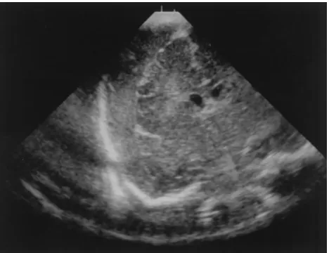
Because of her anemia, an umbilical venous catheter was placed and an exchange transfusion was performed which increased the hematocrit to 43% (0.43). A head ultrasound performed on the first day of life revealed large bilateral subdural hematomas (Fig 1). Computed tomography and magnetic resonance imaging of the cranium were then performed. These scans revealed large bilateral subdural hematomas of varying ages with significant compression of the underlying brain (Figs 2 and 3). Neurosurgery was con-sulted and the hematomas were surgically drained. A coagulopa-thy evaluation, including assays of von Willebrandt’s factor, Fac-tor XIII, and platelet function was normal.
Upon further questioning, baby girl K’s mother admitted that the child’s father had physically assaulted her numerous times throughout the pregnancy. She described approximately 8 beat-ings during the 6 months that she knew she was pregnant. The beatings usually involved her being struck in the face and chest as well as being shoved to the ground. She was also frequently kicked from behind. The mother stated that she was only struck in the abdomen on one occasion, though on one other occasion she struck her abdomen on a piece of furniture as she fell after being pushed. She also describes having vaginal bleeding and abdomi-nal pain throughout the pregnancy that were frequently brought on by a beating. The mother stated that this infant had fewer fetal
Received for publication Apr 1, 1996; accepted Jul 29, 1996.
Address all correspondence to: Robert P. Stephens, MD, 7575 Northcliff Avenue, Suite 301, Brooklyn, OH 44144.
PEDIATRICS (ISSN 0031 4005). Copyright © 1997 by the American Acad-emy of Pediatrics.
Fig 1. Head ultrasound on the first day of life. Coronal ultra-sound image obtained through the anterior fontanel demonstrates mixed echogenicity separating the surface of the brain from the overlying calvarium, consistent with a subdural hematoma. A similar collection was noted on the contralateral side (not shown).
EXPERIENCE AND REASON 619 at Viet Nam:AAP Sponsored on August 30, 2020
www.aappublications.org/news
DOI: 10.1542/peds.99.4.616
1997;99;616
Pediatrics
Samuel O. Sapin
Recognizing Normal Heart Murmurs: A Logic-based Mnemonic
Services
Updated Information &
http://pediatrics.aappublications.org/content/99/4/616
including high resolution figures, can be found at:
References
http://pediatrics.aappublications.org/content/99/4/616#BIBL
This article cites 15 articles, 7 of which you can access for free at:
Permissions & Licensing
http://www.aappublications.org/site/misc/Permissions.xhtml
in its entirety can be found online at:
Information about reproducing this article in parts (figures, tables) or
Reprints
http://www.aappublications.org/site/misc/reprints.xhtml
DOI: 10.1542/peds.99.4.616
1997;99;616
Pediatrics
Samuel O. Sapin
Recognizing Normal Heart Murmurs: A Logic-based Mnemonic
http://pediatrics.aappublications.org/content/99/4/616
located on the World Wide Web at:
The online version of this article, along with updated information and services, is
by the American Academy of Pediatrics. All rights reserved. Print ISSN: 1073-0397.
the American Academy of Pediatrics, 345 Park Avenue, Itasca, Illinois, 60143. Copyright © 1997 has been published continuously since 1948. Pediatrics is owned, published, and trademarked by Pediatrics is the official journal of the American Academy of Pediatrics. A monthly publication, it
at Viet Nam:AAP Sponsored on August 30, 2020
www.aappublications.org/news