GASTRORETENTIVE DRUG DELIVERY SYSTEM: STOMACH SPECIFIC MUCOADHESIVE TABLET
7
0
0
Full text
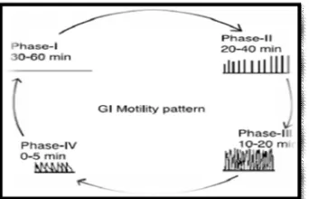
(2) Siddhapara Mihir et al. IRJP 2011, 2 (12), 90-96. Figure 1: Anatomy of the gastrointestinal tract.. The stomach is a J-shaped organ which can be divided into four parts: cardia, fundus, body and antrum. The main function of the stomach is to store and mix food with gastric secretion. It consists of serosa, longitudinal muscle, intermuscular plane, circular muscle, submucosa, lamina propria and epithelium. The stomach has a third muscle layer called as the “oblique muscle layer” situated in the proximal stomach, branching over the fundus and higher regions of the gastric body. The different smooth muscle layers are performing the motor function of the gastrointestinal tract, i.e. gastric emptying and intestinal transit 23. Physiology of the gastrointestinal tract The proximal part is made up of fundus and body. It serves as a reservoir for the materials which remain undigested, whereas the antrum is the main site for mixing motion and acts as a pump for gastric emptying by propelling actions. Gastric emptying occurs during both fasting as well fed states. During the fasting state an interdigestive series of electrical events takes place, which cycles through stomach and intestine every 2 to 3 hours 24. This is called the interdigestive myloelectric cycle or migrating myloelectric cycle (MMC), which further divided into following 4 phases as described by Wilson and Washington 25. (figure 2). Figure 2: A simplified schematic diagram of the interdigestive balanced motility pattern.. Phase I (basal phase) lasts from 40 to 60 minutes with rate contractions. Phase II (preburst phase) lasts for 40 to 60 minutes with intermittent action potential and contractions. As the phase progresses the intensity and frequency also increase gradually. Phase III (burst phase) lasts for 4 to 6 minutes. It includes intense and regular contraction for short period. It is due to this wave that all the undigested material is swept out the stomach down to the small intestine. It is also known as the housekeeper wave. Phase IV lasts for 0 to 5 minutes and occurs between phases III and 1 to 2 consecutive cycles. After the ingestion of a mixed meal, the pattern of contractions changes from fasted to that of fed state. This is also known as digestive motility pattern and comprises continuous contraction as in phase II of fasted state. These contraction result is reducing the size of food particles (to less than 1 mm), which are propelled toward the pylorus in a suspension form. During the fed state onset of MMC is delayed resulting in slowdown of gastric emptying rate 26.. scintigraphy, radiology, endoscopy, ultrasonography, γradiotelemetry and magnetic marker monitoring studies have been applied to determining gastric emptying rates revealed that orally administered controlled release dosage forms are subjected to basically 2 complications that of short gastric residence time and unpredictable gastric emptying rate 27,28. The Mucus Layer Mucus is a translucent and viscid secretion, which forms a thin, continuous gel blanket adherent to mucosal epithelial surface. The mean thickness of this layer varies from about 50-450 μm in humans. It is secreted by the goblet cells lining the epithelia or by special exocrine glands with mucus cells acini. The exact composition of the mucus layer varies substantially, depending on the species, the anatomical location and pathological states. However, it has general composition29, 30. Table no 1: composition of mucous Sr.no 1 2 3. Composition Water Glycoprotein and lipid Minerals salts. % amount 95 0.5-5.0 1. 4. Free proteins. 0.5-1.0. Function of mucus layer The primary functions of the mucus layer are12 Protective: Resulting particularly from its hydrophobic Barrier: The role mucus layer as barrier in tissue absorption of drugs and other substance is well known as it influences the bioavailability of the drugs. Adhesion: Mucus has strong cohesional properties and firmly binds to the epithelial cell surface as continuous layer. Lubrication: an important role of mucus layer is to keep the mucosal membrane moist. Mucoadhesive System Concept Bioadhesive systems are used as a delivery device within the lumen to enhance drug absorption in a site-specific manner. This approach involves the use of bioadhesive polymers, which can adhere to the epithelial surface in the stomach 31. The medications that are included in the category of narrow absorption window drugs are mostly associated with improved absorption at the jejunum and ileum due to their enhanced absorption properties, e.g. large surface area, in comparison to the colon or because of the enhanced solubility of the drug in the stomach as opposed to more distal parts of the gastrointestinal tract 32. It was suggested that compounding narrow absorption window drugs in a unique pharmaceutical DF with gastro retentive properties would enable an extended absorption phase of these drugs. After oral administration, such a stomach-specific mucoadhesive tablets would be retained in the stomach and release the drug there in a controlled and prolonged manner, so that the drug could be supplied continuously to its absorption sites in the upper gastrointestinal tract. This mode of administration would best achieve the known pharmacokinetic and pharmacodynamic advantages of stomachspecific mucoadhesive tablets for these drugs 33. Under certain circumstances prolonging the gastric retention of a delivery system is desirable for achieving greater therapeutic benefit of the drug substance. For example, drugs that are absorbed in the proximal part of the gastrointestinal tract and drugs that are less soluble in or are degraded by the alkaline pH may benefit from prolonged gastric retention. In addition, for local and sustained drug delivery to the stomach and proximal small intestine to treat certain conditions, prolonged gastric retention of the therapeutic moiety may offer numerous advantages including improved bioavailability. INTERNATIONAL RESEARCH JOURNAL OF PHARMACY, 2(12), 2011.
(3) Siddhapara Mihir et al. IRJP 2011, 2 (12), 90-96 and therapeutic efficacy, and possible reduction of dose size. It has been suggested that prolonged local availability of antibacterial agents may augment their effectiveness in treating H. Pylori related peptic ulcers 34, 35. Various gastrointestinal mucoadhesive dosage forms, such as discs, microspheres, and tablets, have been prepared and reported by several research groups36. Adhesion Adhesion can be defined as the bond produced by contact between a pressure-sensitive adhesive and a surface38. The American Society of Testing and Materials has defined it as the state in which two surfaces are held together by interfacial forces which may consist of valence forces, interlocking action, or both38. A bioadhesive is defined as a substance that is capable of interacting with biological materials and being retained on them or holding them together for extended periods of time. According to Good defined bioadhesion as the state in which two materials, at least one biological in nature, are held together for an extended period of time by interfacial forces. It is also defined as the ability of a material (synthetic or biological) to adhere to a biological tissue for an extended period of time 36. In biological systems, four types of bioadhesion can be Distinguished37. 1. Adhesion of a normal cell on another normal cell 2. Adhesion of a cell with a foreign substance 3. Adhesion of a normal cell to a pathological cell 4. Adhesion of an adhesive to a biological substrate Bioadhesive are classified into three types based on phenomenological observation, rather than on the mechanisms of bioadhesion. Type I: Bioadhesion is characterized by adhesion occurring between biological objects without involvement of artificial materials. Cell fusion and cell aggregation are good examples. Type II: Bioadhesion can be represented by cell adhesion onto culture dishes or adhesion to a variety of substances including metals, woods, and other synthetic materials. Type III: Bioadhesion can be described as adhesion of artificial substances to biological Substrates such as adhesion of polymers to skin or other soft tissues 35. Theory Of Mucoadhesions Several bioadhesion theories have been discussed40. Electronic theory It defined as the electron transfer from contact of an adhesive polymer with a glycoprotein network; they form an electrical interface at adhesive polymer and glycoprotein network. Adhesion can produce by attractive forces across the double layer. Absorption theory Absorption theory are defined as they cause after initial contact between two surfaces that is material surface because a force formed between two surfaces, the force is two types of chemical bond that is,. 1. Primary chemical bond of covalent bond: they are high strength so they cause permanent bonds. 2. Secondary chemical bond has types of force of attraction like electrostatic force, Vander Waals forces, hydrogen and hydrophobic bonds. Wetting theory They are only beneficial for liquid bioadhesive systems, analyses adhesive and contact behaviour means they have ability of a liquid or a paste to spread over a biological system. The equation is Wa= Ya + Yb – Yab Here, Wa = work of adhesion = energy/cm2 a and b= biological membrane Work of cohesion equation is Wc = 2YA – (YA+ YAB) Wc = 2YAor YB Bioadhesive material B spreading on a biological substrate A so spreading coefficient that is, SB/A = YA – (YB + YAB) SB/A should be positive for a bioadhesive material to adhere to a biological membrane. Diffusion Theory This theory provides the information the polymer chains and the mucus mix to a sufficient depth to form a semi permanent adhesive bond. The polymer chains penetrate the mucus depends on the diffusion coefficient and the time of contact. Fracture Theory This theory related for difficulty of separation of two surfaces after adhesion, The equation, G = (E e/L)1/2 E = Young’s formula of elasticity e = Fracture energy L= Critical crack length Mucoadhesive Polymers29 Various mucoadhesive polymers are used in gastroretentive Mucoadhesive drug delivery system. There are two classes of mucoadhesive polymer 1) hydrophilic polymer 2) hydrogels In the large classes of hydrophilic polymers containing carboxylic group40, 41 those exhibit best mucoadhesive properties. Poly vinyl pyrrolidone (PVP), Methyl cellulose (MC), Sodium carboxy methyl cellulose (SCMC), hydroxy propyl cellulose (HPC) and other cellulose derivative. Hydrogels those are exhibit the basic characteristics of an hydrogels to swell by absorbing water interacting by means of adhesion with the mucus that covers epithelia. · Anionic group - Carbopol 42, Polyacrylates and their crosslinked modifications. · Cationic groups - Chitosan and its derivatives. · Neutral groups -Eudragit-NE30D etc.. INTERNATIONAL RESEARCH JOURNAL OF PHARMACY, 2(12), 2011.
(4) Siddhapara Mihir et al. IRJP 2011, 2 (12), 90-96 Natural Na alginate Pectin Tragacanth. Table 2: some mucoadhesive polymers 43 Synthetic Biocompatible Polyvinyl alcohol, Polyamides, polycarbonates, Esters of haluronic acid. Polyalkylene glycols, Polyvinyl esters. Polyvinyl acetate. Poly(glycolides). Esters and halides, Polymethacrylic acid, Polymethyl methacrylic acid.. Poly (lactides-co-glycolides), Polycaprolactones.. Ethylene glycol. Biodegradable Poly (lactides). Gelatin Carrageenan. Methylcellulose, Ethylcellulose, Hydroxy - propyl cellulose, Hydroxy propyl methyl –cellulose.. Gum karaya. Sodium carboxymethylcellulose. Gum ghatti. Polyalkyl cynoacrylates, Polyorthoesters, Polyphosphoesters, Polyanhydrides. Polyphosphazenes Chitosan Polyethylene oxide. Characteristics of mucoadhesive polymer 1. The polymer and its degradation products should be nontoxic and should be no absorbable from the GI tract. 2. It should be non-irritant to the mucus membrane. 3. It should preferably form a strong no covalent bond with the mucin–epithelial cell surfaces. 4. It should adhere quickly to most tissue and should possess some site specificity. 5. It should allow easy incorporation of the drug and should offer no hindrance to its release. 6. The polymers must not decompose on storage or during the shelf life of the dosage form. 7. The cost of polymer should not be high so that the prepared dosage form remains competitive. Robinson and his group using the fluorescence technique, concluded that: 1. Cationic and anionic polymers bind more effectively than neutral polymers. 2. Polyanions are better than polycations in terms of binding/ potential toxicity, and further, that water-insoluble polymers give greater flexibility in dosage form design compared with rapidly or slowly dissolving watersoluble polymers. 3. Anionic polymers with sulfate groups bind more effectively than those with carboxylic groups. 4. Degree of binding is proportional to the charge density on the polymer. 5. Highly binding polymers include carboxy methyl cellulose, gelatine, hyaluronic acid, carbopol, and polycarbophyl44. Factors Affecting Mucoadhesion These are the factors which are affecting on mucoadhesion given as below29, 42. 1. Polymer related factors a. Molecular weight b. Concentration of active polymer c. Flexibility of polymer chains d. Special confirmation e. Swelling 2. Environment related factors a. pH of polymer - substrate interface b. Applied strength c. Initial contact time 3. Physiological factors a. Mucin turns over b. Disease state. 1. Polymer-Related Factors Molecular weight The optimum molecular weight for maximum bioadhesion depends upon type of mucoadhesive polymer at issue. It is generally understood that the threshold required for successful bioadhesion is at least 100 000 molecular weight. For example, polyethylene glycol (PEG), with a molecular weight of 20 000, has little adhesive character, whereas PEG with 200 000 molecular weight has improved, and PEG with 400 000 has superior adhesive properties. The fact that mucoadhesiveness improves with increasing molecular weight for linear polymers implies two things: (1) interpenetration is more critical for a low-molecular-weight polymer to be a good mucoadhesive, and (2) entanglement is important for highmolecular-weight polymers. Concentration of active polymer There is an optimum concentration for a mucoadhesive polymer to produce maximum bioadhesion. In highly concentrated system, beyond the optimum level, however, the adhesive strength drops significantly because the coiled molecules become separated from the medium so that the chain available for interpenetration becomes limited. Flexibility of polymer chains Chain flexibility is critical for interpenetration and entanglement. As water soluble polymers become crosslinked, the mobility of an individual polymer chain decreases and thus the effective length of the chain that can penetrate into the mucus layer decreases, which reduces mucoadhesive strength. Spatial conformation Besides molecular weight or chain length, spatial conformation of a molecule is also important. Despite a high molecular weight of 19 500 000 for dextrans, they have adhesive strength similar to that of PEG, with a molecular weight of 200 000. The helical conformation of dextran may shield many adhesively active groups, primarily responsible for adhesion, unlike PEG polymers, which have a linear conformation. Swelling Swelling characteristics are related to the mucoadhesive itself and its environment. Swelling depends on the polymer concentration, the ionic strength, and the presence of water. During the dynamic process of bioadhesion, maximum bioadhesion in vitro occurs with optimum water content. Over hydration will results in the formation of wet slippery mucilage without adhesion. 2. Environment-Related Factors pH of polymer–substrate interface pH can influence the formal charge on the surface of the mucus as well as certain ionizable mucoadhesive polymers. Mucus will have a different charge density depending on pH due to the difference in. INTERNATIONAL RESEARCH JOURNAL OF PHARMACY, 2(12), 2011.
(5) Siddhapara Mihir et al. IRJP 2011, 2 (12), 90-96 dissociation of functional groups on the carbohydrate moiety and the amino acids of the polypeptide backbone. Some studies had shown that the pH of the medium is important for the degree of hydration of cross-linked polycyclic acid, showing consistently increased hydration from pH 4 through pH 7, and then a decrease as alkalinity or ionic strength increases, for example polycarbophil does not show a strong mucoadhesive property above pH 5 because uncharged, rather than ionized, carboxyl group reacts with mucin molecule, presumably through numerous hydrogen bonds. However, at higher pH, the chain is fully extended due to electrostatic repulsion of the carboxyl ate anions. Applied strength To place a solid mucoadhesive system, it is necessary to apply a defined strength. Whatever the polymer, poly (acrylic acid/divinyl benzene) or carbopol 934, the adhesion strength increases with the applied strength or with the duration of its application, up to an optimum. The pressure initially applied to the mucoadhesive tissue contact site can affect the depth of interpenetration. If high pressure is applied for a sufficiently long period of time, polymers become mucoadhesive even though they do not have attractive interactions with mucin. Initial contact time Contact time between the mucoadhesive and mucus layer determines the extent .of swelling and interpenetration of the mucoadhesive polymer chains. More mucoadhesive strength increases as the initial contact time increases. 3. Physiological Factors Mucin turnover The natural turnover of mucin molecules from the mucus layer is important for at least two reasons. Firstly, the mucin turnover is expected to limit the residence time of the mucoadhesives on the mucus layer. No matter how high the mucoadhesive strength, they are detached from the surface due to mucin turnover. The turnover rate may be different in the presence of mucoadhesives, but no information is available on this aspect. Secondly, mucin turnover results in substantial amounts of soluble mucin molecules. These molecules interact with mucoadhesives before they have chance to interact with the mucus layer. Surface fouling is unfavourable for mucoadhesion to the tissue surface. Mucin turnover may depend on the other factors such as the presence of food. The gastric mucosa accumulates secreted mucin on the luminal surface of the tissue during the early stages of fasting. The accumulated mucin is subsequently released by freshly secreted acid or simply by the passage of ingested food; the exact turnover rate of the mucus layer remains to be determined. Disease state The physiochemical properties of the mucus are known to change during disease conditions such as the common cold, gastric ulcers, ulcerative colitis, cystic fibrosis, bacterial, and fungal infections of female reproductive tract, and inflammatory conditions of the eye. The exact structural changes taking place in mucus under these conditions are not clearly understood. If mucoadhesives are to be used in the disease states, the mucoadhesive property needs to be evaluated under the same conditions. Evaluation Of Mucoadhesive Tablets Various evaluation parameter were carried out for mucoadhesive tablets43, 29. General Appearance The general appearance of a tablet, its visual identity and overall “elegance” is essential for consumer acceptance for control of lot-tolot uniformity and general tablet-to-tablet uniformity and for monitoring trouble free manufacturing. The control of the general appearance of a tablet involves the measurement of a number of attributes such as a tablet’s size, shape,. color, presence or absence of an odor, taste, surface texture, physical flaws and consistency43. Hardness Hardness was measured by Monsanto hardness tester and Pfizer tester were used44,45. Friability Twenty tablets were weighed and placed in the Roche friabilator and apparatus was rotated at 25 rpm for 4 minutes. After revolutions the tablets were dusted and weighed again. The percentage friability was measured using the formula43,45, % F = {1-(Wo/W)} ×100 Where, % F = friability in percentage Wo = Initial weight of tablet W = weight of tablets after revolution Weight variation Twenty tablets were randomly selected from each batch and individually weighed. The average weight and standard deviation of 20 tablets was calculated. The batch passes the test for weight variation test if not more than two of the individual tablet weight deviates from the average weight by more than the percentage shown in Table No.3 and none deviate by more than twice the percentage shown43, 46. Table 3: percentage deviation allowed under weight variation test Average weight of tablets Percentage deviation 130 or less. 10. 130-324. 7.5. More than 324. 5. Swelling index Swelling of tablet excipients particles involves the absorption of a liquid resulting in an increase in weight and volume. Liquid uptake by the particle may be due to saturation of capillary spaces within the particles or hydration of macromolecule. The liquid enters the particles through pores and bind to large molecule, breaking the hydrogen bond and resulting in the swelling of particle. The extent of swelling can be measured in terms of % weight gain by the tablet47, 48, 49. Method For each formulation batch, one tablet was weighed and placed in a beaker containing 200 ml of buffer media. After each interval the tablet was removed from beaker and weighed again up to 8 hours. The swelling index was calculated using following formula. Swelling Index (S.I.) = (Wt-Wo)/Wo Where, S.I. = Swelling index Wt = Weight of tablet at time t Wo = Weight of tablet before placing in the beaker. In vitro release study Standard USP or IP dissolution apparatus have been used to study in vitro release profile using both basket and rotating paddle. In vitro release rate study of mucoadhesive tablet of was carried out using the Apparatus 2 (Basket apparatus) method. Place the tablet in a dry basket at the beginning of each test. Lower the Basket before rotation operates the apparatus immediately at 50 rpm. Medium used for release rate study was 900ml 0.1 N HCl during the course of study whole assembly was maintained at 37+0.5 oC. Withdraw a 5 ml of sample at specific time interval and replaced with 5 ml of fresh dissolution medium. The withdrawn samples were dilute with dissolution medium and then filter it with whattman filter paper and assayed. The % release of drug was calculated29, 50.. INTERNATIONAL RESEARCH JOURNAL OF PHARMACY, 2(12), 2011.
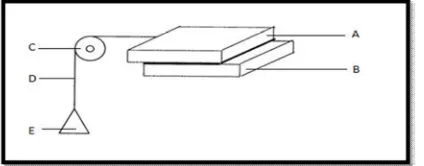
(6) Siddhapara Mihir et al. IRJP 2011, 2 (12), 90-96 In vitro Mucoadhesive strength 1) Shear stress method Two smooth, polished plexi glass blocks were selected; one glass block was fixed with an adhesive on the other glass block which was fixed on to the levelled table. To the upper block a thread was tied and the thread was passed down through a pulley. At the end of the thread a beaker was fixed. The length of the thread from pulley to the beaker was 7 cms. The weight of the beaker was counteracted. The assembly is shown in the figure 3. Solutions of different gums, combination of gums and synthetic polymer were prepared. A fixed volume (0.5ml) of solution of natural gums, their combination and Carbopol 934 P were kept on the centre of the fixed glass block with the help of the pipette, and the second block was placed on the first block and pressed by applying 100 g of weight, so that the drop of synthetic polymer, natural gums and the combination of the gum solutions spreads as a uniform film in between the two blocks. After keeping it for a fixed time intervals of 5, 10, 20 and 30 min, purified was added into the beaker gradually, the weight of purified water just sufficient to pull the upper block or to make it slide down from the base block was recorded. This weight was considered as the adhesive strength i.e. shear stress required to measure the adhesion. Before every experiment care was taken so that no air is entrapped in between the two blocks which might give erratic results. The distance from pulley to the glass block was always same in the observations51.. Figure 3:(The assembly used in the shear stress measurement method) (A)Upper glass plate (B) Lower glass plate (C) Pulley (D) Thread (E) Pan. 2) Wilhelmy plate method In this method small glass plates were coated uniformly by solution of gums, their combination and synthetic polymer to be tested and dried at 60°C. The prepared coated plates were immersed in buffer solution (pH 1.2), for 5, 10, 20, and 30 min at room temperature. The force required to pull the plate out of the solution was determined under constant experimental conditions51.. Figure 4: In vitro mucoadhesive strength measurement apparatus (A)Gastric fluid (pH 1.2) (B) Coated glass plate (C) Pulley (D) Thread (E) Pan. 3) Modified physical balance method The mucoadhesive strength of the tablets was measured on an modified physical balance. The apparatus consist of a modified double beam physical balance in which the right and left pan were with lighter pans. The left side of the balance was made heavier than the right side by placing a 5 g weight on left side pan. Another Teflon block of 3.8 cm diameter and 2 cm height was fabricated. with an upward protrusion of 2 cm height and 1.5 cm diameter on one side. This was kept in the beaker, which was then placed below the left hand set of the balance. The goat gastric mucus membrane was used as the model membrane and pH 1.2 buffer solution was used as the moistening fluid. The goat stomach mucosa was kept in tyrode solution at 37oC for 2 hr. The underlying mucus membrane was separated and washed thoroughly with a pH 1.2 buffer solution. It was then tied to a Teflon-coated glass slide and this slide was fixed over the protrusion in the Teflon block using a thread. The block was then kept in a beaker containing pH 1.2 buffer solution at a level that just touches the membrane so as to moisten the membrane. By keeping a 5 g weight on the right pan that two sides were balanced. The beaker with the Teflon block was kept below the left hand setup of the balance. The tablet was stuck on to the lower side of the left hand side pan. The 5 g weight from the right pan was then removed. This lowered the left pan along with the tablet over the membrane with the weight of 5 g. This was kept undisturbed for 3 min. Then the weight on the right hand side was added in an increment of 0.5 g until the tablet just separates from the membrane surface. The excess weight on the right pan i.e. total weight minus 5 g was taken as the measure of the mucoadhesive strength from the mucoadhesive strength, the force of adhesion was calculated using following formula,52,53,54. Force of adhesion (N) = Mucoadhesive strength/100 × 9.81 In vivo techniques 1. GI Transit using Radio-Opaque Tablets It is a simple procedure involving the use of radio-opaque markers, e.g. barium sulfate, encapsulated in mucoadhesive tablets to determine the effects of mucoadhesive polymers on GI transit time. Feces collection (using an automated faces collection machine) and X-ray inspection provide a non-invasive method of monitoring total GI residence time without affecting normal GI motility. Mucoadhesives labelled with Cr-51, Tc- 99m, In-113m, or I-123 have been used to study the transit of the tablets in the GI tract49. 2. Gamma Scintigraphy Technique Distribution and retention time of the mucoadhesive tablets can be studied using the gamma scintigraphy technique. A study has reported the intensity and distribution of radioactivity in the genital tract after administration of technetium-labelled HYAFF tablets. Dimensions of the stomach part of the sheep can be outlined and imaged using labelled gellan gum, and the data collected are subsequently used to compare the distribution of radiolabeled HYAFF formulations. The retention of mucoadhesive-radiolabeled tablets based on HYAFF polymer was found to be more for the dry powder formulation than for the pessary formulation after 12 h of administration to stomach epithelium. The combination of the sheep model and the gamma scintigraphy method has been proved to be an extremely useful tool for evaluating the distribution, spreading, and clearance of administered stomach mucoadhesive tablets49. Stability study Stability studies were performed according to ICH guidelines. The formulations were stored in room temperature at 25 ± 1°, in hot air oven at 37 ± 1°, and at 60± 1° for a period of 14 weeks55. CONCLUSION Mucoadhesive dosage forms have a high potential of being useful means of delivering drugs to the body, perhaps particularly for topical or local administration where the mechanical trauma experienced by the dosage form may be minimized. The phenomenon of mucoadhesion can be used as a model for the controlled drug delivery approaches for a number of drug candidates. The various advantages of the oral mucoadhesive drug delivery systems like prolongation of the residence time of the drug which in turn increases the absorption of the drug are important factors in the oral bioavailability of many drugs.. INTERNATIONAL RESEARCH JOURNAL OF PHARMACY, 2(12), 2011.
(7) Siddhapara Mihir et al. IRJP 2011, 2 (12), 90-96 REFERENCES 1. World Intellectual Property Organization: An Overview 2007 Edition. 2. Choi BY, Park HJ, Hwang SJ and Park JB. Preparation of alginate beads for floating drug delivery system: effect of CO2 gas-forming agent. International journal of pharmaceutics. 2002; 239: 81-91. 3. Rough N, Buri P, Doelker E. Drug absorption sites in the gastroretentive tract and dosage forms for site- specific delivery. International journal of pharmaceutics. 1996; 136: 117-139. 4. Singh BN and Kim KH. Floating drug delivery systems: an approach to oral controlled drug delivery via gastric retention. Journal of control release. 2000; 63: 235-239. 5. Streubel A, Siepmann J, Bodmeier R. Gastroretentive drug delivery system. Expert Opinion on Drug Delivery. 2006; 3 (2): 217-233. 6. Ali J, Arora S, Khar RK. Floating drug delivery System: A Review. AAPS Pharm Sci Tech. 2005; 06 (03): E372-E390 7. Baumgarter S, Kristl J, Vrecer F. Optimization of floating matrix tablets and evaluation of their gastric residence time. International journal of pharmaceutics. 2000;195:125-135 8. Bulgarelli E, Forni F, Bernabei MT. effect of matrix composition and process conditions on casein-gelatin beads floating properties. International journal of pharmaceutics. 2000; 198: 157-165. 9. Chen J Park K. Synthesis of fast-swelling, Superporous sucrose hydrogels, Carbohydrate polymers. 2000; 41(3): 259-268. 10. Akiyama Y, Nagahara N, Nara E, Kitano M, Yamamoto I, Azuma J, Ogawa Y, et al. Evaluation of oral mucoadhesive microsphere in man on the basis of the pharmacokinetics of man on the basis of the pharmacokinetics of Furosemide and riboflavin, compound with limited gastroretentive absorption sites. Journal of pharmacy and pharmacology. 1998; 50: 159-166. 11. Chickering DE, Jacob JS, Desai TA, Harrison M, Harris WP, Morrell CN, Chaturvedi P, et al. Bioadhesive microspheres: III. An In-vivo transit and bioavailability study of drug-loaded alginate poly( fumaric-co-sebacic anhydride) microspheres, Journal of control release. 1997; 48: 35-46. 12. Kedzierewicz F, Thouvenot P, Lemut J, Etienne A, Hoffman M, Maincent P, et al. Evaluation of peroral silicon dosage forms in human by gamma- scintigraphy, Journal of control release. 1999; 58:195-205. 13. Rouge N, Allemann E. comparative pharmacokinetic study of a floating multipleunit capsule, a high density multiple-unit capsule and an immediate-release tablet containing 25 mg atenolol. Pharmceutica Acta Helbetiae. 1998; 73: 81-87. 14. Cremer K. Drug delivery: Gastro-remaining Dosage forms. Journal of pharmaceutics.1997; 259 (108). 15. Iannucelli V, Coppi G, Bernabei MT, Camerorni R. Air compertment multipleunit system for prolonged gastric residence. Part-I. Formulation study. International journal of pharmaceutics. 1998; 174: 47-54. 16. Manmoham, Shukla TP, Mathur A, Upadhyay N, Sharma S. A review on Gastroretentive drug delivery system: An emerging approach to improve the gastric residence time of solid dosage forms. International journal of pharmaceutical science review and research. June 2011; 8(2): 176-182. 17. Khosla R, Davis SS. THE effect of tablet size on the gastric emptying of nondisintegrant tablets. International journal of pharmaceutics. 1990; 62: R9-R11. 18. Timmermans J. Moes AJ. Factors controlling the buoyancy and gastric retention capabilities of floating matrix capsules: new data for reconsidering the controversy. Journal of Pharmaceutical science. 1994; 83: 18-24. 19. O’Reilly S, Wilson CG, Hardy JG. The influence of food on the gastric emptying of multiparticulate dosage forms. International journal of pharmaceutics.1987; 34: 213-216. 20. Abrahamsson B, Alpsten M, Hugosson M, Jonsson UE, Sundgren M, Svenheden A. Absorption gastrointestinal transit and tablet erosion of felodipine extended release tablets. Pharmaceutical research. 1993; 10: 709-714. 21. Nimmo J, Heading RC, Tothill P, Prescott LF: Pharmacological modification of and metoclopramide on paracetamol absorption. British medical journal. 1973; 1: 587-589. 22. Bennett CE, Hardy JG, Wilson CG. The influence of posture on the gastric emptying of antacids. International journal of pharmaceutics. 1984; 21: 341-347. 23. Helliwell M, The use of bioadhesive in targeted drug delivery within gastrointestinal tract. Advance Drug delivary review. 1993; 11: 221-251. 24. Vantappen GR, Peeters TL, Janssens J. The secretary component of inter digestive migratory motor complex in man. Scandinavian j gastroenterology. 1979; 14: 663-337. 25. Wilson CG, Washington N. The stomach: its role in oral dug delivery. In: rubinstein MH, ed. Physiological pharmaceutical: biological barriers to drug absorption. Chichester, UK: Ellis horwood. 47-70. 26. Desai S, Bolton S. A floating controlled release drug delivery system: in vitro- in vivo evaluation. Pharm Res. 1993; 10: 1321-1325. 27. Wilding IR, Coupe AJ, Davis SS. The role of g-Scintigraphy in oral drug delivery. Adv drug deliv rev. 2001; 46: 103-124.. 28. Weitschies W, Kosch O, Monnikes H, Trahms L. Magnetic marker monitoring: an application of biomagnetic measurement instrumentation and principle for the determination of the gastrointestinal behaviour of magnetically marked solid dosage forms. Adv drug deliv rev. 2005; 57: 1210-1222. 29. Jain NK. Controlled release and novel drug delivery. 1st edition. CBS publishers and distributors, New Delhi. 1997: 353-370. 30. Park H, Robinson JR. Mechanisms of mucoadhesion of polyacrylic acid and hydrogels. Pharmaceutical Research. 1987; 4: 457-64. 31. Moes Aj. Gastroretentive dosage forms. Critical Reviews in therapeutic drug carrier system. 1993; 10: 143-195. 32. Hwang SJ, Park H, Park K. Gastric retentive drug delivery system. Critical reviews in therapeutic drug carrier system. 1998; 15: 243-84. 33. Hoffmann A, Stepensky D, Pharmacodynamic aspect of modes of drug administration for optimization of drug therapy. Critical reviews in therapeutic drug carrier system. 1999; 16: 571-639. 34. Deshpande AA, Rhodes CT, Shah NH, Malick AW. Controlled-release drug delivery systems for prolonged gastric residence: an overview. Drug development and industrial pharmacy. 1996; 22: 31-39. 35. Singh BN, Kim KH. Floating drug delivery systems: an approach to oral controlled drug delivery via gastric retention. Journal of control release. 2000; 63: 235-59. 36. Stockwell AF, Davis SS, Walker SE. In-vitro evaluation of alginate gel systems. Journal of control release. 1986; 3: 167-75. 37. Timmermanns J, Moes A. How well do floating dosage form float. International journal of Pharmaceutics. 1990; 62(3): 207-16. 38. Yeole PG, Khan S, Patel VF. Floating drug delivery systems: need and development. Indian Journal of pharmaceutical science. 2005; 67(3): 265-72. 39. Singh M, Sharma P, Sharma N. Gastroretentive drug delivery system based on natural mucoadhesive polymers: A brief review. Journal of pharmacy research; 4(2): 519-521. 40. Hui HW, Robinson JR. Ocular delivery of progesterone using bioadhesive polymer. International journal of pharmaceutics. 1985; 26: 203-13. 41. Ahuja A, Khar RK, Ali J. Mucoadhesive drug delivery systems. Drug development and industrial pharmacy. 1997; 23 (5): 489-515. 42. Chickering D, Jacob J, Mathiowitz E. poly( fumaric-cosebacic) microsphere as oral drug delivery systems. Biotechnology and Bioengineering. 1996; 52: 96101. 43. Lachman L, Herbert A, Liberman HA, Joseph L, Kangi. The theory and practice of industrial pharmacy. Varghese publishing house. III ed edition 1991: 296-302 44. United state pharmacopoeia. xxiv NF 19, Rockville: united states pharmacopoeia convention. 2000: 2388-89. 45. Deshmukh VN, Jadhav JK, Sakarkar DM. Formulation and evaluation of theophyline anhydrous bioadhesive tablets. Asian journal of pharmaceutics. 2009; 3: 54-58. 46. Gupta A, Garg S, Khar R. Measurement of bioadhesive strength of buccal tablets: design of in vitro assembly. Indian drug. 1992: 30: 152-5. 47. Margeta C, Sachin BS, Debjit B, Bhowmik B, Jayakar B. Formulation and evaluation of controlled release mucoadhesive oral tablet of clarithromycine. Der pharmacia letter: 2009; 1: 83-91. 48. Mathiowitz E, Chickering DE, Lehr CM. Editors. Mucoadhesive drug delivery systems-fundamentals. Novel approaches and development 98. New York. Marcel dekker. 1999:563-99. 49. Rajput G, Majmudar F, Patel j, Thakor R, Rajgor NB. Stomach specific mucoadhesive microsphere as a controlled drug delivery systems. Systematic review in pharmacy. 2010; 1: 36-44. 50. Sonani NG, Hiremath SP, Dasankoppa FS, Jamakandi VG, Steenivas SA. Design and evaluation of gastroretentive mucoadhesive cephalexin tablets. Pharmaceutical development and technology. 2010; 15(2): 178-83. 51. Chanda R, Nath LK, Mahapatra S. Formulation and development of oral mucoadhesive coated terbutaline sulphate tablets using some natural materials extracted from edible fruits available in india. International journal of pharmaceutical science. 2009; 5(1): 3-12. 52. Ali j, Khar R, Ahuja A Karla R. Buccoadhesive erodible disk for treatment of orodental infections design and characterization. International journal of pharmaceutics. 2002; 283: 93-103. 53. Ali J, Khar R, Ahuja A. Effect of polymer loading on drug release and bioadhesion of buccoadhesive carriers for local drug delivery of triaminocolon acetonitride. The eastern pharmacist. 1999; 46 (503): 115-19. 54. Vermani K, Garg S, lourence J. Assembly for in vitro measurement of bioadhesive strength and retention characteristic in simulated vaginal environment. Drug development and industrial pharmacy. 2002; 28 (9): 1133-46. 55. Kulkarni GT, Gosthamarajan K, Suresh B. Stability testing of pharmaceutical product: an overview. Indian journal of pharmaceutical education and research. 2004; 38 (194): 202-20 .. Source of support: Nil, Conflict of interest: None Declared. INTERNATIONAL RESEARCH JOURNAL OF PHARMACY, 2(12), 2011.
(8)
Figure


Related documents
Note that given the “pull” communication model used by dynamiQ to retrieve scenario information, a delay of up to one time interval can occur before user interface actions are
The objectives of this research are to identify the challenges that West African women seafarers face with respect to recruitment on-board ships and further to find ways
In a study that evaluated the properties of phenolic compounds like p-hydroxy benzoic acid, oleuropein, vanillic acid, and protocatechuic acid present in olive
As regard to these recurrent cases, they all have received conservative surgeries before they sought one more treatment at our institutions, whereas no recurrence was found in
The main objective of this work is to analyse to what extent the scientific literature in educational technology in the last 55 years (1960 – 2015), related to experiences on
The remainder of this paper is organized as follows: “ Related work ” section provides an overview of literature related to data sampling methods that address severe class
To adapt to such form of unwrapping using the existing derived map- ping functions, a projection plane that is normal to the optical axis is simply placed some distance away from
