Genes in Tooth Development
V.VIGNESH
1, N. ARAVINDHA BABU
2, N. BALACHANDER
2and L. MALATHI
21Intern, Tagore Dental College and Hospital
2Department of Oral Pathology and Microbiology (COCPAR),
SreeBalaji Dental College & Hospital, Bharath University, Chennai. DOI: http://dx.doi.org/10.13005/bpj/664
(Received: July 25, 2015; accepted: September 10, 2015) ABSTRACT
Teeth form as epithelial appendages such as hairs and glands. During development, reciprocal and sequential epithelial-mesenchymal interactions regulate processes such as proliferation, differentiation and morphogenesis. These interactions are mediated by conserved signaling pathways that are reiteratively used during development of all organs. Mutations in genes encoding molecules in the signaling pathways cause numerous abnormalities in craniofacial bones and teeth including missing or supernumerary teeth, and disturbances in formation of dentin and enamel. This article is regarding the genetic basis of tooth development, methods used to study it, and the genes that have been definitively implicated in the development of human dentition and the brief notice on the deformities caused due to the mutation of the these genes.
Key words: Homeoboxgenes (Hox), MSX, DLX, PAX.
INTRODUCTION
Teeth are vertebrate organs that arise from complex and progressive interactions between an ectoderm, the oral epithelium and an underlying mesenchyme. During their early development, tooth germs exhibit many morphological and molecular similarities with other developing epithelial appendages, such as hair follicles, mammary and salivary glands, lungs, kidneys, etc. Recent advances in molecular genetics have made it possible to identify the exact genes responsible for the development of teeth. This paper is regarding the genetic basis of tooth development, methods used to study it, and the genes that have been definitively implicated human dentition.
History
The inheritance patterns of genes was first described by Gregor Mendel in 18651 and
extensively studied in the early part of the 20th century2 While the traditional Mendelian model of
inheritance is extremely useful in studying traits
caused due to a single gene, it does not give an accurate description of traits caused by multiple genes3 The limitations of the Mendelian model also
meant that the genetic studies often focused on groups with a limited gene pool, such as groups with a history of consanguineous marriage4 or
isolated island populations5 Using these models it
was shown that the initiation of tooth development was controlled by more than one gene6
Experimental studies
In 1913, Bridges showed that genes were located on chromosomes7 but it was not until the
early 1970s that a variety of cytogenetic methods were discovered that produced distinct bands on each chromosome8,9,10 making it possible to give
each gene a specific “genetic address.”11 Even
though the first draft of the entire human genome was completed only in 2001,12 by the 1990s
Suitability for both genetic and embryologic studies, the mouse emerged as the predominant system for contemporary experimental studies on tooth development13 Most of the early genetic analyses
of tooth development, which have formed the basis of our understanding of the genetics of hypodontia, were done by the use of mouse mutations. These could be accomplished by the use of the following methods:
• Knockout mice • Transgenic Mice Knockout mice
In knockout technology, a known gene is selectively targeted for disruption in embryonic stem (ES) cells by the principle of homologous recombination14 Reconstitution of ES cells in
chimeric mice and germ line transmission results in mice which carry a loss-of-function, typically recessive, mutation in a known gene. These mice can then be bred to homozygosity and the phenotypic consequences of the mutation assessed during embryogenesis13 The disadvantage of
knockout mutations was that the gene that is knocked out may have growth implications beyond the development of the tooth resulting in the failure of the embryo to form, a problem that was overcome by the development of conditional knockout systems15
Transgenic mice
In transgenic technology, mutations of the genes responsible for tooth development are chosen and extracted. Next they are injected into fertilized mouse eggs. Embryos are implanted in the uterus of a surrogate mother. The selected genes will be expressed by some of the offspring allowing investigators to study the effects of the gene16
Using mouse models, by late 1990s researchers had managed to identify the genes responsible for mammalian tooth formation17, 18 In
1998, Thomas and Sharpe proposed that the patterning of murine dentition was regulated by a complex interaction of the homeobox genes and termed it the “OdontogenicHomeobox Code.”18 It
was found that the genes responsible for tooth formation comprised of transcription factors, growth factors, and receptors17, 19
A transcription factor (sometimes called a sequence-specific DNA binding factor) is a protein that binds to specific DNA sequences and thereby controls the transfer (or transcription) of genetic information from DNA to mRNA. 4, 20 The MSX21 and
DLX22 families of homeobox genes are examples
of transcription factors that control tooth genesis. A growth factor is a naturally occurring substance capable of stimulating cellular growth, proliferation, and cellular differentiation, matrix metalo proteinase (MMP)23 and fibroblast growth factor
(FGF) being an examples of such factors.
24 Receptors are protein molecules, embedded in
either the plasma membrane or the cytoplasm of a cell, to which one or more specific kinds of signaling molecules may attach. The epithelial growth factor receptor (EGFR) being an example of such a receptor25
Using these mouse models Vieira in 2003 proposed that, “Specific genes were responsible for specific missing teeth” in mice [Table - 1]. In order to know which of these genes to look for in humans and ascertain the location of these genes, oral clefts and syndromic forms of tooth agenesis were proposed as suitable genetic models26
Genes Homeobox
Edward Lewis was the first person to identify the homeotic genes.Homeobox (Hox) genes are a set of genes that determine an organizational pattern in vertebrates. First isolated in the fly Drosophila melanogaster.In mammals, 38 Hox genes have been identified which reside in four main chromosomal clusters, termed Hoxa, Hoxb, Hoxc, and Hoxd, and define 13 paralogous groups.Homeobox (Hox) genes are a set of genes that determine an organizational pattern in vertebrates27 First isolated in the fly Drosophila
melanogaster, the temporal and spatial control of Hox gene expression is essential for correct patterning of many animals28 Other vertebrate
genes, such as the Msx gene family, also contain homeoboxes, but since these are dispersed to different chromosomal locations in the genome, they are referred to as “homeobox genes” rather than “Hox genes.”29 Remarkably, the relative order
HOM-C, a paralogous relationship which permits the mammalian Hox genes to be grouped into the 13 discrete groups30
MSX
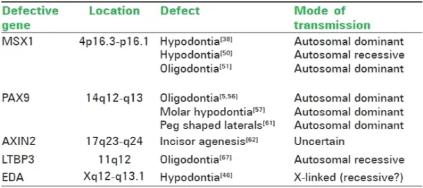
MSX1 (muscle segment), initially called homeobox 7 (HOX7), is a non-clustered homeobox protein which is located on the small arm of chromosome 4 with the genetic address 4p16.3-p16.131 This gene encodes a member of the muscle
segment homeobox gene family. The encoded protein functions as a transcriptional repressor during embryogenesis through interactions with components of the core transcription complex and other homeoproteins32 The gene has been shown
to have a considerable role in tooth development. MSX2 – expressed by precursors of maxillary and mandibular bone, Meckel’s cartilage and the tooth germs. At bud stage MSX2 is detectable in vestibular lamina and both dental epithelium and mesenchyme and later in cap stage in enamel knot and vestibular epithelium
DLX
DLX is another gene that is also responsible in the tooth development. They are the distal less gene. The interaction of the dlx and the MSX gene help in the tooth development. MSX and DLX have opposite transcription factors i.e. when the MSX functions as a suppressor the DLX functions as an activator. MSX and DLX genes participate in the tooth development by reciprocal epithelial-mesenchymal interaction.as the epithelium of the prospective oral cavity thickens to form the dental lamina the expression of the msx2 localizes. Activation of MSX1, MSX2, DLX1, DLX2
in dental mesenchyme in response to BMP4 and FGF signals form the overlying epithelium. PAX
PAX9 is a member of the paired box (PAX) family of transcription factors. The PAX 9 gene is located on the long arm of chromosome 14 and has the genetic address 14q12-q1333 These genes
play critical roles during fetal development and cancer growth34 The PAX9 gene was first shown to
be associated with autosomal dominant, non-syndromic, familial oligodontia. Since then several novel mutations in the gene have been discovered in families around the world35, 36, 37 {TABLE-2}. In
addition, a recent study has also found that families with affected benign hereditary chorea show a deletion of the PAX9 gene resulting in oligodontia. Peg-shaped lateral incisors and other forms of microdontia have long been known to be mild forms of hypodontia. PAX9 mutations have been associated with both hypodontia and a generalized reduction of the size of the teeth.
Table 1: Genes responsible for specific missing teeth in mice
Defective gene Type of tooth agenesis
Dlx-1 and Dlx-2 Upper molars
Activin bA, activin Incisors and lower molars receptors IIA and IIB,
Smad 2
Fgf8 All but lower incisors
Msx1 All
Pax9 All
AXIN2 or axis inhibitor protein 2 is a gene located on the long arm of chromosome 17 with a genetic address of 17q23-q24. The association of the gene to tooth agenesis was first found in a Finnish family with a predisposition for colorectal cancer. It has been shown that the AXIN2 mutations may also be responsible for sporadic forms of incisor agenesis. The mode of transmission of hypodontia due to defects in the AXIN2 gene has not been definitively proved.
LTBP3 (latent transforming growth factor beta binding protein 3) is a gene that modulates the bioavailability of TGF-beta. Located on the long arm of chromosome 11, it has the genetic address 11q12. A study on a Pakistani family with a history of consanguineous marriage found that a mutation in the LTBP3 gene causes an autosomal recessive form of familial oligodontia.
EDA (ectodysplasin 1) is a gene located at Xq12-q13.1 that has been linked to X-linked recessive ectodermal dysplasia. A study of Chinese families with non syndromic X-linked hypodontia showed that a Thr338Met mutation of the EDA gene was responsible for the congenital absence of maxillary and mandibular central incisors, lateral incisors, and canines, with the high possibility of persistence of maxillary and mandibular first permanent molars.
The proximal maxilla develop, does not express Hox genes and tooth development is controlled by local interactions involving non-Hoxhomeobox and other transcriptional regulators .At embryonic days 9–11, the oral epithelium initiates tooth development by signalling through
generic molecules including FGFs (Fibroblast Growth Factors), BMPs (Bone Morphogenetic Proteins), Wnts and Shh (Sonic hedgehog) to the underlying neural crest-derived mesenchyme.
Wnt genes 4, 6,10a and10b, which are expressed in the presumptive dental epithelium at this stage, are likely candidates for these Wnt signals involved in the dental lamina to bud stage transition. Binding of Wnts to their receptors causes formation in the nucleus of active transcription complexes between b-catenin and Lef1, a member of the LEF/TCF family of DNA binding proteins that activates Wnt target gene expression. Wntsignaling is also required for the bud-to-cap transition
Therefore the jaw is divided into a tooth-forming LHX-positive domain and a non-tooth-formingGSC-positive domain. In mice, in which GSC has been knocked out, the teeth form normallybut the supporting skeletal structures in the aboral region are absent – thus defining the Rostral-caudal patterning.Overall called OdontogenicHomeobox Code.
CONCLUSION
With the many studies conducted and the experimental analysis done the basic genes involved in the tooth development include the homeobox,MSX, DLX, PAX group. Any mutation in the above genes can result in tooth related deformity or can alter the development. Even now studies are being conducted on identifying the genes for tooth development and the search will go on.
REFERENCES
1. Mendel JG.
VersucheüberPlflanzenhybridenVerhandlungen des naturforschendenVereines in Brünn, Bd. IV für das Jahr, 1865.
2. Bowler PJ. The Mendelian Revolution: The Emergence of Hereditarian Concepts in Modern Science and Society. Baltimore, MD: Johns Hopkins University Press; 1989. 3. Lorentz CP, Wieben ED, Tefferi A, Whiteman
DA, Dewald GW. Primer on medical
genomics part I: History of genetics and sequencing of the human genome. Mayo ClinProc; 77: 1395-6 (2002).
4. Bridges CB. Direct proof through non-disjunction that the sex-linked genes of Drosophila are borne on the X-chromosome. Science; 40: 107-9 (1914).
(1952).
6. Huskins CL. On the inheritance of an anomaly of human dentition, J Hered; 21: 279-82 (1930).
7. Eidelman E, Chosack A, Rosenzwei KA. Hypodontia: Prevalence amongst Jewish Populations of different or igin. Am J PhysAnthropol; 39: 129-33 (1973).
8. Caspersson T, Zech L, Modest EJ. Fluorescent labeling of chromosomal DNA: Superiority of quinacrine mustard to quinacrine. Science; 170: 762 (1970). 9. Seabright M. A rapid banding technique for
human chromosomes. Lancet; 2: 971-2 (1971).
10. Davies KE. The application of DNA recombinant technology to the analysis of the human genome and genetic disease. Hum Genet; 58: 351-7 (1981).
11. International Human Genome Sequencing Consortium. Initial sequencing and analysis of the human genome. Nature; 409: 860-921 (2001).
12. Maas R, Bei M. The Genetic control of early tooth development. Crit Rev Oral Biol Med; 8:4 (1997).
13. Capecchi MR. Targeted gene replacement. SciAmer; 270: 52-9 (1994).
14. Gu H, Marth JD, Orban PC, Mossmann H, Rajewsky K. Deletion of a DNA polymerase beta gene segment in T cells using cell type-specific gene targeting. Science, 265: 103-6 (1994).
15. Kist R, Greally E, Peters H. Derivation of a mouse model for conditional inactivation of Pax9. Genesis; 45: 460-4 (2007).
16. Line SR. Molecular morphogenetic fields in the development of human dentition. J TheoretBiol; 211: 67-75 (2001).
17. Thomas BL, Sharpe PT. Patterning of the murine dentition by homeobox genes. Eur J Oral Sci; 106: 48-54 (1998).
18. Francis-West PH, Robson L, Evans DJ. Craniofacial Development: The tissue and molecular interactions that control development of the head. In: Beck F, Kriz W, Marani E, Sano Y, Schoenwolf GC, Zilles K, editors. Advances in Anatomy Embryology and Cell Biology. New York: Springer-Verlag; 2003.
19. Latchman DS. Transcription factors: An overview. Int J Biochem Cell Biol; 29: 1305-12 (1997).
20. Satokata I, Ma L, Ohshima H, Bei M, Woo I, Nishizawa K, et al. Msx2 deficiency in mice causes pleiotropic defects in bone growth and ectodermal organ formation. Nat Genet; 24: 391-5 (2000).
21. Qiu M, Bulfone A, Ghattas I, Meneses JJ, Christensen L, and Sharpe PT, et al. Role of the Dlxhomeobox genes in roximodistal patterning of the branchial arches: Mutations of Dlx-1, Dlx-2, and Dlx-1 and -2 alter morphogenesis of proximal skeletal and soft tissue structures derived from the first and second arches. DevBiol; 185:165-84 (1997). 22. Peres RC, Line SR. Analysis of MMP-9 and TIMP-2 gene promoter polymorphisms in individuals with hypodontia. Braz Dent J; 16: 231-6 (2005).
23. Menezes R, Letra A, Ruff J, Granjeiro JM, Vieira AR. Studies of genes in the FGF signaling pathway and oral clefts with or without dental anomalies. Am J Med Genet Am; 146:1614-7 (2008).
24. Vieira AR. Oral clefts and syndromic forms of tooth agenesis as models for genetics of isolated tooth agenesis. J Dent Res; 82: 162-5 (2003).
25. McGinnis W, Krumlauf. Homeobox genes and axial patterning. Cell; 68: 283-302 (1992).
26. Arte S, Nieminen P, Pirinen S, Thesleff I, Peltonen L. Gene defect in hypodontia: Exclusion of the EGF, EGFR and FGF -3 as candidate genes. J Dent Res; 75: 1346-52 (1996).
27. Soshnikova N, Duboule D. Epigenetic regulation of vertebrate Hox genes A dynamic equilibrium Epigenetics; 4: 537-40 (2009).
28. Hunt P, Whiting J, Nonchev S, Sham M, Marshal H, Graham A, et al. The branchialHox code and its implications for gene regulation, patterning of the nervous system and head evolution. Development;( Suppl 2):63-77 (1991)
30. Campbell K, Flavin N, Ivens A, Robert B, Buckingham M, Williamson R. The human homeobox gene HOX7 maps to 4p16.1 and is deleted in Wolf-Hirschhorn syndrome patients. Am J Hum Genet; 45(suppl):A179 (1989).
31. Blin-Wakkach C, Lezot F, Ghoul-Mazgar S, Hotton D, Monteiro S, Teillaud C, et al. Endogenous Msx1 antisense transcript: In vivo and in vitro evidences, structure, and potential involvement in skeleton development in mammals. Proc Nat AcadSci; 98: 7336-41 (2001).
32. Stapleton P, WeighA, Urbanek P, Kozmik Z, Busslinger M. Chromosomal localization of seven PAX genes and cloning of a novel family member, PAX-9. Nat Genet; 3:292-8 (1993).
33. Peters H, Neubuser A, Kratochwil K, Balling R. Pax9-deficient mice lack pharyngeal pouch derivatives and teeth and exhibit craniofacial and limb abnormalities. Genes Dev; 12: 2735-47 (1998).
34. Kist R, Watson M, Wang X, Cairns P, Miles C, Reid DJ, et al. Reduction of Pax9 gene dosage in an allelic series of mouse mutants causes hypodontia and oligodontia. Hum Mole Genet ; 14:3605-17 (2005).
35. Kapadia H, Frazier-Bowers S, Ogawa T, D’Souza RN. Molecular characterization of a novel PAX9 missense mutation causing posterior tooth agenesis. Eur J Hum Genet; 14: 403-9 (2006).