ISSN: 2372-5060 (Print), 2372-5079 (Online) Copyright © The Author(s). All Rights Reserved. Published by American Research Institute for Policy Development DOI: 10.15640/ijhs.v5n3a2 URL: https://doi.org/10.15640/ijhs.v5n3a2
Effect of Dry Cupping on Vascular Function among Young Individuals
Arturo A. Arce-Esquivel
1, Brandon J. Warner
2, Diana M. Gallegos
3& S. Andrew Cage
4Abstract
Background: Cupping is most widely used in China as part of traditional Chinese medicine. Cupping is an
ancient technique used in treating pain and various disorders. It has been suggested that cupping promotes hyperemia (i.e., increased blood flow). Thus, this study aimed to assess the effect of a single dry cupping treatment on microvascular function among healthy young individuals.
Methods: Eleven apparently healthy young individuals (age: 22 ± 1.43 years) participated in this study. Dry
cupping treatment was performed applying two plastic cups on the non-dominant arm of each participant. Before and after a 10-minute cupping treatment, microvascular function (fingertip Digital Thermal Monitoring of vascular reactivity) was evaluated.
Results: Following the 10-minute cupping treatment, the individuals experienced a significant 36% increase
in Vascular Reactivity Index (VRI) from baseline (2.60 ± 0.40 to 3.53 ± 0.42, p < 0.05). Participants experienced no complications as a result of the intervention.
Conclusion: These findings demonstrated that in healthy individuals, dry cupping treatment, was capable of
increasing microvascular function, specifically VRI. This study underlies the role of cupping treatment in promoting vascular function improvements.
Keywords: Vascular Function; Cupping; Hyperemia; Blood Flow
1. Introduction
Cupping is a traditional Chinese medicine therapy that dates back at least 2,000 years. Cupping is a treatment used in the realm of folk medicine and by clinicians in several cultures. Cupping is most widely used in China as part of traditional Chinese medicine. It is mostly used in Asian and Middle Eastern countries, and it has been reported to reduce pain (Kim, Lee, Lee, Boddy, & Ernst, 2011; Lee, Kim, & Ernst, 2011) as well as a host of other symptoms (Cao, Li, & Liu, 2012; Yoo & Tausk, 2004). Interestingly, a Korean survey in 2006 showed that 93.5% of 6708 oriental physicians used cupping treatments in their clinical fields (Han, Kim, Lee, Shin, & Choi, 2006). The technique involves a plastic, bamboo, or glass cup to create suction on the skin over an acupuncture point, painful area, or a reflex zone. There are two types of cupping. In wet cupping the skin is lacerated so that blood from the dermal microcirculation is drawn into the cup. Whilst, dry cupping pulls the skin into the cup without drawing blood, negative pressure on the skin would act as an irritant to subcutaneous tissues (Lee et al., 2011). The skin is dragged into the cupping glass, resulting in rubor and heat at the affected area with increased perfusion (Teut et al., 2012). The use of cupping is generally safe based on long-term clinical trials. In clinical practice cupping is regularly observed to bring about pain relief and to increase a patient’s general feeling of wellbeing (Lauche et al., 2011). For instance, dry cupping treatments seemed to be effective in dismissing chronic non-specific neck pain (Lauche et al., 2011).
1 Department of Health and Kinesiology, The University of Texas at Tyler, Tyler, Texas. 2 Grand Canyon University, Phoenix, Arizona.
3 AT Still University, Mesa, Arizona.
4
In the United States, there has been a gradual increase in the use of cupping and other types of complementary and integrative remedies (Eisenberg et al., 1998). For instance, in a study among pediatric patients with chronic pain, the authors reported that cupping and acupuncture treatments were pleasant and helpful for pain conditions (Kemper et al., 2000). Furthermore, it has been reported that cupping treatment is used to reduce musculoskeletal inflammation and pain, and may consequently increase physical performance during sport activity (Musumeci, 2016). Nonetheless, whether or not cupping therapy effectively enhances athletes’ performance is still questionable and needs further study.
Although cupping treatment may be effective for reducing pain (Kim et al., 2011; Lee et al., 2011); the mechanism of cupping treatment is still unclear. The postulated modes of actions include the interruption of blood circulation and congestion as well as inhibiting the inflammatory extravasations (escaping of bodily fluids such as blood) from the tissues (Yoo & Tausk, 2004). Cupping stimulates an increase in subcutaneous blood flow and further stimulation of the autonomic nervous system, promoting pain relief (Yoo & Tausk, 2004). Researchers have suggested that placement of cups on selected acupoints on the skin promotes hyperemia (i.e., increased blood flow), which results in a therapeutic effect (Cao et al., 2012). The endothelium, a thin single sheet of endothelial cells, is a metabolically active layer that coats the inner surface of blood vessels and acts as an interface between the circulating blood and the vessel wall. The increased blood flow is mediated by different vasodilators, where nitric oxide (NO) is a critical mediator of vascular homeostasis. Furthermore, this increase in local blood circulation might improve skin blood flow (Liu, Piao, Meng, & Wei, 2013). Other investigators have suggested that the mechanisms of action include boosting immunity and promoting the anti-inflammatory process through the removal of oxidants and reduction of oxidative stress (Mehta & Dhapte, 2015). Nevertheless, we are unaware of any study that has examined the effect of dry cupping on peripheral microcirculation. Thus, the purpose of this study aimed to assess the effect of a single dry cupping treatment on microvascular function among healthy young individuals.
2. Methods
2.1. Experimental Design
The proposed study was a controlled design to determine the effectiveness of a single dry cupping treatment. Each participant was examined twice, before and after a 10-minute cupping treatment, for the measurement of microvascular function.
2.2. Study Participants
Eleven apparently healthy young college students were recruited to participate in the study. The majority of participants were recreationally trained. Individuals with manifestations of cardiovascular, metabolic, orthopedic or neurological disease or were on any medication, or dietary supplements (including vitamins and antioxidants), or who used tobacco, which could affect the results of this study were excluded. Following explanation of all the details of the study, each participant signed an informed consent approved by the Institutional Review Board.
2.3. Study procedures and assessment
2.3.1. Cupping Treatment
Figure 1. Application of the adaptable glass cups on the non-dominant arm.
2.3.2. Vascular Function
The fingertip digital thermal monitoring (DTM), a non-invasive test, of vascular reactivity was used as previously described by us (Arce-Esquivel et al., 2016) and others (Ahmadi et al., 2011). All DTM measurements were performed in a quiet, dimly lit room with a controlled ambient temperature between 23 and 25°C. The studies were conducted after an overnight fast of at least 10 hours (water was permitted) and abstinence from tobacco, alcohol, caffeine, vasoactive medications, exercise, high-fat foods, and vitamin C. The measurements were obtained with the subjects seated for 20 minutes of rest. Segmental pressure cuffs were positioned around the forearms. Disposable finger probes were attached to the index fingers of both hands. DTM of both hands was obtained during 5-minute stabilization, 5-minute cuff inflation, and 5-minute deflation using an automated, operator independent protocol (VENDYS, Endothelix Inc., Houston, TX, USA). The right upper arm cuff (i.e., occluded arm) was rapidly inflated to 50 mmHg above systolic pressure for 5 minutes and then rapidly deflated to invoke reactive hyperemia distally. Thermal changes during a 5-minute arm-cuff-induced reactive hyperemia test were monitored continuously in the fingertip of both the occluded (i.e., right) and the non-occluded (i.e., left) arms using VENDYS software. Finally, the area under the temperature curve was used to determine the vascular reactivity index (VRI) which assessed the difference in response in microvascular function before and after cupping therapy.
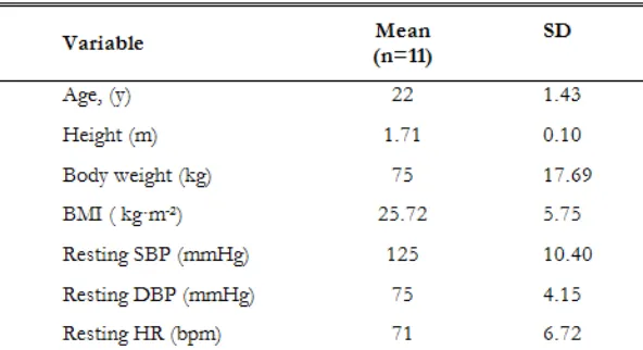
Table 1. Participant Characteristics
The repeatability of DTM is excellent and can be used as a reproducible and operator-independent test for non-invasive measurement of vascular function (Ahmadi et al., 2011; McQuilkin et al., 2009). Indeed, DTM of vascular Participants Characteristics. reactivity appears to be an appropriate test to evaluate vascular reactivity in clinical and research settings (Ahmadi et al., 2011; Zeb et al., 2013).
2. 4. Statistical analysis
All values are means ± standard deviation (SD) or ± standard error (SE) when appropriate. Analyses was performed using Student t-test for paired samples with Wilcoxon matched-pairs signed rank test. All statistical analyses were performed with GraphPad Prism version 6.0a (GraphPad Software, Inc. CA, USA). The level of significance was set at p < 0.05.
3. Results
A total of 11 young healthy college students participated in all aspects of this study. There were 9 women and 2 men. The average age was 22 ± 1.43 years. The oldest participant tested was 24 years old, and the youngest was 19 years old. None of the participants reported any problem following cupping treatment. The participants’ characteristics are presented in Table 1.
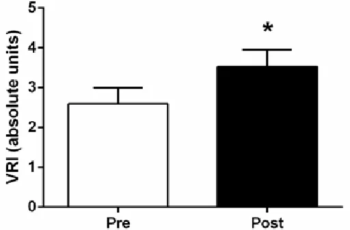
The average values for the microvascular function assessments, before and after dry cupping treatment, are presented in Table 2 and Figure 2. The average baseline VRI was 2.60 ± 0.40. Following the 10-minute cupping treatment, VRI (i.e., microvascular function) increased significantly. Indeed, cupping therapy promoted a 36% increase from baseline (2.60 ± 0.40 to 3.53 ± 0.42, p < 0.05).
Table 2. Vascular Assessments
Values are means ± SE. VRI, vascular reactivity index. * p < 0.05 for Pre and Post comparison at the measured characteristic.
4. Discussion
The purpose of this study was to assess the effects of a single dry cupping treatment on microvascular function among healthy young individuals. These preliminary findings indicated that dry cupping treatment is capable of significantly increasing microvascular function (i.e., vascular reactivity) among the participants.
The DTM is a non-invasive technique to study vascular reactivity (i.e., microvascular function) in humans. Briefly, a standard arm-cuff promotes a temporary occlusion of blood flow in the arm. During the cuff occlusion, the lack of blood flow (i.e., ischemia) elicits a microvascular dilative response. Upon releasing the cuff, blood flow rushes into the forearm and hand, not only restoring baseline flow but also resulting in an overshoot (i.e., reactive hyperemia). The reactive hyperemia promotes shear stress in the larger arteries to dilate and accommodate the increased blood flow. In general, the study of blood flow responses following occlusion indicates that younger, fitter, and healthier individuals exhibit greater flow responses, suggesting better vascular function. Reductions in the hyperemic response, a hallmark feature of impaired vascular function, are observed during DTM. Indeed, the VRI provides a non-invasive “window” to the cardiovascular system allowing the detection, measurement, and monitoring of vital patient information such as overall cardiovascular health. The VRI is reported in absolute units using a vascular reactivity scale: a) < 1 (“poor”), b) 1 – 2 (“intermediate”), and c) > 2 (“good”) (VENDYS, Endothelix Inc., Houston, TX, USA). The average baseline VRI was 2.60 ± 0.40. These vascular assessments placed the participants in the “good” category of vascular reactivity (> 2) before the treatment.
The baseline VRI reported here is clearly higher than the one we found among person with disabilities, where microvascular function was compromised (Arce-Esquivel et al., 2016). Interestingly, following cupping treatment the participants experienced a significant increase (36% increase) in their VRI (3.53 ± 0.42, p < 0.05). Our findings indicate that younger and healthier individuals were able to exhibit greater flow responses following cupping treatment.
There is evidence that cupping treatment may help to promote hemodynamic activity. Indeed, Li et al., (Li, Li, Lin, & Li, 2017), using Near-infrared spectroscopy, quantified the concentration changes of hemoglobin during a single 5-minute dry cupping treatment session among healthy individuals. These investigators reported an elevation in oxy-hemoglobin and a decline in deoxy-hemoglobin during the 5-minute treatment. They concluded that these findings induced an oxygen elevation (i.e., oxygen uptake) in the local tissue, which may have mediated the positive therapeutic cupping effects (Li et al., 2017).
It has been speculated that mechanically, cupping increases blood circulation, whereas physiologically it activates the immune system and stimulates the mechanosensitive fibers, thus leading to a reduction in pain (Rozenfeld & Kalichman, 2016). For instance, Tham et al., (Tham, Lee, & Lu, 2006) demonstrated that in a soft tissue model that the soft tissue directly under the rim of the cup compresses, while the periphery tenses. The tensile stresses appear to be greater in the bulb-shaped region under the center of the cup, extending down to the muscle layer. However, these forces reach their maximum on a very small area of the skin layer near to and just inside the cup’s rim. The stretching of the skin, into the cup, was the result of the applied vacuum. In addition, several authors have hypothesized that cupping increases the circulation surrounding the treated area, thus enabling toxins trapped deep in the soft tissue layers to rise to the body surface (Yoo & Tausk, 2004). The existence of high tensile stresses inside the cup is believed to cause a severe dilation of the capillaries (Tham et al., 2006).
5. Conclusion
In conclusion, the present findings demonstrated that in young healthy college students, cupping treatment is capable of increasing microvascular function. Considering that decreased vascular function is associated with many chronic conditions and/or disabilities, noninvasive alternative protocols, like cupping, might be a potential therapeutic tool for those populations. This study underlies the potential use of cupping treatment in promoting vascular function improvements. Finally, cupping is an inexpensive and low-risk alternative therapeutic modality.
References
Ahmadi, N., McQuilkin, G. L., Akhtar, M. W., Hajsadeghi, F., Kleis, S. J., Hecht, H., Budoff, M. (2011). Reproducibility and variability of digital thermal monitoring of vascular reactivity. Clin Physiol Funct Imaging,
31(6), 422-428.
Arce-Esquivel, A. A., Ballard, J. E., Haas, B. K., Hermanns, M. L., Rizer, C. A., Kimmel, G. T., & Wang, Y. T. (2016). Effect of tai chi on vascular function among patients with peripheral neuropathy. J Heart Cardiol, 2(4), 1-6. Cao, H., Li, X., & Liu, J. (2012). An updated review of the efficacy of cupping therapy. PLoS ONE, 7(2), e31793. Eisenberg, D. M., Davis, R. B., Ettner, S. L., Appel, S., Wilkey, S., Van Rompay, M., & Kessler, R. C. (1998). Trends
in alternative medicine use in the United States, 1990-1997: results of a follow-up national survey. Jama,
280(18), 1569-1575.
Han, C. H., Kim, S. W., Lee, S. D., Shin, M. S., & Choi, S. M. (2006). Telephone survey for grasping clinical actual state of blood letting therapeutics in Korea. J. Korean Acupunct. Moxibustion Soc., 23, 177-187.
Kemper, K. J., Sarah, R., Silver-Highfield, E., Xiarhos, E., Barnes, L., & Berde, C. (2000). On pins and needles? Pediatric pain patients' experience with acupuncture. Pediatrics, 105(4 Pt 2), 941-947.
Kim, J. I., Lee, M. S., Lee, D. H., Boddy, K., & Ernst, E. (2011). Cupping for treating pain: a systematic review. Evid
Based Complement Alternat Med, 2011, 467014.
Lauche, R., Cramer, H., Choi, K. E., Rampp, T., Saha, F. J., Dobos, G. J., & Musial, F. (2011). The influence of a series of five dry cupping treatments on pain and mechanical thresholds in patients with chronic non-specific neck pain--a randomised controlled pilot study. BMC Complement Altern Med, 11, 63.
Lee, M. S., Kim, J. I., & Ernst, E. (2011). Is cupping an effective treatment? An overview of systematic reviews. J
Acupunct Meridian Stud, 4(1), 1-4.
Li, T., Li, Y., Lin, Y., & Li, K. (2017). Significant and sustaining elevation of blood oxygen induced by Chinese cupping therapy as assessed by near-infrared spectroscopy. Biomed Opt Express, 8(1), 223-229.
Liu, W., Piao, S., Meng, X., & Wei, L. (2013). Effects of cupping on blood flow under skin of back in healthy human.
World J Acupuncture - Moxibustion, 23, 50-52.
McQuilkin, G. L., Panthagani, D., Metcalfe, R. W., Hassan, H., Yen, A. A., Naghavi, M., & Hartley, C. J. (2009). Digital thermal monitoring (DTM) of vascular reactivity closely correlates with Doppler flow velocity. Conf
Proc IEEE Eng Med Biol Soc, 2009, 1100-1103.
Mehta, P., & Dhapte, V. (2015). Cupping therapy: A prudent remedy for a plethora of medical ailments. J Tradit
Complement Med, 5(3), 127-134.
Musumeci, G. (2016). Could cupping therapy be used to improve sports performance? J Funct Morphol Kinesiol, 1(4), 373-377.
Rozenfeld, E., & Kalichman, L. (2016). New is the well-forgotten old: The use of dry cupping in musculoskeletal medicine. J Bodyw Mov Ther, 20(1), 173-178.
Teut, M., Kaiser, S., Ortiz, M., Roll, S., Binting, S., Willich, S. N., & Brinkhaus, B. (2012). Pulsatile dry cupping in patients with osteoarthritis of the knee - a randomized controlled exploratory trial. BMC Complement Altern
Med, 12, 184.
Tham, L. M., Lee, H. P., & Lu, C. (2006). Cupping: from a biomechanical perspective. J Biomech, 39(12), 2183-2193. Yoo, S. S., & Tausk, F. (2004). Cupping: East meets West. Int J Dermatol, 43(9), 664-665.
Zeb, I., Ahmadi, N., Molnar, M. Z., Li, D., Shantouf, R., Hatamizadeh, P., Budoff, M. J. (2013). Association of coron ary artery calcium score and vascular dysfunction in long-term hemodialysis patients. Hemodial Int, 17(2),