R E S E A R C H A R T I C L E
Open Access
Altered Platelets
’
morphological parameters in
children with type 1 diabetes
–
a case-control
study
Beata Malachowska, Bartlomiej Tomasik, Agnieszka Szadkowska, Anna Baranowska-Jazwiecka, Olga Wegner,
Wojciech Mlynarski
*and Wojciech Fendler
Abstract
Background:Platelet hyperreactivity is a factor which contributes towards increased risk of cardiovascular events in adults with type 2 diabetes (T2DM). However, little is known about platelets’disturbances among children with type 1 diabetes (T1DM). The aim of the study was to investigate whether platelets’morphology or function are altered in children with type 1 diabetes, potentially predisposing them to cardiovascular events in the future.
Methods:The study group consisted of 389 children with T1DM during the 2008–2010 period. Patients with acute diabetes complications and ongoing infections were excluded from the study. An equinumerous (N = 389), age and sex-matched control group was assembled from children undergoing routine, minor surgical procedures in the same hospital. Platelet: count (PLT), mean volume (MPV), distribution width (PDW) and platelet large cell ratio (P-LCR) as well as HbA1c levels were measured. For statistical analysis we used Chi-square tests, the student’s t-test, one-way analysis of variance (ANOVA), the Pearson’s correlation coefficient and linear regression models in order to adjust for covariates. Results:MPV, PDW and P-LCR were significantly higher among children with diabetes in comparison with the control group (MPV 10.47+/−0.85 fL vs 10.23+/−0.94 fL, p = 0.0007; PDW 12.09+/−1.80% vs 11.66+/−1.90%, p = 0.0032; P-LCR 28.21+/−6.15% vs 26.29+/−6.38%, p < 0.0001). PLT however, were shown to be similar (263.55+/−60.04 vs 268.77+/−65.78 103/μl; p = 0.5637). In both cases and controls age was inversely correlated with platelet count (for study group: r =−0.30, p < 0.0001; for control group: r =−0.34, p < 0.0001), positively correlated with MPVs (r = 0.20, p < 0.0001; r = 0.26, p < 0.0001), PDW (r = 0.25, p < 0.0001 and r = 0.24, p < 0.0001) and P-LCR (r = 0.26, p < 0.0001; r = 0.26, p < 0.0001). After adjustment for confounding factors, higher platelet counts were associated with poorer metabolic control (beta = 0.20; 0.0001).
Conclusions:Platelets of paediatric patients with T1DM show morphological evidence of hyperreactivity (higher MPV, PDW and P-LCR), while poorer metabolic control increases their number potentially predisposing the patients to future cardiovascular events.
Keywords:Platelets, Mean platelet volume, Type 1 diabetes, Children
Background
Type 1 diabetes mellitus (T1DM) is the most common metabolic disease among children, adolescents and young adults and its incidence rate is still rapidly increasing [1]. As the onset of the disease occurs in early life, the afflicted are at great risk of developing cardiovascular disease as a complication of diabetes. Those long-term complications are the leading cause of premature mortality in this group
of patients [2,3]. The majority of ischemic events - occur due to intravascular thrombosis. This is a state in which subtle equilibrium between pro- and antithrombotic mech-anisms is disrupted and the balance is shifted to favour platelet aggregation and adhesion [4]. The main abnormal-ity observed in diabetic platelets is their hypersensitivabnormal-ity to agonists which leads to their hyperreactivity. It has been shown that platelets obtained from patients suffering from diabetes expressed augmented adhesiveness and ag-gregation both spontaneous and in response to stimulat-ing agents [5]. Currently available reports have shown
* Correspondence:[email protected]
Department of Paediatrics, Oncology, Haematology and Diabetology, Medical University of Lodz, 36/50 Sporna St., 91-738 Lodz, Poland
unequivocally that increased platelet activity is associated with an elevated frequency of vascular complications in adult patients with type 2 diabetes (T2DM) [4,6]. Based on these observations large clinical trials were undertaken and resulted in the introduction of the antiplatelet drugs as a form of primary prevention in those patients [4].
Mean platelet volume (MPV) is considered as a marker of platelet function. This is based on the fact that the larger platelets are younger, contain more dense granules and thus produce more thromboxane A2[7]. In vitro studies showed that increased MPV was associated with greater aggregation in response to ADP and collagen [8]. Larger platelets are more sensitive to platelets’stimulants are thus are more rapidly recruited to thrombus formation [9]. Tak-ing into consideration aforementioned facts platelet size is thought to be indirect indicator of platelet activity and thus an important factor in micro- and macrovascu-lar diabetes complications [10]. Higher MPV was noted in people with both T1DM and T2DM and these alterations are connected with metabolic control [10,11]. However the influence of metabolic control on platelet morphologic parameters, especially in patients with T1DM, is still unclear and several studies have yielded ambiguous data [12,13]. Studies concerning the link between platelet morphology or function alteration and diabetes were most often conducted on adult populations with T2DM. Our study was aimed to compare morphological platelet pa-rameters–as the indirect indicators of platelet activity -between children with T1DM and their healthy peers. Additionally, we wanted to correlated platelet morphology parameters with glycated haemoglobin level.
Methods
Study group
The study group were children with T1DM treated with insulin in the Department of Paediatrics, Oncology, Haematology and Diabetology between years 2008– 2010. The study was approved by the institutional Ethics Committee of the Medical University of Lodz and car-ried out in compliance with the Helsinki Declaration. Written parental consent for participation of their chil-dren in the study was obtained. All patients were treated intensively with multiple daily insulin injections (MDI) or continuous subcutaneous insulin infusions (CSII). Criteria for inclusion were set as: complete blood count and HbA1c measurement performed on the same day, duration of diabetes longer than 6 months and age lower than 18 years. Patients with: known monogenic dia-betes, acute complications of diabetes (severe hypoglycaemia, diabetic ketoacidosis – defined according to International Society of Pediatric and Adolescent Diabetes Guidelines 2014 [14,15]), ongoing infectious disease or children treated with lipid-lowering or non-steroid anti-inflammatory agents were excluded from the study group. Acute infection
in the control group was ruled out on the basis on elevated CRP protein (>5 mg/L) results or elevated leukocyte count (>11.000/μl). Patients with PLT (platelet counts) higher than 500 or lower than 100 (103/μl) were excluded from the study. In the study group acute complications of dia-betes or infection were excluded on the basis of medical records from our Department.
Control group
The control group constituted of non-diabetic, healthy children with blood count results who underwent minor, planned surgical procedures in our hospital in 2008– 2010. Patients with infection or comorbidities, which may affect significantly complete blood count parame-ters, were excluded from the study. The control group was age- and sex-matched to the study group.
Data collection
Platelets morphology parameters e.g. platelet count (PLT), mean platelet volume (MPV), platelet distribution width (PDW) and platelet large cell ratio (P-LCR) were extracted from routinely performed complete blood count results. The blood counts were performed withPentra XLR (Horiba ABX Ltd., Warsaw, Poland).
HbA1c measurements were performed with the VARIANT device (Bio-Rad Laboratories, Hercules, CA, USA). Results of HbA1c measurements were in line with NGSP (National Glycohaemoglobin Standardization Program) guide-lines on HbA1c measurement standardization as meeting the DCCT standard (http://www.ngsp.org/docs/methods. pdf ).
Statistical analysis
Results
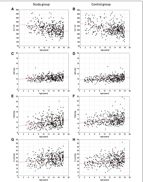
Baseline characteristics of the study groups were shown in Table 1. The study and control groups did not differ in terms of age and sex. In both groups age was nega-tively correlated with patient’s platelet counts (for study group: r =−0.30, p < 0.0001; for control group: r =−0.34, p < 0.0001), positively correlated with MPVs (r = 0.20, p < 0.0001; r = 0.26, p < 0.0001), PDW (r = 0.25, p < 0.0001 and r = 0.24, p < 0.0001) and with P-LCR (r = 0.26, p < 0.0001; r = 0.26, p < 0.0001) (Figure 1).
Comparison of the study and control group
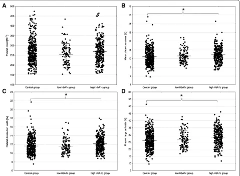
Mean platelet volume was significantly higher among chil-dren with diabetes in comparison with the control group (MPV 10.47+/−0.85 fL vs. 10.23+/−0.94 fL, p = 0.0007). Additionally, platelet distribution width was higher in the in study group than in controls (PDW 12.09+/−1.80% vs. 11.66+/−1.90%, p = 0.0032). This could be associated with greater population of the larger platelets in diabetic chil-dren, which were more numerous in the study group (PLC-R 28.21+/−6.15% vs. 26.29+/−6.38%, p < 0.0001). We did not find a significant difference in PLT between study and control group (263.55+/−60.04 103/uL vs. 268.77+/−65.78 103/uL; p = 0.5637) (Table 2). Although the differences became visible when the group with T1DM was divided into well and poorly controlled disease and then compared with control group (below 6.5% of Hb1Ac for good metabolic control according to American Diabetes Association standards 2014 [16]). Surprisingly PLT was the lowest in patients with good metabolic con-trol (255.08+/−53.93 103/uL), PLT was similar in poorly controlled patients and control group (269.84+/−60.08 103/uL vs 271.39+/−63.39 103/uL respectively; p ANOVA = 0.0563). In case of other platelet parameters (MPV, PDW, P-LCR) the lowest values were observed in the control group while the highest in the poorly controlled subgroup of patients with T1DM (Figure 2). This suggested that well-controlled diabetic patients have platelet morphology parameters similar to healthy children.
Influence of metabolic control on platelet morphology
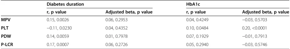
To evaluate the influence of HbA1c on platelet parameters we built multivariate regression models to adjust correla-tions for patient’s age, sex, diabetes duration and diabetes treatment type (Table 3). After adjustment for covariates, only PLT was shown to positively correlate with metabolic control of diabetes (B = 0.20; p < 0.0001). All platelet pa-rameters showed significant correlations with the duration of diabetes. However this effect was due to correlation of diabetes duration with patients’age.
Discussion
Our study showed that children with T1DM have ele-vated platelet volume, wider platelet size distribution width and increased percentage of large platelets. We also found that platelet count was significantly associated with metabolic control. The positive correlation between PLT and HbA1c remained significant after adjustment for con-founding factors.
Very little is known about correlation of platelet mor-phologic parameter with metabolic control of the T1DM in children. A study by Pirgon et al. focused on relation between MPV and HbA1c, but no significant correlation was found [17]. The authors found also that MPV was higher in the group of patients with T1DM in compari-son with healthy children, which is in agreement with the results of our study. Both studied populations were in similar age (7.9+/−4.2 years and control group was 7.4+/−3.2 years). The study however was conducted on limited number of patients (N = 56) and the results were not adjusted for patients’age and sex. In our study MPV in patients with diabetes was only slightly increased (10.47+/−0.85 fL vs. 10.23+/−0.94), but we cannot rule out that this might have clinical implications. Studies conducted on patients with T2DM found significant re-lationship between micro- and macrovascular complica-tions and MPV [18,19]. This had lead us to hypothesize that even a small increase of MPV might reflect a higher propensity for vascular complications in patients with dia-betes than healthy peers. In our study the highest MPV was observed in poorly controlled patients which supports abovementioned hypothesis.
Our recent ongoing study provided information that higher PLT among children with T1DM was connected with shorter time of thrombus formation (unpublished data). Taken into consideration that PLT was found to be independently associated with HbA1c we suppose that poorly controlled T1DM in children have increased plate-let activity.
The underlying cause of the observed differences may be related to several factors, most notably platelet function or their turnover. Diabetes mellitus is a prothrombotic state characterized by platelet hyperreactivity, hyperag-gregability, enhanced thrombinogenesis and reduced Table 1 Clinical characteristics of the studied and control
groups
Study group Control group p value
Boys/All (%) 222/389 (57.07%) 207/389 (53.21%) 0.2794
Age [years] 12.76+/−3.81 12.33+/−4.34 0.1467
Treatment type [CSII/all (%)]
231/389 (59.38%) --- NA
Diabetes duration [years]
5.03+/−355 --- NA
HbA1c [%]/[mmol/mol]
7.54+/−1.49/58.9 +/−11.64
--- NA
fibrinolysis regardless diabetes type [5]. Platelets in pa-tients with diabetes are more active than in healthy popu-lation. Even in the absence of vascular injuries, the platelets have greater expression of the glycoprotein IIb/ IIIa receptor, which is the final common pathway of plate-let activation. A recent study concluded that advanced gly-cation end products might be the source of altered platelet function [20]. Thus platelets in patients with dia-betes respond to even subthreshold stimuli which leads to faster exhaustion and consumption and, in turn, ends in
releasing fresh, hyperreactive platelets [21]. Therefore higher MPV, connected with platelet activity, is often con-sidered as a hallmark of impaired thrombopoiesis in dia-betes mellitus [9,22]. It has been shown that platelet population in patients with diabetes exhibits pronounced bimodality i.e. extreme dimensions with an increased number of very large and very small platelets [23]. This was reflected by higher PDW values observed in our patients. The platelet size is also connected with its life time – small platelets are regarded as older, having Table 2 Comparison of platelet parameters between the study groups
Study group Control group p p adjusted
for age and sex Mean+/−Std. Dev. Mean+/−Std. Dev.
MPV [fL] 10.47+/−0.85 10.23+/−0.94 0.0003 0.0007
PLT [103/uL] 263.55+/
−60.04 268.77+/−65.78 0.2482 0.5637
PDW [%] 12.09+/−1.80 11.66+/−1.90 0.0012 0.0032
P-LCR [%] 28.21+/−6.15 26.29+/−6.38 <0.0001 <0.0001
MPV–mean platelet volume; PLT–platelet count; PDW–platelet distribution width; P-LCR–platelet large cell ratio.
undergone many episodes of activation whereas large platelets are considered younger and more active [24]. Thus, bimodal distribution of platelet size may be indica-tive of increased platelet turnover. We observed the high-est proportion of large, most active platelets in poorly controlled patients with T1DM.
One could also hypothesise that the observed alter-ations are the result of primary disorder of platelet pre-cursors, megakaryocytes. Some authors have stated that peripheral platelets are activated in newly diagnosed or even prediabetic patients which could mean that every-thing is determined at the level of progenitor cells [25]. Many studies report that cytokines such as interleukin-3, 6 and 11 influence megakaryocyte ploidy and may lead to production of more reactive platelets [26,27]. This idea is supported by higher levels of proinflammatory IL-6 in children with T1DM than in healthy peers [28]. Moreover, megakaryocyte functions might be impaired in case of long lasting low glucose levels which are present in well controlled patients and that might result in reduced plate-let production. This hypothesis might explain why lower PLT counts were found in the subgroup of patients with low HbA1c levels.
The major clinical question is whether one should coun-teract increased platelet activity in paediatric patients with T1DM. Maybe good metabolic control in those patients is sufficient to prevent cardiovascular complications and no additional treatment is needed. However recent type re-ports do not support this hypothesis. In a metaanalysis performed on 2254 patients with 1 diabetes, intensive gly-cemic control did not affect all-cause mortality or cardio-vascular mortality [29]. Nevertheless, we still wait for final answer from DCCT/EDIC which even after 30 years of data collection does not provide a conclusive result [30].
It should be stated that our study has several limita-tions. Metabolic control was assessed by HbA1c, which corresponds to average glycaemia from the three preceding months. However, platelet lifetime is about 7 to 10 days, which may have affected the observed correlations. One could thus suggest to use a substance such as fructosamine with a shorter half-life time period instead. Unfortunately this marker was not routinely used in our Department throughout the study period. Although we excluded
patients with severe infections there is still a possibil-ity that minor infections could affect the platelet pa-rameters. Additionally patients from the control group underwent minor scheduled surgical procedures and the stress conditions connected with this situation might affect the platelet parameters. Another issue which should be taken into consideration in this age group is the matter of pubescence. We countered that by adjusting for pa-tients’ age and sex, but individual levels and potential impact of sex hormone levels on megakaryocytes and platelets are unknown to us.
We believe that metabolic control may be connected with platelet parameters and functions in patients with T1DM. The abovementioned platelet alterations might be associated with diabetes complications particularly because this group of patients is exposed for hypergly-caemia, oxidative stress and proinflammatory state from the early years. The results of our study support the the-ory that platelet function is altered even in young pa-tients with diabetes potentially contributing to increased cardiovascular risk later on.
Conclusions
Despite the abovementioned limitations to our knowledge our study is the firstly major report presenting the impact of T1DM on commonly measured platelet parameters in the paediatric population. Further studies are needed as it remains to be evaluated whether antiplatelet drugs may help to safeguard patients with T1DM against future car-diovascular diabetes complications.
Abbreviations
ANOVA:analysis of variance; Beta: partial correlation coefficient; CRP: C-reactive protein; CSII: continuous subcutaneous insulin infusion; DCCT/EDIC: Diabetes Control and Complications Trial/Epidemiology of Diabetes Interventions and Complications; HbA1c: glycated haemoglobin; MDI: multiple daily injections; MPV: mean platelet volume; NA: not applicable; PLT: platelet count; PDW: platelet distribution width; P-LCR: platelet large cell ratio.
Competing interest
The authors declare that they have no competing interest.
Authors’contributions
BM, BT, ABJ, OW and AS collected the data. BM and BT participated in study design and drafted the manuscript. BM performed data analysis. WF conceived of the study and together with WM participated in its design and
Table 3 Multivariate regression model results testing for the association of platelet parameters and metabolic control
Diabetes duration HbA1c
r, p value Adjusted beta, p value r, p value Adjusted beta, p value
MPV 0.15, 0.0026 0.06, 0.2953 0.04, 0.4249 −0.03, 0.5703
PLT −0.11, 0.0230 0.04, 0.4352 0.10, 0.0484 0.20, <0.0001
PDW 0.14, 0.0059 0.01, 0.7978 0.07, 0.1929 −0.01, 0.7913
P-LCR 0.17, 0.0007 0.06, 0.2726 0.05, 0.2940 −0.03, 0.5746
coordination and prepared the final version of the manuscript. All authors read and approved the final manuscript.
Funding
BM, ABJ, WM and WF received financial support from the TEAM project financed from the Innovative Economy Operational Program and coordinated by the Foundation for Polish Science. ABJ received financial support from Ministry of Science and Higher Education (DI 2011 022641).
Received: 1 September 2014 Accepted: 26 March 2015
References
1. Jarosz-Chobot P, Polanska J, Szadkowska A, Kretowski A, Bandurska-Stankiewicz E, Ciechanowska M, et al. Rapid increase in the incidence of type 1 diabetes in Polish children from 1989 to 2004, and predictions for 2010 to 2025. Diabetologia. 2011;54(3):508–15.
2. Nathan DM. Long-term complications of diabetes mellitus. N Engl J Med. 1993;328(23):1676–85.
3. Krishnan S, Short KR. Prevalence and significance of cardiometabolic risk factors in children with type 1 diabetes. J Cardiometab Syndr. 2009;4(1):50–6. 4. Colwell JA, Nesto RW. The platelet in diabetes: focus on prevention of
ischemic events. Diabetes Care. 2003;26(7):2181–8.
5. Watala C. Blood platelet reactivity and its pharmacological modulation in (people with) diabetes mellitus. Curr Pharm Des. 2005;11(18):2331–65. 6. Greisenegger S, Endler G, Hsieh K, Tentschert S, Mannhalter C, Lalouschek W. Is
elevated mean platelet volume associated with a worse outcome in patients with acute ischemic cerebrovascular events? Stroke. 2004;35(7):1688–91. 7. Hekimsoy Z, Payzin B, Ornek T, Kandogan G. Mean platelet volume in Type
2 diabetic patients. J Diabetes Complications. 2004;18(3):173–6. 8. Smith NM, Pathansali R, Bath PM. Platelets and stroke. Vasc Med.
1999;4(3):165–72.
9. Tschoepe D, Roesen P, Esser J, Schwippert B, Nieuwenhuis HK, Kehrel B, et al. Large platelets circulate in an activated state in diabetes mellitus. Semin Thromb Hemost. 1991;17(4):433–8.
10. Glassman AB. Platelet abnormalities in diabetes mellitus. Ann Clin Lab Sci. 1993;23(1):47–50.
11. Sharpe PC, Trinick T. Mean platelet volume in diabetes mellitus. Q J Med. 1993;86(11):739–42.
12. Lattanzio S, Santilli F, Liani R, Vazzana N, Ueland T, Di Fulvio P, et al. Circulating dickkopf-1 in diabetes mellitus: association with platelet activation and effects of improved metabolic control and low-dose aspirin. J Am Heart Assoc. 2014; doi: 10.1161/JAHA.114.001000. 13. Singer J, Weissler Snir A, Leshem-Lev D, Rigler M, Kornowski R, Lev EI. Effect
of intensive glycemic control on platelet reactivity in patients with long-standing uncontrolled diabetes. Thromb Res. 2014;134(1):121–4.
14. Wolfsdorf JI. The International Society of Pediatric and Adolescent Diabetes guidelines for management of diabetic ketoacidosis: do the guidelines need to be modified? Pediatr Diabetes. 2014;15(4):277–86.
15. Ly TT, Maahs DM, Rewers A, Dunger D, Oduwole A, Jones TW. Assessment and management of hypoglycemia in children and adolescents with diabetes. Pediatr Diabetes. 2014;15 Suppl 20:180–92.
16. Standards of medical care in diabetes-2014. Diabetes Care. 2014;37(Suppl 1):S14-80. 17. Pirgon O, Tanju IA, Erikci AA. Association of mean platelet volume between
glucose regulation in children with type 1 diabetes. J Trop Pediatr. 2009;55(1):63–4.
18. Unubol M, Ayhan M, Guney E. The relationship between mean platelet volume with microalbuminuria and glycemic control in patients with type II diabetes mellitus. Platelets. 2012;23(6):475–80.
19. Ulutas KT, Dokuyucu R, Sefil F, Yengil E, Sumbul AT, Rizaoglu H, et al. Evaluation of mean platelet volume in patients with type 2 diabetes mellitus and blood glucose regulation: a marker for atherosclerosis? Int J Clin Exp Med. 2014;7(4):955–61.
20. Rubenstein DA, Maria Z, Yin W. Combined incubation of platelets and endothelial cells with glycated albumin: Altered thrombogenic and inflammatory responses. Diab Vasc Dis Res. 2014;11(4):235–42. 21. Sobol AB, Watala C. The role of platelets in diabetes-related vascular
complications. Diabetes Res Clin Pract. 2000;50(1):1–16.
22. Tschope D, Langer E, Schauseil S, Rosen P, Kaufmann L, Gries FA. Increased platelet volume–sign of impaired thrombopoiesis in diabetes mellitus. Klin Wochenschr. 1989;67(4):253–9.
23. Watala C, Boncler M, Pietrucha T, Trojanowski Z. Possible mechanisms of the altered platelet volume distribution in type 2 diabetes: does increased platelet activation contribute to platelet size heterogeneity? Platelets. 1999;10(1):52–60.
24. Wong T, Pedvis L, Frojmovic M. Platelet size affects both micro- and macro-aggregation: contributions of platelet number, volume fraction and cell surface. Thromb Haemost. 1989;62(2):733–41.
25. Winocour PD. Platelet turnover in advanced diabetes. Eur J Clin Invest. 1994;24 Suppl 1:34–7.
26. Debili N, Masse JM, Katz A, Guichard J, Breton-Gorius J, Vainchenker W. Effects of the recombinant hematopoietic growth factors interleukin-3, interleukin-6, stem cell factor, and leukemia inhibitory factor on the megakaryocytic differentiation of CD34+ cells. Blood. 1993;82(1):84–95. 27. Brown AS, Hong Y, de Belder A, Beacon H, Beeso J, Sherwood R, et al.
Megakaryocyte ploidy and platelet changes in human diabetes and atherosclerosis. Arterioscler Thromb Vasc Biol. 1997;17(4):802–7. 28. Ryba-Stanislawowska M, Skrzypkowska M, Mysliwska J, Mysliwiec M. The
serum IL-6 profile and Treg/Th17 peripheral cell populations in patients with type 1 diabetes. Mediators Inflamm. 2013;2013:205284.
29. Kahler P, Grevstad B, Almdal T, Gluud C, Wetterslev J, Vaag A, et al. Targeting intensive versus conventional glycaemic control for type 1 diabetes mellitus: a systematic review with meta-analyses and trial sequential analyses of randomised clinical trials. BMJ Open. 2014;4(8):e004806. 30. Lachin JM, Orchard TJ, Nathan DM. Update on cardiovascular outcomes at
30 years of the diabetes control and complications trial/epidemiology of diabetes interventions and complications study. Diabetes Care. 2014;37(1):39–43.
Submit your next manuscript to BioMed Central and take full advantage of:
• Convenient online submission
• Thorough peer review
• No space constraints or color figure charges
• Immediate publication on acceptance
• Inclusion in PubMed, CAS, Scopus and Google Scholar
• Research which is freely available for redistribution